


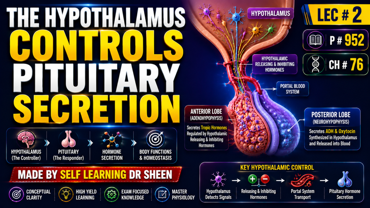
- Almost all pituitary hormone secretion is controlled by hormonal or nervous signals from the hypothalamus.
- If the pituitary is removed from its normal position beneath the hypothalamus and transplanted elsewhere, secretion of most pituitary hormones falls to very low levels, except prolactin.
- Posterior pituitary secretion is controlled by nerve signals coming from the hypothalamus and ending in the posterior pituitary.
- Anterior pituitary secretion is controlled by hypothalamic releasing and inhibitory hormones.
- These hypothalamic hormones travel to the anterior pituitary through tiny blood vessels called hypothalamic-hypophysial portal vessels (Fig. 76.4).
- In the anterior pituitary, these releasing and inhibitory hormones act on glandular cells to control hormone secretion.
- The hypothalamus receives information from many parts of the nervous system, so different body signals can change pituitary secretion.
- Pain signals can reach the hypothalamus and influence its activity.
- Strong depressing or exciting thoughts can also send signals to the hypothalamus.
- Smell-related signals, whether pleasant or unpleasant, can strongly affect the hypothalamus directly and through the amygdaloid nuclei.
- Changes in blood levels of nutrients, electrolytes, water, and hormones can also excite or inhibit different parts of the hypothalamus.
- Therefore, the hypothalamus acts as an integration center for information about the body’s internal condition and uses this information to control important pituitary hormone secretions.
KEY CONCEPT
Body signals → hypothalamus → controls pituitary secretion.
- Posterior pituitary: hypothalamus → nerve signals → hormone secretion.
- Anterior pituitary: hypothalamus → releasing/inhibitory hormones → portal vessels → hormone secretion.
CONCEPTUAL EXAMPLES
- Pain → hypothalamus → changes pituitary control.
- Blood nutrient/water changes → hypothalamus → adjusts pituitary secretion.
- Hypothalamus → portal vessels → anterior pituitary.
- Hypothalamus → nerve signals → posterior pituitary.
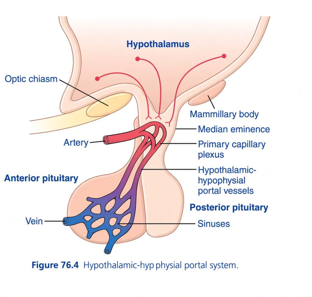
HYPOTHALAMIC-HYPOPHYSIAL PORTAL BLOOD VESSELS OF THE ANTERIOR PITUITARY GLAND
- The anterior pituitary has a rich blood supply with many capillary sinuses among its glandular cells.
- Almost all blood entering these sinuses first passes through a capillary bed in the lower hypothalamus.
- Blood then travels through the hypothalamic-hypophysial portal vessels to the anterior pituitary.
- The median eminence is the lowest part of the hypothalamus and connects with the pituitary stalk (Fig. 76.4).
- It acts as the functional link between the hypothalamus and anterior pituitary.
- Small arteries enter the median eminence, and small vessels return to its surface and join together to form the hypothalamic-hypophysial portal vessels.
- These vessels travel down the pituitary stalk and supply the anterior pituitary sinuses.
- Special hypothalamic neurons produce releasing and inhibitory hormones that control anterior pituitary secretion.
- Their nerve fibers extend to the median eminence and tuber cinereum.
- Unlike most CNS nerve endings, these endings mainly release hormones into tissue fluid rather than transmit signals to another neuron.
- The hormones immediately enter the portal blood system and are carried directly to the anterior pituitary sinuses.
- These hypothalamic hormones control the secretion of anterior pituitary hormones.
- For most anterior pituitary hormones, releasing hormones are the main controllers.
- For prolactin, a hypothalamic inhibitory hormone probably provides greater control.
- TRH → TSH release
- CRH → ACTH release
- GHRH → GH release
- GHIH (somatostatin) → GH inhibition
- GnRH → LH + FSH release
- PIH (dopamine) → prolactin inhibition (Table 76.2)
- The hypothalamus also has a hormone that stimulates prolactin secretion and possibly other hormones that inhibit anterior pituitary hormones.
- Most hypothalamic hormones are released from nerve endings in the median eminence before reaching the anterior pituitary.
- Electrical stimulation of the median eminence can excite these nerve endings and cause release of essentially all hypothalamic hormones.
- However, the neuron cell bodies producing these nerve endings are located in specific areas of the hypothalamus or closely related basal brain areas.
KEY CONCEPT
Hypothalamus → releasing/inhibitory hormones → median eminence → portal blood vessels → anterior pituitary → controlled hormone secretion.
CONCEPTUAL EXAMPLES
- TRH → anterior pituitary → TSH
- CRH → anterior pituitary → ACTH
- GHRH → GH ↑; somatostatin → GH ↓
- GnRH → LH + FSH
- Dopamine (PIH) → prolactin ↓
PHYSIOLOGICAL FUNCTIONS OF GROWTH HORMONE
- Most anterior pituitary hormones, except growth hormone (GH), mainly act by stimulating target glands such as the thyroid, adrenal cortex, ovaries, testes, and mammary glands.
- GH is different because it acts directly on almost all tissues of the body.
GROWTH HORMONE PROMOTES GROWTH OF MANY BODY TISSUES
- GH is also called somatotropic hormone or somatotropin.
- It is a small protein containing 191 amino acids in one chain, with a molecular weight of 22,005.
- GH promotes growth in almost all tissues capable of growing.
- It increases cell size and mitosis, producing more cells and promoting differentiation of certain cells, including bone growth cells and early muscle cells.
- In rats given daily GH injections, growth was markedly greater than in rats not given GH, both during early life and even after adulthood (Fig. 76.5).
- During early development, GH increased the size of all organs proportionately.
- After adulthood, most bones stop lengthening, but many soft tissues can continue growing.
- Once the epiphyses of long bones unite with their shafts, further lengthening of the bones cannot occur, although other body tissues can continue to grow throughout life.
KEY CONCEPT
GH → ↑ cell size + ↑ mitosis → ↑ number of cells + tissue growth.
CONCEPTUAL EXAMPLES
- GH during growth → bones and many tissues grow.
- After epiphyseal fusion → long bones cannot lengthen further.
- After adulthood → many soft tissues can still grow.
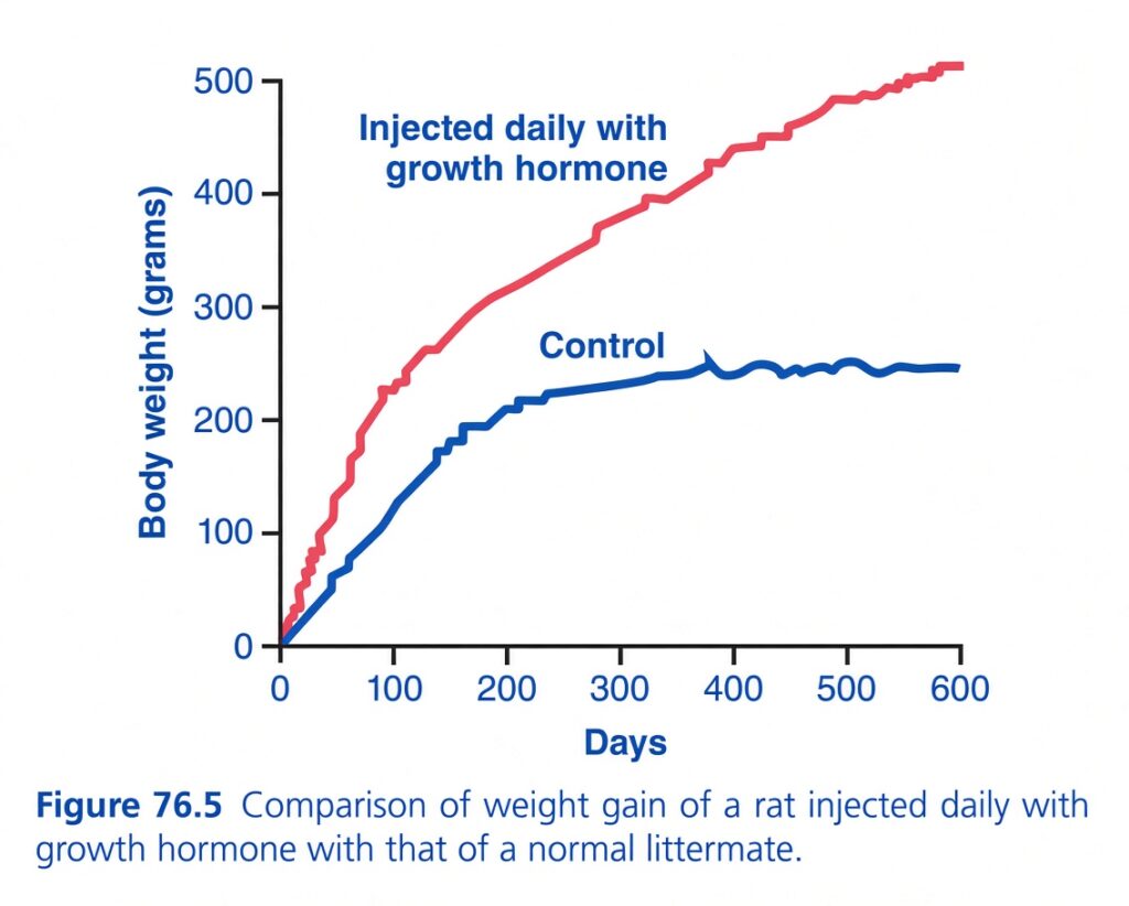
Growth Hormone & Rat Body Weight Graph
🎯 BIG IDEA
The graph compares two rats over ~600 days:
🔴 Red = rat injected daily with growth hormone (GH)
🔵 Blue = normal control rat
Result:
👉 Daily GH makes the rat much larger/heavier.
📊 Understand the axes
➡️ X-axis = Days
Shows how much time has passed.
0 → 100 → 200 → 300 → 400 → 500 → 600 days
⬆️ Y-axis = Body weight (grams)
0 → 100 → 200 → 300 → 400 → 500 g
So we’re asking:
“As the rat gets older, how does its body weight change?”
🔴 RED LINE = Daily Growth Hormone
At the beginning:
~0 g
Then GH is injected every day.
The rat’s weight rises rapidly:
- Around 100 days → ~230 g
- Around 200 days → ~310 g
- Around 300 days → ~380 g
- Around 400 days → ~430 g
- Around 600 days → ~510 g
🔥 The rat keeps gaining much more weight.
🔵 BLUE LINE = Control / Normal Rat
This rat does NOT receive extra GH.
Its weight increases naturally:
- Around 100 days → ~120 g
- Around 200 days → ~210 g
- Around 300 days → ~230 g
- Around 400 days → ~245 g
- Around 600 days → ~245 g
Eventually, its weight levels off.
🧠 WHY DO THE LINES BEHAVE DIFFERENTLY?
🔴 GH-treated rat
GH ↑
↓
Protein synthesis & growth ↑
↓
Growth of tissues/body ↑
↓
Body weight ↑↑
🔵 Normal rat
Normal GH levels → normal growth → eventually reaches its adult body size.
⭐ MOST IMPORTANT THING IN THE GRAPH
Notice:
🔵 Control
Weight rises → plateaus around 240–250 g
🔴 GH
Weight rises → continues increasing → ~500+ g
So:
Extra growth hormone keeps promoting growth and increases body weight far beyond the normal adult level.
⚠️ Don’t make this mistake
Body weight ≠ only muscle.
GH promotes growth of multiple tissues and also influences protein, fat, and metabolism.
So the graph simply demonstrates the overall growth-promoting effect of GH, not that every gram gained is muscle.
🧠 10-SECOND MEMORY TRICK
🔵 Normal GH → normal growth → plateau
🔴 Extra GH every day → enhanced growth → much higher body weight
🔥 One-line exam concept:
Chronic growth hormone administration markedly increases body growth and body weight compared with a normal control animal.
GROWTH HORMONE HAS SEVERAL METABOLIC EFFECTS
- Besides promoting growth, GH has important metabolic effects (Fig. 76.6).
- GH:
- ↑ protein synthesis in most cells.
- ↑ fatty-acid release and use for energy.
- ↓ glucose utilization throughout the body.
- Overall, GH increases body protein, decreases fat stores, and conserves carbohydrates.
- GH promotes protein deposition through several actions:
- ↑ amino-acid entry into cells → more amino acids available for protein synthesis.
- ↑ RNA translation at ribosomes → more protein is produced, even without increased amino-acid concentration.
- Over 24–48 hours, GH ↑ DNA transcription → ↑ RNA → ↑ protein synthesis; this may be its most important long-term action.
- GH also ↓ protein and amino-acid breakdown, partly because fatty acids are used for energy and therefore spare protein.
- Overall, GH ↑ amino-acid uptake and protein synthesis while ↓ protein breakdown.
- GH enhances fat utilization for energy by:
- Releasing fatty acids from adipose tissue → ↑ fatty acids in body fluids.
- Increasing conversion of fatty acids to acetyl-CoA and their use for energy.
- Therefore, GH causes the body to use fat preferentially over carbohydrates and proteins.
- Fat utilization takes several hours, whereas increased protein synthesis can begin within minutes.
- Increased fat use together with increased protein building produces greater lean body mass.
- Excess GH can have a ketogenic effect:
- Excessive fat mobilization → liver forms large amounts of acetoacetic acid → ketosis.
- Excessive fat mobilization can also cause fatty liver.
- GH decreases carbohydrate utilization by:
- ↓ glucose uptake by skeletal muscle and adipose tissue.
- ↑ glucose production by the liver.
- ↑ insulin secretion.
- These effects occur because GH causes insulin resistance, reducing insulin’s ability to promote glucose uptake and use and to inhibit glucose production by the liver.
- The result is ↑ blood glucose → compensatory ↑ insulin secretion.
- Therefore, GH has diabetogenic effects, and excessive GH can produce metabolic changes similar to type 2 diabetes.
- Patients with acromegaly from excess GH are usually lean with little visceral fat, whereas patients with type 2 diabetes are often overweight with excessive visceral fat.
- Increased lipolysis and blood fatty acids, along with fat accumulation in the liver, can reduce the sensitivity of the liver and skeletal muscle to insulin.
Necessity of Insulin and Carbohydrate for the Growth-Promoting Action of Growth Hormone
- GH cannot produce normal growth in animals without a pancreas.
- GH also cannot produce growth when carbohydrates are excluded from the diet.
- Therefore, effective GH action requires adequate insulin and adequate carbohydrate availability.
- Insulin and carbohydrates provide energy needed for growth and also support other growth processes.
- Insulin is especially important because it helps transport some amino acids into cells, similar to its effect on glucose transport.
KEY CONCEPT
GH → ↑ protein synthesis + ↓ protein breakdown + ↑ fat use + ↓ glucose use → growth, more lean tissue, less fat, and carbohydrate conservation.
CONCEPTUAL EXAMPLES
- GH + amino acids → ↑ protein synthesis → ↑ tissue growth.
- GH → ↑ lipolysis → fatty acids used for energy → protein is spared.
- GH → insulin resistance → ↓ glucose utilization → ↑ blood glucose → ↑ insulin.
- Excess GH → excessive fat mobilization → ketosis + fatty liver.
- GH + adequate insulin + carbohydrates → effective growth.
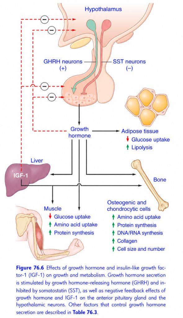
This Guyton Figure 76.6 is basically the complete story of Growth Hormone (GH): who controls it, where it acts, and how it causes growth + metabolic effects.
🧠 BIG IDEA — UNDERSTAND THE WHOLE FIGURE FIRST
Think of GH as having 2 major jobs:
🏗️ 1. Make the body GROW
Mainly through IGF-1
🔥 2. Change METABOLISM
Especially:
⭐ THE WHOLE PATHWAY IN ONE LINE
Hypothalamus → GHRH → Pituitary → GH → Liver → IGF-1 → Growth
But there is another hormone from the hypothalamus:
Somatostatin (SST) ⛔ → inhibits GH
And finally:
GH + IGF-1 ⛔ → negative feedback
1️⃣ HYPOTHALAMUS = THE CONTROL CENTER 🧠
At the top is the:
Hypothalamus
It controls GH secretion from the anterior pituitary.
It sends TWO opposite signals:
🟢 GHRH = GO
GHRH = Growth Hormone-Releasing Hormone
It stimulates GH secretion.
Think:
GHRH says: “Pituitary, release GH!”
🔴 SST = STOP
SST = Somatostatin
It inhibits GH secretion.
Think:
Somatostatin says: “STOP releasing GH!”
Easy memory:
GHRH = GO
SST = STOP
2️⃣ ANTERIOR PITUITARY = GH FACTORY 🏭
GHRH stimulates cells of the anterior pituitary.
⬇️
They release:
Growth Hormone (GH)
So:
GHRH → ↑ GH
while:
SST → ↓ GH
3️⃣ GH NOW TRAVELS THROUGH THE BLOOD 🩸
GH leaves the pituitary and acts on several tissues.
Look at the figure.
GH goes toward:
- Liver
- Muscle
- Adipose tissue
- Bone
And the effects are different in each.
4️⃣ LIVER = IGF-1 FACTORY 🏭
One of the most important arrows:
GH → Liver
The liver responds by producing:
IGF-1
IGF-1 = Insulin-like Growth Factor-1
Think:
GH tells the liver: “Make IGF-1.”
Then:
IGF-1 → growth-promoting effects
This is why IGF-1 is a major mediator of GH’s growth-promoting effects.
🦴 5️⃣ BONE = GROWTH
Look at the right side.
GH/IGF-1 act on:
Osteogenic and chondrogenic cells
These are cells involved in bone and cartilage growth.
The figure shows:
⬆️ Amino acid uptake
⬆️ Protein synthesis
⬆️ DNA/RNA synthesis
⬆️ Collagen
⬆️ Cell size
⬆️ Cell number
So the overall result is:
🦴 MORE GROWTH
🧠 EASY WAY TO UNDERSTAND BONE EFFECTS
GH/IGF-1 basically tell growth-related cells:
“Take in building materials → make proteins → make DNA/RNA → make collagen → grow bigger → make more cells.”
Therefore:
GH/IGF-1 = BUILD + DIVIDE + GROW
💪 6️⃣ MUSCLE = BUILD PROTEIN
Look at the muscle section.
GH causes:
🔴 ↓ Glucose uptake
🟢 ↑ Amino acid uptake
🟢 ↑ Protein synthesis
The most important concept:
GH encourages muscle to use amino acids for protein building.
So:
Amino acids ↑ → protein synthesis ↑ → muscle growth
🍞 WHY DOES GLUCOSE UPTAKE DECREASE?
This is a very important metabolic effect of GH.
GH tends to make muscle use less glucose.
So:
GH → ↓ glucose uptake by muscle
This helps shift energy use toward fat.🧈 7️⃣ ADIPOSE TISSUE = BURN FAT
Look at the adipose tissue on the upper right.
GH causes:
🔴 ↓ Glucose uptake
🟢 ↑ Lipolysis
Lipolysis = breakdown of stored fat
So:
GH → fat breakdown ↑
Think:
GH tells fat cells: “Release your stored fat!”
This provides fatty acids that can be used as an energy source.
🔥 VERY IMPORTANT METABOLIC PATTERN
GH generally:
↓ Glucose use
and
↑ Fat use
and
↑ Protein synthesis
So remember:
GH = FAT-BURNING + PROTEIN-BUILDING + GLUCOSE-SAVING
8️⃣ NEGATIVE FEEDBACK ⛔
Now look at the red dashed lines with minus signs.
These are extremely important.
They mean:
Negative feedback
As GH and IGF-1 increase, they send signals back to the:
- Anterior pituitary
- Hypothalamus
to reduce further GH secretion.
Think:
“We have enough GH/IGF-1. Stop making so much!”
So:
GH ↑ → feedback inhibition ⛔
IGF-1 ↑ → feedback inhibition ⛔
🔄 WHY IS NEGATIVE FEEDBACK NEEDED?
Without feedback, GH could keep increasing.
The body wants balance.
So:
GH rises → effects occur → GH/IGF-1 rise → brain/pituitary are told to reduce GH → GH falls
This keeps the system controlled.
🧩 NOW DECODE EVERY ARROW
🧠 TOP
Hypothalamus
⬇️
GHRH (+) → stimulates pituitary
SST (−) → inhibits pituitary
🏭 PITUITARY
GHRH stimulates:
GH secretion
🩸 GH GOES TO TISSUES
GH → Liver
⬇️
IGF-1
GH → Muscle
⬇️
↓ glucose uptake
↑ amino acid uptake
↑ protein synthesis
GH → Adipose tissue
⬇️
↓ glucose uptake
↑ lipolysis
GH/IGF-1 → Bone/cartilage cells
⬇️
↑ amino acid uptake
↑ protein synthesis
↑ DNA/RNA synthesis
↑ collagen
↑ cell size
↑ cell number🎯 THE MOST IMPORTANT CONCEPT: GH vs IGF-1
This is where many students get confused.
GH is the hormone released by the pituitary.
IGF-1 is produced mainly by the liver in response to GH.
Think:
GH = COMMANDER
IGF-1 = MAJOR GROWTH EXECUTOR
GH gives the growth signal, and IGF-1 mediates many of the actual growth-promoting effects.
🏆 SUPER-SIMPLE STORY
Imagine your body is a construction company.
Hypothalamus = CEO 🧠
It decides whether to increase or decrease GH.
GHRH = “START construction!” 🟢
Somatostatin = “STOP construction!” 🔴
Pituitary = GH factory 🏭
It releases GH.
GH = Construction manager 👷
It tells tissues what to do.
Liver = IGF-1 factory 🏭
GH tells liver:
“Make IGF-1.”
IGF-1 = Growth worker 🧑🔧
It helps tissues grow.
Amino acids = bricks 🧱
Protein synthesis = building 🏗️
DNA/RNA synthesis = making more workers 📋
Cell size + number ↑ = Bigger tissue 📈
🚨 HIGH-YIELD EXAM TRAPS
❌ GHRH inhibits GH
✅ GHRH stimulates GHomatostatin stimulates GH
✅ Somatostatin inhibits GH
❌ GH only causes growth
✅ GH has growth + metabolic effects
❌ GH increases glucose uptake in muscle
✅ GH decreases glucose uptakeGH decreases fat breakdown
✅ GH increases lipolysis GH decreases protein synthesis
✅ GH increases protein synthesis
❌ IGF-1 stimulates GH release
✅ IGF-1 participates in negative feedback
🧠 10-SECOND MEMORY MAP
GHRH → GH → IGF-1 → GROWTH
And:
SST → ↓ GH
GH also causes:
Muscle
↓ glucose uptake + ↑ amino acid uptake + ↑ protein synthesis
Fat
↓ glucose uptake + ↑ lipolysis
Bone
↑ amino acids + ↑ protein + ↑ DNA/RNA + ↑ collagen + ↑ cell size + ↑ cell number
Finally:
GH + IGF-1 → negative feedback ⛔
🔑 THE ONE SENTENCE TO MEMORIZE
“GHRH turns GH ON, somatostatin turns GH OFF; GH promotes protein building and fat breakdown, stimulates the liver to make IGF-1, and GH/IGF-1 promote growth while providing negative feedback.”
GROWTH HORMONE STIMULATES CARTILAGE AND BONE GROWTH
- GH promotes protein deposition and growth in almost all tissues, but its most obvious effect is growth of the skeletal frame.
- GH increases bone growth by:
- ↑ protein deposition in chondrocytes and osteogenic cells.
- ↑ reproduction of these cells.
- Converting chondrocytes into osteogenic cells, which helps form new bone.
- Long bones grow in length at the epiphyseal cartilage.
- GH first promotes new cartilage formation, which is then converted into new bone.
- This elongates the bone shaft and pushes the epiphyses farther apart.
- During late adolescence, the epiphyseal cartilage is progressively used up.
- When it disappears, the shaft and epiphysis fuse, so further lengthening of the long bones cannot occur.
- Bones can also increase in thickness:
- Osteoblasts deposit new bone on the surfaces of older bone.
- Osteoclasts remove old bone.
- When deposition > resorption, bone thickness increases.
- GH strongly stimulates osteoblasts, so bones can continue becoming thicker throughout life, especially membranous bones.
- For example, GH can stimulate jaw growth after adolescence, causing forward protrusion of the chin and lower teeth.
- Skull bones can also become thicker, producing bony protrusions over the eyes.
GROWTH HORMONE RECEPTOR SIGNALING
- The GH receptor (GHR) is a cytokine receptor found in almost all tissues, with especially high levels in the liver, muscle, fat, and kidneys.
- GHR has:
- An extracellular GH-binding domain
- One membrane-spanning domain
- An intracellular domain (Fig. 76.7)
- The intracellular domain contains JAK2, a tyrosine kinase of the Janus kinase (JAK) family and the main signal transducer for GH.
- GH binds GHR → ↑ JAK2 activity → protein phosphorylation → STAT phosphorylation.
- Phosphorylated STAT proteins enter the nucleus, bind DNA, and produce GH-specific gene expression.
- GHR/JAK2 activation also recruits other signaling pathways involving IRS proteins and Shc adapter proteins.
- These pathways work together to produce the complex effects of GH in target cells.
- GH signaling can also be reduced by SOCS proteins, which disrupt the JAK-STAT pathway and cause resistance to GH.
- In kidney failure or critical illness, inflammatory cytokines can increase SOCS proteins and therefore impair GH signaling.
KEY CONCEPT
GH → GHR → JAK2 → STAT phosphorylation → STAT enters nucleus → DNA/gene expression → GH effects.
CONCEPTUAL EXAMPLES
- GH → epiphyseal cartilage → new cartilage → new bone → ↑ long-bone length.
- Epiphyseal cartilage disappears → bone fusion → no further long-bone lengthening.
- GH → osteoblasts → ↑ bone deposition → ↑ bone thickness.
- GH → GHR/JAK2/STAT → gene expression → cellular response.
- ↑ SOCS → ↓ JAK-STAT signaling → GH resistance.
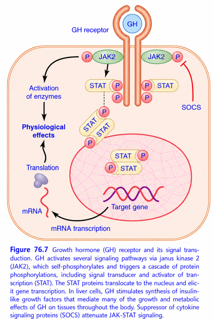
This figure looks complicated, but the main story is actually very simple:
🧠 BIG IDEA
This figure explains HOW Growth Hormone (GH) talks to a cell and changes what the cell does.
The entire pathway:
GH → GH receptor → JAK2 → STAT → nucleus → gene transcription → mRNA → protein → physiological effect
And the cell has a brake:
SOCS ⛔ → inhibits JAK-STAT signaling
🚀 FIRST: THE 10-SECOND VERSION
Think of GH as a boss giving instructions.
GH = boss 📢
↓
GH receptor = receives the order 📞
↓
JAK2 = activates the message ⚡
↓
STAT = carries the message to DNA 📄
↓
Nucleus = reads the instruction 🧠
↓
mRNA = copies the instruction ✍️
↓
Protein = gets manufactured 🏭
↓
Physiological effect = cell does something
And:
SOCS = STOP/BRAKE
1️⃣ GH BINDS TO GH RECEPTOR
At the top:
GH
binds to:
GH receptor
The receptor is located in the cell membrane.
Important:
GH does not need to enter the cell.
It gives its message from outside.
2️⃣ JAK2 = THE SIGNALING SWITCH ⚡
On the inside of the GH receptor are proteins called:
JAK2
JAK2 = Janus kinase 2
When GH binds its receptor, JAK2 becomes activated.
Then JAK2 adds phosphate groups to proteins.
This is called:
Phosphorylation
In the figure, the red circles marked P represent phosphate groups.
3️⃣ JAK2 ACTIVATES STAT
Now look at:
STAT
STAT = Signal Transducer and Activator of Transcription
Don’t worry about the long name.
Think:
STAT = message carrier that ultimately tells DNA what to do.
JAK2 phosphorylates STAT.
So:
JAK2 → STAT phosphorylation STAT PAIRS UP
The phosphorylated STAT proteins come together as a pair.
This is called:
STAT dimerization
You don’t necessarily need to obsess over the word, but understand the concept:
Two activated STAT molecules join together.
Now they are ready to enter the nucleus.
5️⃣ STAT ENTERS THE NUCLEUS 🧠
The STAT pair moves into the:
Nucleus
Why?
Because the nucleus contains:
DNA / genes
STAT interacts with target genes.
Think:
STAT is carrying GH’s instruction to the cell’s instruction book (DNA).
6️⃣ TARGET GENE IS ACTIVATED 🧬
Inside the nucleus:
STAT → target gene
The target gene is switched on.
This causes:
mRNA transcription
In simple language:
The DNA instruction is copied into mRNA.
7️⃣ mRNA LEAVES THE NUCLEUS 📄
The newly produced:
mRNA
moves into the cytoplasm.
Think:
DNA = original recipe book
mRNA = photocopy of one recipe
The photocopy can now be taken to the protein-making machinery.
8️⃣ TRANSLATION = MAKE THE PROTEIN 🏭
The figure shows:
Translation
Translation uses the mRNA instructions to make:
Protein
So:
mRNA → protein
That protein can then produce the:
Physiological effects of GH
🎯 THE COMPLETE CHAIN
Memorize this:
GH
↓
GH receptor
↓
JAK2
↓
STAT phosphorylation
↓
STAT dimer
↓
Nucleus
↓
Target gene
↓
mRNA
↓
Translation
↓
Protein
↓
Physiological effect
That’s basically every major arrow in this figure.
🧠 WHAT DOES “PHOSPHORYLATION” MEAN HERE?
You’ll see many red:
P
These represent phosphate groups.
JAK2 adds phosphate groups to proteins.
This changes their activity.
So:
JAK2 = phosphate-adding activator
Very simple.
🛑 NOW THE SOCS PART
Look at the right side:
SOCS
SOCS stands for:
Suppressor of Cytokine Signaling
Its job is to reduce JAK-STAT signaling.
Think:
JAK2 = accelerator 🚗💨
SOCS = brake 🛑
When the signal becomes strong enough, SOCS helps prevent excessive signaling.
So:
SOCS → ↓ JAK-STAT signaling
🔄 WHY DOES THE CELL NEED SOCS?
Because the cell doesn’t want GH signaling to remain permanently ON.
Without a brake:
GH → JAK2 → STAT → genes → proteins
could continue excessively.
SOCS helps shut the pathway down.
That’s another example of negative regulation/feedback.
🧩 WHAT DOES THE LEFT SIDE OF THE FIGURE MEAN?
You see:
Activation of enzymes
and:
Physiological effects
The figure is showing that GH signaling doesn’t only work through gene transcription.
GH can activate several signaling pathways, producing different effects.
So don’t think:
“GH only activates STAT.”
Instead:
🏭 WHY DOES GH AFFECT THE LIVER?
The caption gives an important point:
In liver cells:
GH → stimulates synthesis of IGF-1
IGF-1 then mediates many of GH’s:
- growth effects
- metabolic effects
So connect this figure to your previous Figure 76.6:
Previous figure:
GH → Liver → IGF-1 → Growth
This figure explains HOW GH produces intracellular signaling that can lead to those effects.
🔗 CONNECT FIGURE 76.6 + 76.7
This is extremely useful.
Figure 76.6 asked:
“WHAT does GH do?”
Answer:
Growth + metabolic effectsigure 76.7 asks:
“HOW does GH signal inside the cell?”
Answer:
GH → receptor → JAK2 → STAT → genes → proteins → effects
So:
76.6 = EFFECTS
76.7 = MECHANISM
🧠 ELI5 STORY
Imagine a school.
GH = Principal 📢
Principal gives an instruction.
GH receptor = Reception desk 📞
Receives the instruction.
JAK2 = Messenger coordinator ⚡
Activates the message.
STAT = Messenger 🏃
Carries the instruction into the office.
Nucleus = Principal’s instruction room 🧠
Contains the DNA.
Target gene = Specific instruction 📖
STAT activates it.
mRNA = Photocopy 📄
Copies the instruction.
Translation = Factory 🏭
Uses the photocopy to make a protein.
Protein = Worker 👷
Performs the actual job.
Physiological effect = Result 🎯
The cell changes its behavior.
SOCS = Security guard 🛑
Says:
“Enough! Stop the signal.”
🚨 HIGH-YIELD EXAM TRAPS
❌ GH enters the nucleus
✅ GH binds the membrane receptor.
❌ JAK2 enters the nucleus
✅ STAT enters the nucleus.
❌ STAT directly makes protein
✅ STAT activates target gene transcription → mRNA → protein.
❌ mRNA is translated into DNA
✅ mRNA is translated into protein.
❌ SOCS stimulates JAK2
✅ SOCS suppresses JAK-STAT signaling.❌ GH receptor itself is an enzyme
✅ The receptor associates with JAK2, which provides kinase activity.
🏆 ULTRA-SHORT REVISION
GH → Receptor → JAK2 → STAT → Nucleus → Gene → mRNA → Protein → Effect
And:
SOCS = brake
🔑 Golden sentence:
“GH binds its receptor, activates JAK2, JAK2 phosphorylates STAT, STAT enters the nucleus and activates target genes, producing mRNA and proteins that create GH’s physiological effects; SOCS acts as the brake.”
