- Gangliosidoses are lysosomal storage diseases in which gangliosides accumulate, mainly in the brain.
- They occur because of deficiency of lysosomal enzymes needed to break down these glycolipids.
- Depending on the stored ganglioside, they are divided into:
- GM1 gangliosidoses
- GM2 gangliosidoses
- Tay-Sachs disease is the most common gangliosidosis.
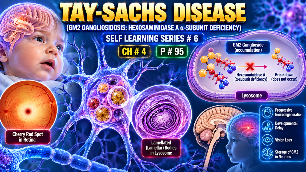
- It is caused by loss-of-function mutations affecting the α-subunit of hexosaminidase A.
- Hexosaminidase A is required to degrade GM2 ganglioside.
- Therefore: ↓ Hexosaminidase A → failure of GM2 degradation → GM2 accumulation, especially in the brain
- More than 100 mutations have been identified.
- Most mutations interfere with:
- proper protein folding
- normal intracellular transport
- Because of a founder effect, Tay-Sachs disease is more common in people of Ashkenazi Jewish ancestry.
- In this population, the estimated carrier frequency is about 1 in 30.
- Ashkenazi Jews originated mainly from Eastern and Central Europe and form most of the Jewish population in the United States.
- Heterozygous carriers can be detected by:
- measuring hexosaminidase activity in serum
- DNA sequencing
KEY CONCEPT
- Tay-Sachs = Hexosaminidase A α-subunit defect → GM2 ganglioside cannot be degraded → GM2 accumulates mainly in the brain.
- Increased prevalence in Ashkenazi Jewish populations.
- Carrier detection → enzyme assay or DNA sequencing.
CONCEPTUAL EXAMPLES
- Hexosaminidase A is defective → lysosome cannot remove GM2 → GM2 progressively accumulates.
- GM2 accumulates mainly in neural tissue → brain is the major affected site.
- Healthy person with one mutant allele → may be identified as a carrier by enzyme testing or DNA analysis.
Pathogenesis
- In Tay-Sachs disease, absence of hexosaminidase A → GM2 ganglioside accumulates in many tissues.
- Although GM2 may accumulate in the heart, liver, spleen, and nervous system, the main clinical effects come from involvement of:
- CNS neurons
- autonomic neurons
- retina
- GM2 accumulates within:
- neurons
- axons
- glial cells
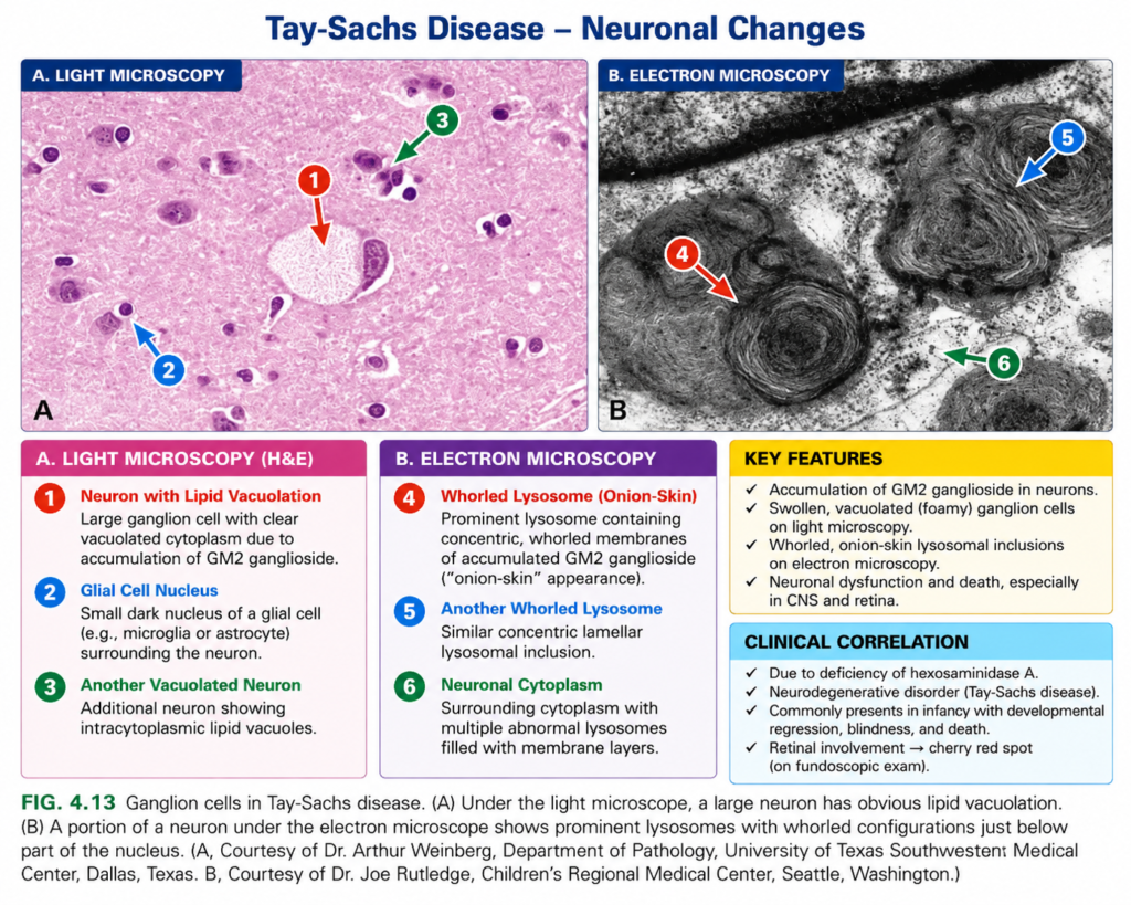
- Affected cells become swollen and sometimes foamy (Fig. 4.13A).
- Electron microscopy shows onion-skin–like whorled membranes inside lysosomes (Fig. 4.13B).
- These changes occur throughout:
- brain and spinal cord
- peripheral nerves
- autonomic nervous system
- retina
- In the retina:
- surrounding ganglion cells become swollen and pale
- the relatively unaffected central macula appears as a contrasting cherry-red spot.
- The exact mechanism of neuronal injury is not fully known.
- Many mutant proteins are misfolded → activate the unfolded protein response.
- If chaperones cannot stabilize the abnormal enzyme:
misfolded protein → proteasomal degradation → less enzyme activity → toxic substrate accumulation in neurons. - This has led to trials of molecular chaperone therapy:
- synthetic chaperones enter the brain
- bind mutant protein
- improve folding
- may restore enough enzyme activity to improve cell function
- In the common acute infantile form:
- 3–6 months → motor weakness begins
- then → progressive neurologic impairment
- blindness develops
- neurologic dysfunction becomes increasingly severe
- death usually occurs by 2–3 years
Niemann-Pick Disease Types A and B
- Types A and B are caused by deficiency of acid sphingomyelinase.
- This enzyme normally breaks down:
Sphingomyelin → ceramide + phosphorylcholine
- Enzyme deficiency → sphingomyelin accumulation.
- These disorders are more common in individuals of Ashkenazi Jewish ancestry.
- The acid sphingomyelinase gene is preferentially expressed from the maternal chromosome because the paternal gene is epigenetically silenced.
- Type A:
- severe sphingomyelinase deficiency
- sphingomyelin accumulates in macrophages and neurons
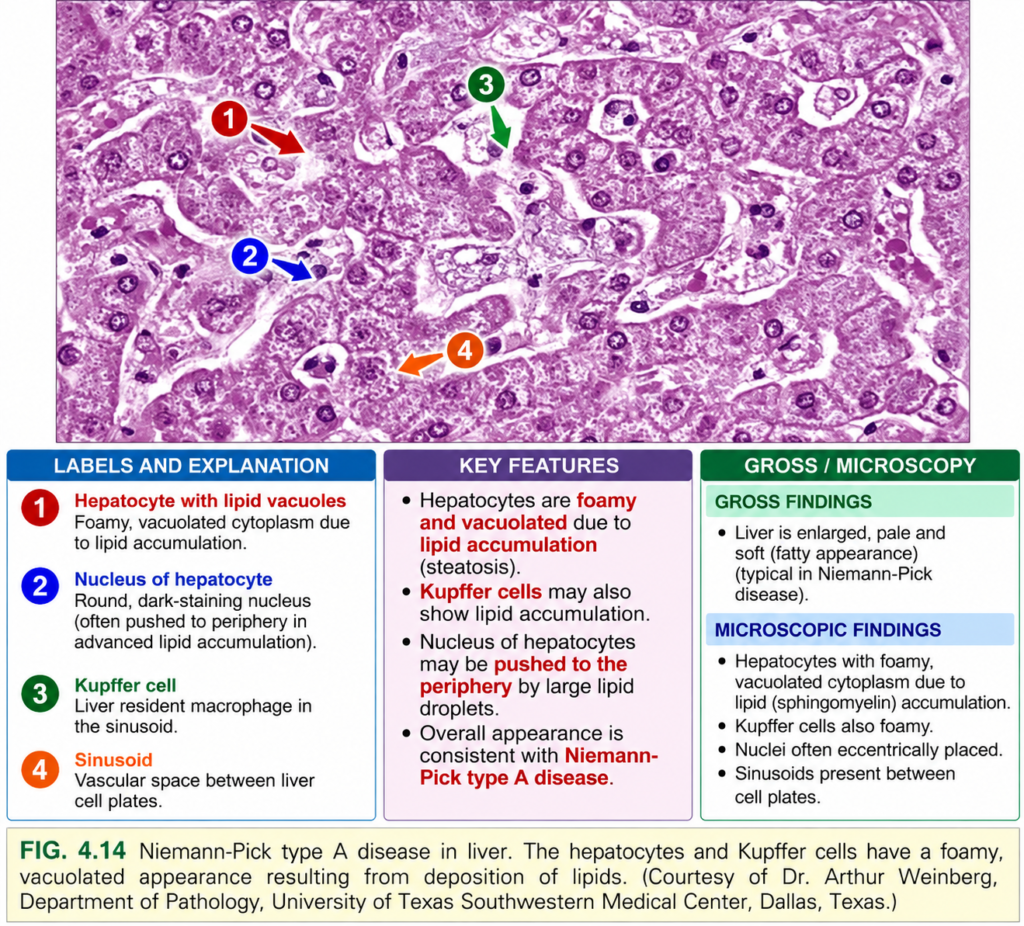
- macrophages become filled with lipid → foamy cytoplasm (Fig. 4.14)
- electron microscopy shows lysosomes with concentric membranous structures called “zebra bodies”
- Organs most affected because of abundant phagocytic cells:
- spleen
- liver
- bone marrow
- lymph nodes
- lungs
- Splenomegaly may be marked.
- CNS involvement is also severe:
- neurons become enlarged and vacuolated from lipid accumulation
- disease presents in infancy with:
- massive organomegaly
- severe neurologic deterioration
- death usually occurs within the first 3 years
- Type B:
- mutant sphingomyelinase retains some activity
- causes organomegaly
- usually no neurologic manifestations
- Diagnosis can be made by:
- measuring sphingomyelinase activity in leukocytes
- molecular genetic testing
KEY CONCEPT
- Tay-Sachs → hexosaminidase A deficiency → GM2 accumulation, especially in neurons → neurodegeneration + cherry-red spot.
- Niemann-Pick A/B → acid sphingomyelinase deficiency → sphingomyelin accumulation.
- Type A → severe enzyme loss → organomegaly + severe CNS disease.
- Type B → residual enzyme activity → organomegaly with little/no neurologic disease.
CONCEPTUAL EXAMPLES
- Hexosaminidase A absent → GM2 fills neurons → cells swell → progressive neurologic decline in Tay-Sachs.
- Retina becomes pale around the macula → central macula stands out as a cherry-red spot.
- Severe sphingomyelinase deficiency → lipid-filled macrophages + neuronal storage → Niemann-Pick type A.
- Partial sphingomyelinase activity → storage mainly in organs without major CNS damage → Niemann-Pick type B.