


1. LEARNING OBJECTIVES
At the end of this topic, students should be able to:
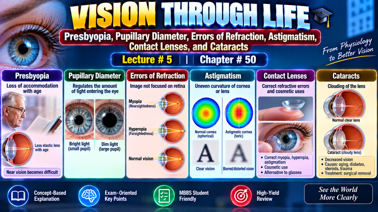
- Explain presbyopia and pupillary diameter.
- Differentiate the major errors of refraction.
- Describe their optical correction.
- Explain contact lenses and cataracts.
2. INTRODUCTION
Vision depends on proper focusing of light on the retina.
The provided text explains:
- loss of accommodation with age,
- effects of pupil size,
- normal and abnormal refraction,
- correction of refractive errors,
- contact lenses,
- and cataracts.
3. CONCEPT IN SIMPLE AND EASIEST WORDS
A. PRESBYOPIA — LOSS OF ACCOMMODATION BY THE LENS
As a person becomes older:
Lens becomes larger and thicker.
The lens also becomes much less elastic.
One reason is the progressive denaturation of lens proteins.
Because the lens becomes less elastic:
Its ability to change shape decreases with age.
The power of accommodation also progressively decreases.
Accommodation with Age
- In a child → about 14 diopters
- At 45–50 years → less than 2 diopters
- At 70 years → almost 0 diopters
After this stage, the lens remains almost completely unable to accommodate.
This condition is called:
Presbyopia
After presbyopia develops:
Each eye remains focused at an almost constant distance.
This distance depends on the physical characteristics of that person’s eyes.
The eyes can no longer accommodate properly for both:
- near vision
- far vision
Therefore, an older person may need bifocal glasses.
Bifocal Glasses
Upper segment → focused for far vision
Lower segment → focused for near vision, such as reading
B. PUPILLARY DIAMETER
The major function of the iris is to control the amount of light entering the eye.
During darkness
Iris → increases the amount of light entering the eye
During daylight
Iris → decreases the amount of light entering the eye
The reflexes controlling this mechanism are considered in Chapter 52.
Amount of Light Entering Through the Pupil
The amount of light entering the eye through the pupil is proportional to:
Area of pupil
or:
Square of pupil diameter
The human pupil can become approximately:
1.5 mm → smallest
and:
8 mm → largest
Because the pupillary aperture changes, the quantity of light entering the eye can change approximately:
30-fold
DEPTH OF FOCUS OF THE LENS SYSTEM
The depth of focus increases when pupillary diameter decreases.
Fig. 50.11 shows two otherwise identical eyes with different pupil sizes.
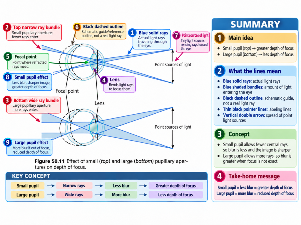
Upper Eye
The pupillary aperture is small.
Lower Eye
The pupillary aperture is large.
There are two small point sources of light in front of each eye.
Light from each source:
passes through pupil → focuses on retina
Therefore, in both eyes:
Retina sees two spots of light in perfect focus.
If the Retina Moves Out of Focus
If the retina moves forward or backward from the correct focal position:
In the eye with the small pupil
The size of each light spot changes only a little.
In the eye with the large pupil
The size of each light spot becomes much larger.
This larger spot becomes a:
Blur Circle
Therefore:
Small pupil → greater depth of focus
Large pupil → shallower depth of focus
Great Depth of Focus
When the lens system has a great depth of focus:
- the retina can move considerably from the focal plane, or
- lens strength can change considerably,
and the image can still remain nearly sharply focused.
Shallow Depth of Focus
When depth of focus is shallow:
Even a small movement of the retina away from the focal plane → marked blurring
Greatest Possible Depth of Focus
The greatest depth of focus occurs when:
The pupil is extremely small.
With a very small aperture:
Almost all light rays pass through the center of the lens.
The centralmost rays are always in focus.
C. ERRORS OF REFRACTION
1. EMMETROPIA — NORMAL VISION
Fig. 50.12
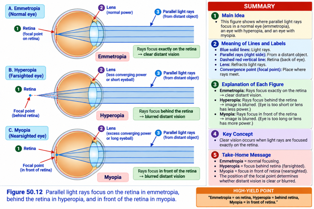
An eye is called normal or emmetropic when:
Parallel light rays from distant objects → focus sharply on the retina
while:
Ciliary muscle is completely relaxed.
Therefore:
Emmetropic eye → sees distant objects clearly with relaxed ciliary muscle
For close objects:
Ciliary muscle must contract → appropriate accommodation occurs
2. HYPEROPIA — FARSIGHTEDNESS
Hyperopia means:
Farsightedness
It is usually caused by:
Eyeball being too short
or occasionally:
Lens system being too weak
When the lens system is relaxed:
Parallel light rays are not bent enough to come to focus by the time they reach the retina.
How the Eye Compensates
To overcome this problem:
Ciliary muscle contracts → lens becomes stronger
This increases accommodation.
Therefore, a farsighted person may focus distant objects on the retina by using accommodation.
If only a small amount of accommodative power is used for distant vision:
More accommodative power remains available.
Therefore:
Closer objects can also be focused
until:
Ciliary muscle reaches its limit of contraction.
Hyperopia in Old Age
In old age:
Lens becomes presbyopic → accommodation decreases
Therefore, a farsighted person may become unable to accommodate enough even for:
Distant objects
and even less for:
Near objects
3. MYOPIA — NEARSIGHTEDNESS
Myopia means:
Nearsightedness
When the ciliary muscle is completely relaxed:
Light rays from distant objects → focus in front of the retina
Myopia is usually caused by:
Eyeball being too long
or:
Too much refractive power in the lens system
Why the Eye Cannot Correct Distant Vision in Myopia
The eye cannot decrease lens strength below the strength present when:
Ciliary muscle is completely relaxed.
Therefore:
Myopic person cannot focus distant objects sharply on the retina.
What Happens When the Object Comes Closer?
As the object moves nearer:
Eventually its image can be focused.
If the object comes still closer:
Accommodation can be used to maintain clear focus.
Therefore, a myopic person has a definite limiting:
Far Point
for clear vision.
D. CORRECTION OF MYOPIA AND HYPEROPIA
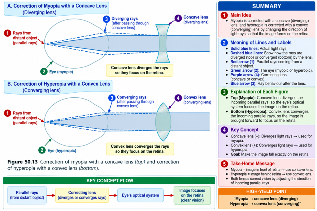
Myopia
In myopia:
Refractive power is too great.
This excessive refractive power can be neutralized by placing:
A concave spherical lens
in front of the eye.
The concave lens:
Diverges light rays.
Hyperopia
In hyperopia:
Lens system is too weak.
It can be corrected by adding refractive power using:
A convex lens
in front of the eye.
Finding Correct Lens Strength
The strength of the required concave or convex lens is usually found by:
Trial and error
A lens is tried.
Then:
- a stronger lens, or
- a weaker lens
is tested until the lens giving the best visual acuity is found.
E. ASTIGMATISM
Astigmatism is a refractive error.
In astigmatism:
Image in one plane → focuses at one distance
while:
Image in the plane at right angles → focuses at another distance
Astigmatism usually results from:
Too much curvature of the cornea in one plane
Egg Example
An astigmatic lens can be compared with:
The surface of an egg lying sideways to incoming light.
The curvature along the long axis of the egg is:
Less
than the curvature along the short axis.
Therefore:
Different planes have different curvatures.
DIFFERENT REFRACTION IN ASTIGMATISM
Because curvature in one plane is less than in the other:
Light rays in one plane → bend less
while:
Light rays in the other plane → bend more
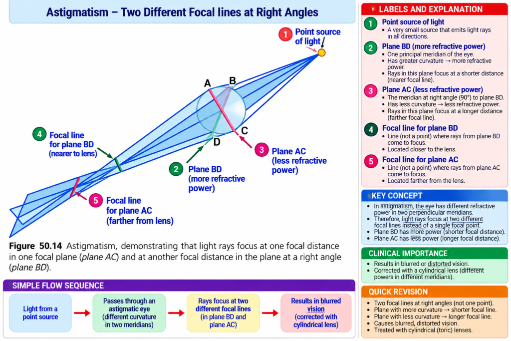
Vertical Plane BD
The light rays are refracted greatly because:
Vertical curvature is greater.
Horizontal Plane AC
The light rays are not bent as much because:
Horizontal curvature is less.
Therefore:
All rays do not reach one common focal point.
Rays passing through one plane focus:
Far in front of rays passing through the other plane.
WHY ACCOMMODATION CANNOT CORRECT ASTIGMATISM
During accommodation:
Curvature of the eye lens changes approximately equally in both planes.
But in astigmatism:
Each plane requires a different degree of accommodation.
Therefore:
Accommodation cannot compensate for astigmatism.
Without glasses:
A person with astigmatism never sees in completely sharp focus.
F. CORRECTION OF ASTIGMATISM WITH A CYLINDRICAL LENS
An astigmatic eye may be considered to have:
Two cylindrical lenses
that have:
- different strengths
- positions at right angles to each other
First Step
A spherical lens is selected by:
Trial and error
This corrects the focus in:
One of the two planes.
Second Step
An additional:
Cylindrical lens
is used to correct the error in the remaining plane.
Two things must be determined:
- Axis
- Strength
of the cylindrical lens.
FINDING THE CYLINDRICAL AXIS
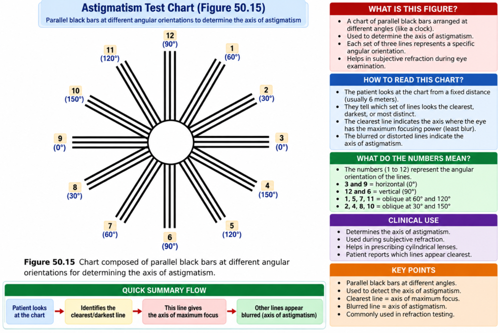
Parallel black bars are used.
Some bars are:
- vertical
- horizontal
- at different angles
Various spherical lenses are placed in front of the astigmatic eye.
A lens may make:
One set of parallel bars sharply focused
while:
Bars at right angles remain fuzzy.
The axis of the out-of-focus cylindrical component is:
Parallel to the fuzzy bars.
Finding Correct Cylindrical Lens Strength
After finding the axis:
- stronger,
- weaker,
- positive,
- or negative
cylindrical lenses are tried.
The cylindrical lens axes are placed:
In line with the out-of-focus bars.
This continues until:
All crossed bars are seen with equal clarity.
Then a special lens can be prepared combining:
Spherical correction + cylindrical correction at the correct axis
G. CORRECTION WITH CONTACT LENSES
Contact lenses can be made of:
- glass
- plastic
They fit closely against:
The anterior surface of the cornea.
They are held in place by:
A thin layer of tear fluid
between:
Contact lens and anterior eye surface
SPECIAL FEATURE OF CONTACT LENSES
A contact lens almost completely nullifies:
Refraction that normally occurs at the anterior surface of the cornea.
Why?
The tears between the contact lens and the cornea have a refractive index:
Almost equal to that of the cornea.
Therefore:
Anterior surface of cornea → no longer has a significant optical role
Instead:
Outer surface of contact lens → plays the major optical role
Therefore:
Refraction by contact lens surface substitutes for normal corneal refraction.
CONTACT LENSES IN KERATOCONUS
This property is especially important when the refractive error is caused by:
An abnormally shaped cornea.
An example is:
Keratoconus
In keratoconus:
Cornea is odd-shaped and bulging.
Without a contact lens:
Bulging cornea → severe abnormality of vision
and ordinary glasses may not correct the vision satisfactorily.
With a contact lens:
Abnormal corneal refraction is neutralized
and:
Normal refraction by the outer surface of the contact lens substitutes for it.
OTHER ADVANTAGES OF CONTACT LENSES
Advantage 1
The contact lens:
Turns with the eye
and therefore provides:
A broader field of clear vision than glasses.
Advantage 2
The contact lens has:
Little effect on the size of the object being seen.
In contrast:
Lenses about 1 cm in front of the eye affect image size
in addition to correcting focus.
H. CATARACTS — OPAQUE AREAS IN THE LENS
Cataracts are especially common in:
Older people
A cataract is:
A cloudy or opaque area, or areas, in the lens.
Early Stage
Proteins in some lens fibers become:
Denatured
Later Stage
The same proteins:
Coagulate → form opaque areas
instead of the normal:
Transparent protein fibers
TREATMENT OF SEVERE CATARACT
If a cataract blocks light transmission enough to seriously impair vision:
Lens can be surgically removed.
After removing the lens:
Eye loses a large amount of refractive power.
This refractive power must be replaced.
It can be replaced by:
A powerful convex lens in front of the eye
but usually:
An artificial plastic lens is implanted in the eye
in place of the removed lens.
5. FIGURE / TABLE NUMBERS
Figure numbers have been mentioned once at their relevant concepts:
- Fig. 50.11 — Depth of focus and pupil diameter
- Fig. 50.12 — Emmetropia and refractive errors
- Fig. 50.13 — Correction of myopia and hyperopia
- Fig. 50.14 — Astigmatic refraction
- Fig. 50.15 — Parallel bars for determining astigmatic axis
No table number appears in the provided text.
6. MATHEMATICAL EQUATION / FORMULA
The provided text states:
Amount of light entering the eye ∝ Area of pupil
and:
Pupil area ∝ Diameter²
Therefore:
Light entering eye ∝ Diameter²
The pupil diameter can change from:
1.5 mm → 8 mm
Using the relationship given in the text:
\left(\frac{8}{1.5}\right)^2
]
[
(5.33)^2
]
[
\approx 28.4
]
This is approximately the:
30-fold change
stated in the provided text.
Easy Concept
Pupil diameter increases → pupil area increases greatly → much more light enters the eye.
7. KEY CONCEPTS WITH EXAMPLES
Key Concept 1 — Presbyopia
Age increases → lens elasticity decreases → accommodation decreases
Example
Accommodation changes from approximately:
14 D in child → <2 D at 45–50 years → almost 0 D at 70 years
Key Concept 2 — Pupil Size
Small pupil → greater depth of focus
Example
When the retina moves away from the focal plane:
Small pupil → small change in light spot
whereas:
Large pupil → large blur circle
Key Concept 3 — Emmetropia
Distant parallel rays → sharply focused on retina with relaxed ciliary muscle
Key Concept 4 — Hyperopia
Eyeball too short or lens too weak
Example:
Accommodation can increase lens strength to help focus distant objects.
Key Concept 5 — Myopia
Eyeball too long or refractive power too great
Example:
Distant rays focus in front of retina.
Key Concept 6 — Astigmatism
Different curvature in different planes → different focal distances
Example:
Vertical plane BD bends light more than horizontal plane AC.
Key Concept 7 — Contact Lens
Contact lens almost neutralizes anterior corneal refraction.
Example:
This is particularly important in:
Keratoconus
Key Concept 8 — Cataract
Lens proteins denature → coagulate → opaque areas form
8. CLINICAL NOTE / IMPORTANCE
Presbyopia
Older people may require bifocal glasses for both far and near vision.
Hyperopia
Correction requires additional refractive power using a convex lens.
Myopia
Correction requires a concave spherical lens that diverges light rays.
Astigmatism
Correction requires:
Spherical correction + cylindrical correction with proper axis and strength
Keratoconus
Contact lenses can neutralize abnormal corneal refraction when glasses do not satisfactorily correct the vision.
Cataract
When light transmission becomes seriously impaired, the lens can be surgically removed and its refractive power replaced.
9. HIGH-YIELD POINTS
- Presbyopia = loss of accommodation with aging.
- Lens accommodation becomes nearly 0 D at age 70 years.
- Small pupil = greater depth of focus.
- Emmetropia = distant rays focus on retina with relaxed ciliary muscle.
- Hyperopia = eyeball too short or lens system too weak.
- Hyperopia → convex lens.
- Myopia = eyeball too long or refractive power too great.
- Myopia → concave lens.
- Myopic eye has a limiting far point.
- Astigmatism = different focal distances in different planes.
- Astigmatism requires a cylindrical lens.
- Contact lens nearly neutralizes refraction at the anterior corneal surface.
- Keratoconus = odd-shaped, bulging cornea.
- Cataract = cloudy or opaque area in the lens.
10. COMMON STUDENT MISTAKES
Mistake 1
Thinking presbyopia means increased accommodation.
Correct: Accommodation progressively decreases with age.Mistake 2
Thinking a large pupil gives greater depth of focus.
Correct: A small pupil gives greater depth of focus.
Mistake 3
Confusing hyperopia and myopia.
Hyperopia → eyeball too short / lens too weak
Myopia → eyeball too long / refractive power too great
Mistake 4
Reversing corrective lenses.
Myopia → concave lens
Hyperopia → convex lens
Mistake 5
Thinking accommodation can correct astigmatism.
Correct: Accommodation changes lens curvature approximately equally in both planes, while the two planes in astigmatism require different degrees of correction.
Mistake 6
Thinking a contact lens has no special effect on corneal refraction.
Correct: It almost completely neutralizes normal refraction at the anterior corneal surface.
11. QUICK REVISION
Presbyopia
Age ↑ → lens elasticity ↓ → accommodation ↓
Small Pupil
Pupil size ↓ → depth of focus ↑
Emmetropia
Distant rays → retina
Hyperopia
Eyeball too short / lens weak → convex lens
Myopia
Eyeball too long / refractive power high → rays focus in front of retina → concave lens
Astigmatism
Different curvature → different focal distances → cylindrical correction
Contact Lens
Neutralizes anterior corneal refraction
Cataract
Lens proteins denature and coagulate → opacity
12. SUMMARY
With increasing age, the lens becomes larger, thicker, and less elastic. Its ability to change shape and accommodate decreases. When accommodation becomes almost completely lost, the condition is called presbyopia.
The iris controls the amount of light entering the eye. The quantity of light depends on pupil area and therefore on the square of pupil diameter. A small pupil produces greater depth of focus.
In emmetropia, parallel rays from distant objects focus sharply on the retina while the ciliary muscle is relaxed.
In hyperopia, the eyeball is usually too short or the lens system is too weak. A convex lens provides additional refractive power.
In myopia, the eyeball is usually too long or refractive power is too great. Distant rays focus in front of the retina. A concave lens neutralizes excessive refractive power.
In astigmatism, different planes have different curvatures, so light in different planes focuses at different distances. Correction requires spherical and cylindrical lens correction.
Contact lenses almost neutralize the normal refraction at the anterior surface of the cornea. This is particularly important in an abnormally shaped cornea such as keratoconus.
A cataract is a cloudy or opaque area in the lens. Lens proteins first denature and later coagulate to form opaque areas. Severe cataract can be treated by surgical removal of the lens and replacement of its refractive power.
13. CLINICALLY CONCEPTUAL 3 MCQs
MCQ 1
A 70-year-old person has almost completely lost the ability of the lens to change its shape. The eyes can no longer properly adjust their focus for both near and far vision. Which condition best explains this finding?
A. Hyperopia
B. Myopia
C. Astigmatism
D. Presbyopia
E. Cataract
Correct Answer: D. Presbyopia
Explanation
With aging:
Lens becomes larger and thicker → elasticity decreases → ability to change shape decreases → accommodation decreases.
The power of accommodation decreases from about 14 diopters in a child to almost 0 diopters at age 70 years.
When the lens becomes almost totally nonaccommodating, the condition is called presbyopia.
MCQ 2
A person has difficulty seeing distant objects clearly. When the ciliary muscle is completely relaxed, rays coming from distant objects are focused in front of the retina. Which correction is most appropriate according to the provided text?
A. Convex spherical lens
B. Concave spherical lens
C. Cylindrical lens only
D. Bifocal lens only
E. Artificial plastic lens
Correct Answer: B. Concave spherical lens
Explanation
Focusing of distant light rays in front of the retina describes myopia.
Myopia is usually caused by:
Eyeball being too long
or:
Too much refractive power in the lens system.
A concave spherical lens diverges light rays and neutralizes the excessive refractive power.
MCQ 3
A person has an odd-shaped, bulging cornea causing such severe visual abnormality that ordinary glasses do not correct the vision satisfactorily. Which option described in the provided text is most useful?
A. Bifocal glasses
B. Convex spectacle lens
C. Concave spectacle lens
D. Contact lens
E. Cylindrical lens alone
Correct Answer: D. Contact lens
Explanation
The provided text describes an odd-shaped, bulging cornea as keratoconus.
A contact lens is useful because:
Tear fluid between the contact lens and cornea has a refractive index almost equal to the cornea.
Therefore:
Anterior corneal refraction is almost neutralized
and:
The outer surface of the contact lens provides the important refractive surface.
This allows the abnormal corneal refraction to be replaced by the refraction of the contact lens.
14. FINAL EXAM CONCEPT
Remember the complete flow:
Aging → Presbyopia
Small pupil → Greater depth of focus
Normal eye → Emmetropia
Short eye / weak lens → Hyperopia → Convex lens
Long eye / excessive refractive power → Myopia → Concave lens
Unequal curvature in different planes → Astigmatism → Cylindrical lens
Abnormal corneal shape → Contact lens may neutralize corneal refraction
Lens protein denaturation + coagulation → Cataract
Reference Guyton Physiology Textbook 15th Edition Page 641| Chapter 50
