


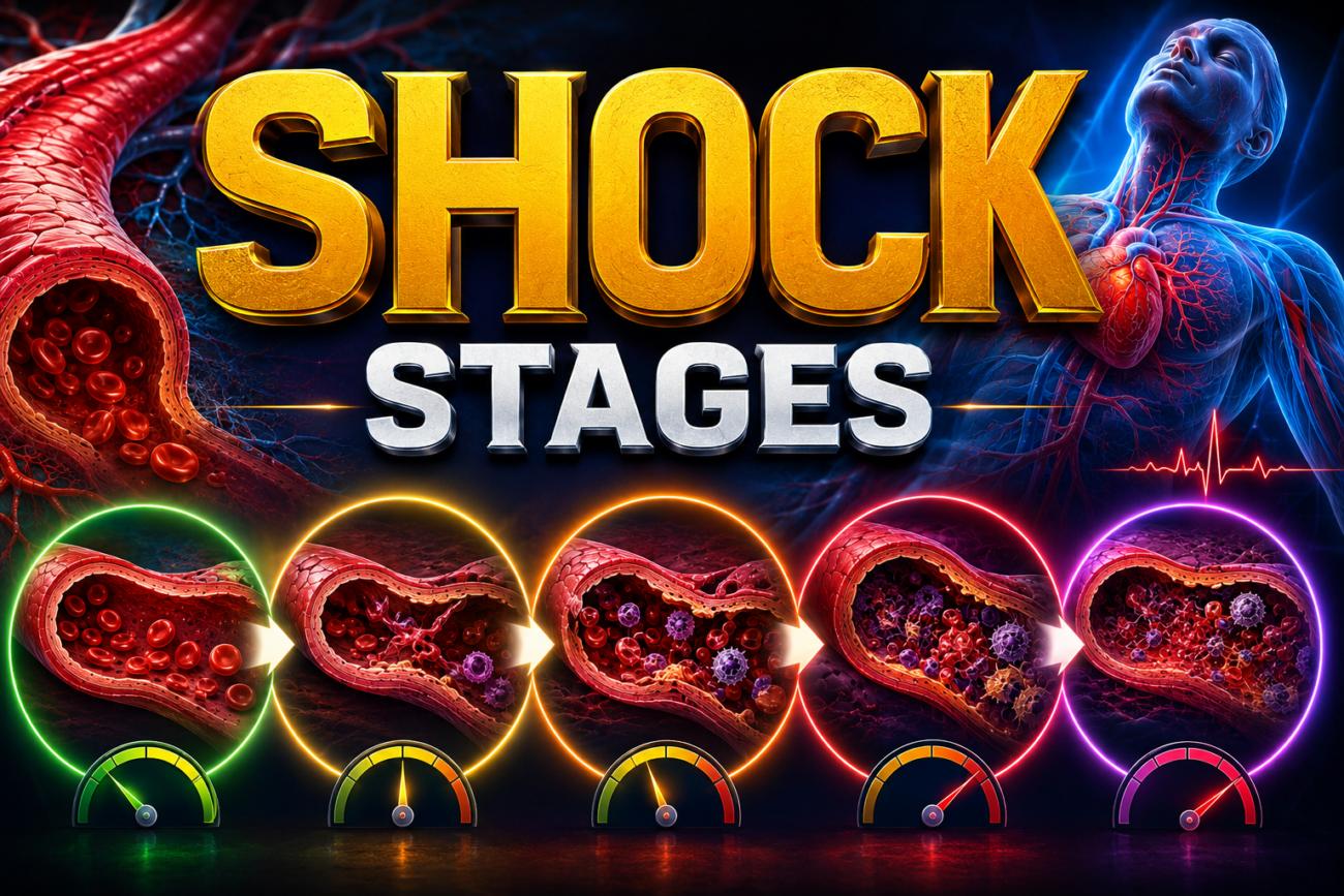
- Shock = inadequate tissue perfusion caused by:
- ↓ cardiac output, or
- ↓ effective circulating blood volume.
- Poor tissue perfusion → cellular hypoxia.
- Early injury is reversible, but prolonged shock → irreversible tissue injury → organ failure → death.
- Shock may occur with severe:
- hemorrhage
- trauma or burns
- myocardial infarction
- pulmonary embolism
- microbial sepsis
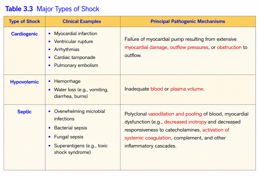
- The three main types are listed in Table 3.3:
- Cardiogenic shock:
- Heart pump fails → ↓ cardiac output → ↓ tissue perfusion.
- Causes:
- myocardial infarction
- ventricular arrhythmia
- cardiac tamponade
- pulmonary embolism causing outflow obstruction
- Hypovolemic shock:
- Loss of blood or plasma volume → ↓ circulating volume → ↓ cardiac output.
- Examples:
- severe hemorrhage
- major fluid loss from burns
- Septic shock:
- Usually triggered by microbial infection with severe systemic inflammatory response syndrome (SIRS).
- SIRS may also occur with burns, trauma, or pancreatitis.
- Massive inflammatory mediator release →
arterial vasodilation + vascular leakage + venous pooling. - These changes → ↓ tissue perfusion → cellular hypoxia → metabolic disturbances → organ dysfunction.
- If severe and persistent → organ failure → death.
- Less common forms:
- Neurogenic shock → loss of vascular tone after anesthesia or spinal cord injury.
- Anaphylactic shock → IgE-mediated hypersensitivity → systemic vasodilation + increased vascular permeability.
KEY CONCEPT
- Shock = inadequate perfusion → cellular hypoxia.
- Cardiogenic = pump failure.
- Hypovolemic = volume loss.
- Septic = inflammatory vasodilation + vascular leak + venous pooling.
- Neurogenic = loss of vascular tone.
- Anaphylactic = IgE-mediated vasodilation + vascular leakage.
- Persistent shock → irreversible injury → organ failure → death.
CONCEPTUAL EXAMPLES
- Myocardial infarction → weak cardiac pump → cardiogenic shock.
- Severe bleeding → major blood-volume loss → hypovolemic shock.
- Severe bacterial infection → massive inflammatory response → septic shock.
- Spinal cord injury → loss of vascular tone → neurogenic shock.
- Severe allergic reaction → widespread vasodilation and vascular leak → anaphylactic shock.
Pathogenesis of Septic Shock
- Septic shock accounts for about 2% of hospital admissions in the United States; about 50% of affected patients require ICU care.
- More than 750,000 cases/year occur, and mortality remains about 20%–30%.
- Increasing incidence is related to more critically ill and immunocompromised patients and more multidrug-resistant organisms.
- Septic shock is most commonly triggered by:
- Gram-positive bacteria
- then Gram-negative bacteria
- then fungi
- Microbial products activate macrophages, neutrophils, dendritic cells, endothelial cells, and complement → complex inflammatory responses → septic shock + multiorgan dysfunction (Fig. 3.19).
- Inflammatory and counterinflammatory responses:
- Microbial components are recognized by innate immune receptors:
- TLRs → recognize microbial PAMPs
- G-protein–coupled receptors → recognize bacterial peptides
- C-type lectin receptors → recognize fungal components
- Activation → release of TNF, IL-1, type I interferon, IL-12, IL-18 and other inflammatory mediators.
- Markers such as C-reactive protein and procalcitonin increase.
- Reactive oxygen species and prostaglandins are also produced.
- These mediators activate endothelium → ↑ adhesion molecules + further cytokine/chemokine production.
- Complement activation produces:
- C3a + C5a → anaphylatoxins
- C5a → chemotaxis
- C3b → opsonization
- Microbial products can also activate coagulation through factor XII and altered endothelial function.
- Widespread thrombin activation can further increase inflammation.
- Microbial components are recognized by innate immune receptors:
- With time, the initial excessive inflammation may trigger immunosuppression.
- Patients may therefore alternate between hyperinflammatory and immunosuppressed states.
- Proposed mechanisms include:
- shift from Th1 → Th2 responses
- ↑ anti-inflammatory mediators such as IL-10
- lymphocyte apoptosis
- immunosuppressive effects of apoptotic cells
- Endothelial activation and injury:
- Inflammation activates endothelium → ↑ adhesion molecules.
- Cytokines loosen endothelial junctions → vascular leakage → protein-rich tissue edema.
- Edema interferes with nutrient delivery and waste removal.
- Endothelium also produces more NO and other vasoactive mediators → vascular smooth-muscle relaxation → systemic hypotension.
- Induction of a procoagulant state:
- Severe coagulation abnormalities can cause DIC in up to half of septic patients.
- Proinflammatory cytokines:
- ↑ tissue factor
- ↓ anticoagulant factors such as TFPI, thrombomodulin, and endothelial protein C receptor
- ↑ plasminogen activator inhibitor-1 → ↓ fibrinolysis (Fig. 3.10)
- Vascular leak + edema → slower small-vessel blood flow → stasis.
- Stasis → less removal of activated clotting factors.
- Result:
↑ thrombin → fibrin-rich microthrombi → further ↓ tissue perfusion. - In severe DIC, platelets and coagulation factors are consumed → thrombosis + bleeding occur together.
- Metabolic abnormalities:
- Severe sepsis → insulin resistance + hyperglycemia.
- TNF, IL-1, glucagon, growth hormone, glucocorticoids, and catecholamines → ↑ gluconeogenesis.
- Proinflammatory cytokines also:
- ↓ insulin release
- ↑ insulin resistance
- Sepsis initially increases glucocorticoid production, but later adrenal insufficiency may occur.
- Adrenal necrosis may occur in Waterhouse-Friderichsen syndrome.
- Cellular hypoxia → ↓ oxidative phosphorylation → ↑ lactate → lactic acidosis.
- Organ dysfunction:
- Hypotension + edema + small-vessel thrombosis → ↓ oxygen and nutrient delivery.
- Cellular hypoxia and mitochondrial damage → impaired oxygen use.
- Cytokines can ↓ myocardial contractility → ↓ cardiac output.
- Vascular leak and endothelial injury may cause acute respiratory distress syndrome.
- Progressive injury may cause failure of the:
- kidneys
- liver
- lungs
- heart
- Severe multiorgan failure → death.
- Outcome depends on:
- extent and virulence of infection
- immune status
- other diseases
- amount and pattern of mediator production
- Standard treatment focuses on:
- antibiotics for infection
- intravenous fluids
- pressors
- supplemental oxygen
to maintain blood pressure and reduce tissue hypoxia.
- Superantigens can produce a similar syndrome, such as toxic shock syndrome.
- They activate many T lymphocytes → massive cytokine release → rash → vasodilation → hypotension → shock → death.
KEY CONCEPT
Microbial infection → innate immune activation → massive inflammatory mediators → endothelial leak + vasodilation + coagulation → hypotension + microthrombi + cellular hypoxia → multiorgan failure
- Inflammation → cytokine storm.
- Endothelial injury → vascular leak + hypotension.
- Coagulation → microthrombi ± DIC.
- Metabolic disturbance → hyperglycemia + lactic acidosis.
- Final result → multiorgan dysfunction and possible death.
CONCEPTUAL EXAMPLES
- Bacterial infection → TLR activation → TNF/IL-1 release → systemic inflammation.
- Cytokines loosen vessels → fluid leaves circulation → edema + low blood pressure.
- ↑ Tissue factor + ↓ anticoagulants → tiny fibrin clots form throughout vessels → DIC.
- Poor tissue perfusion → cells switch toward lactate production → lactic acidosis.
- Persistent hypotension + microthrombi → kidney, liver, lung, and heart dysfunction → multiorgan failure.
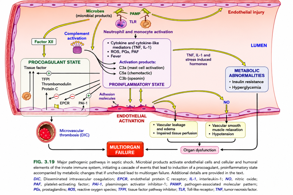
FIG. 3.19 — PATHOGENESIS OF SEPTIC SHOCK
🧠 Simplest concept
Microbes enter blood/tissues → immune system becomes massively activated → inflammation + clotting + leaky/dilated vessels + metabolic problems → poor organ blood flow → MULTIORGAN FAILURE
1️⃣ TOP — Microbes start everything
🟠 Microbial products = PAMPs
PAMP = Pathogen-Associated Molecular Pattern
= recognizable pieces of microbes.
Examples: bacterial LPS, cell-wall components.
PAMP → TLR
🟠 PAMP attaches to TLR (Toll-like receptor) on:
- 🔵 neutrophils
- 🟤 monocytes/macrophages
⬇️
Neutrophil + monocyte activation
These immune cells now become highly active.
2️⃣ Activated immune cells → PROINFLAMMATORY STATE
They release:
🔥 TNF + IL-1
Major inflammatory cytokines.
→ activate more inflammatory cells
→ activate endothelium
→ cause fever
⚡ ROS
Reactive oxygen species
→ can damage cells/endothelium.
🧪 PGs
Prostaglandins
→ inflammation, fever, vascular effects.
🧪 PAF
Platelet-activating factor
→ promotes inflammation and platelet activity.
Easy flow:
Microbe → TLR → leukocytes → TNF/IL-1 → massive inflammation
3️⃣ Complement activation
Microbes also activate the complement system.
C3a
→ mast-cell activation
→ inflammation ↑
C5a
→ chemotaxis
= attracts neutrophils to the site.
C3b
→ opsonization
= coats microbes so phagocytes can eat them more easily.
🧠 Memory:
C3a = inflammation
C5a = calls neutrophils
C3b = coats bacteria
4️⃣ 🩷 ENDOTHELIAL ACTIVATION / INJURY
The purple-pink cells lining the vessel = endothelium.
Microbial products + TNF/IL-1 activate and injure it.
Activated endothelium expresses:
🔴 Adhesion molecules
→ leukocytes stick to the vessel wall
→ inflammation becomes stronger.
This forms a vicious cycle:
Inflammation → endothelial activation → more inflammation
5️⃣ Endothelium becomes LEAKY
Yellow arrow downward:
Endothelial injury → increased vascular permeability
So fluid escapes from blood into tissues.
➡️ Vascular leakage → edema
At the same time:
➡️ less blood remains effectively circulating
➡️ tissue perfusion falls
Result:
Edema + poor perfusion → organ dysfunction
6️⃣ NO → vasodilation → hypotension
Activated endothelium produces excess:
NO = nitric oxide
NO relaxes vascular smooth muscle.
↑ NO → vessel dilation → ↓ blood pressure
⬇️
Hypotension
→ organs receive less blood
→ organ dysfunction
🧠 Memory:
NO = vessel relaxer
7️⃣ LEFT — PROCOAGULANT STATE
Sepsis makes blood clot too much
↑ Tissue factor
Green + means increased procoagulant activity.
Tissue factor ↑ → coagulation cascade ↑ → thrombin/fibrin ↑
Normal anticoagulants decrease
The figure shows reduction of:
- TFPI = tissue factor pathway inhibitor
- Thrombomodulin
- Protein C
- EPCR = endothelial protein C receptor
🔴 − signs = decreased anticoagulant protection
So:
More clotting + less anticoagulation = thrombosis
8️⃣ PAI-1 increases
PAI-1 = plasminogen activator inhibitor-1
Green + indicates increase.
PAI-1 blocks fibrinolysis.
So:
↑ PAI-1 → ↓ clot breakdown → fibrin clots remainFactor XII + coagulation
Microbial/inflammatory activation can also promote coagulation pathways, including Factor XII activation.
⬇️
This further favors a:
PROCOAGULANT STATE
🔟 Result → DIC
Many tiny clots form throughout small vessels.
🔴 Small vessel full of clot = microvascular thrombosis
DIC
Disseminated Intravascular Coagulation
Think:
Sepsis → widespread tiny clots → small vessels blocked → organs lose blood supply
⬇️
Multiorgan failure
1️⃣1️⃣ Metabolic abnormalities
TNF, IL-1 and stress hormones cause:
Insulin resistance
Cells respond poorly to insulin.
⬇️
Hyperglycemia
Blood glucose rises.
These metabolic disturbances further impair cell and organ function.
🎨 COLOR / ARROW GUIDE
- 🩷 Purple-pink lining = endothelium
- 🟠 Microbial shapes = PAMPs
- 🔵/tan immune cells = neutrophils & monocytes
- 🔴 Activated endothelial region = endothelial activation
- 🟨 Yellow arrows = important activating/pathogenic pathways
- ⚫ Black arrows = downstream consequences
- 🟢 + = increased activity
- 🔴 − = decreased activity
⭐ The 4 big problems in septic shock
1. 🔥 Inflammation
PAMP → TLR → TNF, IL-1, complement
2. 🩸 Excess coagulation
↑ Tissue factor + ↓ Protein C/TFPI + ↑ PAI-1 → DIC
3. 💧 Vascular dysfunction
Endothelial injury → leakage + edema + ↓ perfusion
4. 📉 Vasodilation
↑ NO → hypotension
All converge on:
ORGAN DYSFUNCTION → MULTIORGAN FAILURE
🧠 Fastest exam recall
SEPSIS = Inflammation + DIC + Leaky vessels + Vasodilation
Microbes → PAMP/TLR → TNF/IL-1 → endothelial activation → clotting + leakage + NO-mediated hypotension → poor perfusion → multiorgan failure.
Stages of Shock
- Shock is progressive and may lead to death if the underlying cause is not corrected.
- In septic shock, the exact mechanism of death is still not fully understood.
- Cellular necrosis is usually limited.
- Death commonly follows multiple organ failure.
- In hypovolemic and cardiogenic shock, progression is better understood.
- Unless the initial injury is immediately fatal, shock usually develops through three stages:
- Initial nonprogressive stage:
- Compensatory reflexes maintain blood pressure and perfusion of vital organs.
- Important mechanisms include:
- baroreceptor reflexes
- catecholamine release
- antidiuretic hormone release
- renin–angiotensin–aldosterone activation
- generalized sympathetic stimulation
- Final effects:
tachycardia + peripheral vasoconstriction + renal fluid retention. - Skin becomes cool and pale because of cutaneous vasoconstriction.
- In early septic shock, skin may instead be warm and flushed because of vasodilation.
- Coronary and cerebral vessels remain relatively open → blood is redirected mainly to the heart and brain.
- Progressive stage:
- Persistent poor perfusion → widespread tissue hypoxia.
- Lack of oxygen → aerobic metabolism is replaced by anaerobic glycolysis.
- Anaerobic glycolysis → ↑ lactic acid → metabolic acidosis.
- Acidosis reduces arteriolar vasomotor response → arterioles dilate.
- Vasodilation → blood pools in microcirculation → ↓ cardiac output.
- Endothelial ischemic injury may develop → can contribute to DIC.
- Continued hypoxia → vital organs begin to fail.
- Irreversible stage:
- Cellular and tissue injury becomes so severe that survival is impossible even if blood pressure is corrected.
- Lysosomal enzymes leak from damaged cells → further cell injury.
- Myocardial contractility worsens.
- Ischemic bowel may allow intestinal bacteria to enter the blood → bacteremia may worsen shock.
- Renal ischemia commonly causes renal failure.
- The downward spiral continues → death.
KEY CONCEPT
- Stage 1: compensated → body maintains vital organ perfusion.
- Stage 2: progressive → hypoxia → anaerobic glycolysis → lactic acidosis → vasodilation → worsening perfusion.
- Stage 3: irreversible → severe cellular injury + organ failure → death.
CONCEPTUAL EXAMPLES
- Blood loss → sympathetic activation → tachycardia + vasoconstriction → early compensated shock.
- Continued low perfusion → tissues make lactate → acidosis → vessels dilate → progressive shock.
- Prolonged severe shock → kidney failure + weak heart + bowel injury → irreversible shock.
MORPHOLOGY
- The tissue changes in shock are mainly due to hypoxic injury caused by:
- hypoperfusion
- microvascular thrombosis
- Any organ may be affected, but the most commonly involved are:
- brain
- heart
- kidneys
- adrenal glands
- gastrointestinal tract
- Fibrin thrombi may form in many tissues.
- They are especially easy to see in kidney glomeruli.
- In the adrenal cortex, stored lipids decrease because they are increasingly used for steroid synthesis during stress.
- In hypovolemic shock after hemorrhage, the lungs are relatively resistant to hypoxic injury.
- However, sepsis or trauma can cause diffuse alveolar damage → called “shock lung.”
- If the patient survives, many affected tissues may recover completely.
- Important exceptions are:
- neuronal loss
- cardiomyocyte loss
- These cells do not recover well once irreversibly damaged.
KEY CONCEPT
- Shock morphology mainly reflects:
hypoperfusion + microvascular thrombosis → hypoxic tissue injury. - Most affected organs → brain, heart, kidneys, adrenals, GI tract.
- Kidney glomeruli commonly show fibrin thrombi.
- Sepsis/trauma → diffuse alveolar damage = shock lung.
- Most tissues may recover, but lost neurons and cardiomyocytes do not.
CONCEPTUAL EXAMPLES
- Poor blood flow to kidney → glomerular microthrombi + ischemic injury.
- Severe stress → adrenal cortex uses more lipid for steroid production → lipid depletion.
- Sepsis → diffuse lung injury → shock lung.
- Surviving patient → many tissues recover, but dead brain and heart muscle cells remain lost.
Hemodynamic Disorders — whole chapter Summary
Imagine the circulatory system as a huge network of roads and pipelines carrying blood, oxygen, nutrients, and fluid to every tissue of the body. Normally, blood stays inside vessels, flows smoothly, forms a clot only when a vessel is injured, and delivers enough oxygen to keep tissues alive. Hemodynamic disorders occur when any part of this balance is disturbed.
Edema — When Fluid Leaves the Blood Vessels
The story begins when too much fluid moves from the blood vessels into the surrounding tissues. This abnormal accumulation of fluid is called edema.
Edema fluid may be either a transudate or an exudate. A transudate is usually protein-poor and occurs without inflammation, whereas an exudate is protein-rich and is usually associated with inflammation.
Noninflammatory edema can develop through several mechanisms. If the pressure inside blood vessels becomes too high, as in heart failure, fluid is pushed outward into the tissues. This is called increased hydrostatic pressure.
Edema can also occur when plasma proteins, especially albumin, become too low. Albumin normally pulls water back into the circulation by maintaining colloid osmotic pressure. Albumin may decrease because the liver does not make enough of it, as in severe liver disease or protein malnutrition, or because albumin is lost through the kidneys, as in nephrotic syndrome.
Another cause is lymphatic obstruction. Normally, lymphatic vessels remove excess tissue fluid. If lymphatics become blocked by fibrosis, tumors, surgery, or other causes, fluid accumulates in the tissues.
The kidneys can also cause edema by retaining too much sodium and water, such as in renal failure.
In inflammatory conditions, the mechanism is different. Inflammation makes blood vessels more permeable, allowing both fluid and proteins to escape into tissues, producing a protein-rich exudate.
So the basic concept is:
More fluid leaving vessels or less fluid returning to vessels → edema.
Hemostasis — How the Body Stops Bleeding
Now imagine that a blood vessel is damaged. Blood begins to escape, so the body immediately activates a protective process called hemostasis, meaning stopping bleeding.
Hemostasis occurs in two closely connected stages: primary hemostasis and secondary hemostasis.
Primary Hemostasis — Making the Platelet Plug
When the vessel wall is injured, the underlying extracellular matrix becomes exposed. One important protein present there is von Willebrand factor, or vWF.
Platelets circulating in the blood recognize the damaged area. Their GpIb receptors attach to vWF, allowing the platelets to stick to the site of injury.
This platelet adhesion causes the platelets to become activated. Activated platelets change shape, release the contents of their granules, and change the structure of their membrane receptors.
One particularly important receptor is GpIIb/IIIa. After platelet activation, GpIIb/IIIa binds fibrinogen. Fibrinogen acts like a bridge connecting one platelet to another.
Therefore:
Vessel injury → vWF exposed → platelet GpIb binds vWF → platelet activation → GpIIb/IIIa binds fibrinogen → platelet aggregation.
This produces the temporary primary hemostatic plug.
Secondary Hemostasis — Strengthening the Plug With Fibrin
The platelet plug alone is not strong enough. The body therefore activates the coagulation cascade.
Damaged tissue exposes tissue factor, which starts coagulation. Important clotting factors include factors VII, IX, X, V, VIII, II, and fibrinogen.
The central enzyme of this process is thrombin.
Thrombin converts soluble fibrinogen into fibrin. Fibrin forms strong strands around the platelet plug and creates a stable secondary hemostatic plug.
Thrombin also activates factor XIII, which cross-links fibrin strands and makes the clot stronger. It also promotes platelet contraction, helping the clot tighten.
So:
Tissue factor → coagulation cascade → thrombin → fibrinogen becomes fibrin → stable clot.
However, clotting must remain limited to the damaged area. Otherwise, the entire circulation could thrombose.
The body prevents excessive clotting by washing away activated clotting factors, removing them through the liver, restricting coagulation reactions to phospholipid surfaces on activated platelets, using anticoagulant molecules such as thrombomodulin on normal endothelium, and activating fibrinolysis through molecules such as tissue plasminogen activator, or tPA.
Thus, normal hemostasis is a balance between:
Making enough clot to stop bleeding but not so much clot that blood vessels become blocked.
Thrombosis — When Clotting Happens Inside a Vessel
Sometimes this normal clotting system becomes abnormal and a clot forms inside an intact blood vessel. This abnormal clot is called a thrombus, and the process is called thrombosis.
Three major factors promote thrombosis. Together, they form Virchow’s triad:
Endothelial injury + abnormal blood flow + hypercoagulability.
First, the endothelium may be damaged by hypertension, inflammation, toxins, metabolic abnormalities, or atherosclerosis.
Second, abnormal blood flow may occur. Blood may become stagnant, called stasis, or flow chaotically, called turbulence. This can occur in aneurysms, abnormal heart chambers, or around atherosclerotic plaques.
Third, the blood itself may have an increased tendency to clot, known as hypercoagulability. This may be inherited, such as factor V Leiden, or acquired, such as during prolonged bed rest or other clinical conditions.
Once a thrombus forms, several things can happen. It may grow larger, called propagation. It may dissolve, called resolution. It may become incorporated into the vessel wall, called organization. Or part of it may break away and travel through the circulation, called embolization.
The major danger of thrombosis is interruption of blood flow.
Therefore:
Thrombus → vessel blockage → reduced blood supply → tissue injury.
Or:
Thrombus breaks off → embolus → blockage at a distant site.
Embolism — When Material Travels and Blocks a Distant Vessel
An embolus is any solid, liquid, or gaseous material carried through the bloodstream from one place to another, where it becomes lodged and blocks a vessel.
Most emboli are pieces of thrombi and are therefore called thromboemboli.
Pulmonary Embolism
A pulmonary embolus usually begins as a deep vein thrombosis in the lower limb.
A piece of the thrombus breaks off and travels through:
Leg veins → inferior vena cava → right side of heart → pulmonary arteries.
The consequences depend on the size, number, and location of the emboli.
A small embolus may cause few symptoms. A larger embolus may cause pulmonary hemorrhage or pulmonary infarction. A massive pulmonary embolus can suddenly obstruct pulmonary blood flow, causing acute right-sided heart failure and sudden death.
Systemic Embolism
Systemic emboli travel through the arterial circulation.
They commonly originate from thrombi inside the heart, particularly mural thrombi, diseased heart valves, aortic aneurysms, or atherosclerotic plaques.
They can travel to organs such as the brain, kidneys, spleen, intestines, and lower limbs.
Whether tissue dies depends partly on whether that tissue has an alternative blood supply.
Fat Embolism
After severe bone fractures or crushing injuries, fat droplets from bone marrow can enter the circulation.
These fat emboli can damage the lungs and brain, producing respiratory insufficiency and neurologic abnormalities.
Amniotic Fluid Embolism
During childbirth, amniotic fluid may rarely enter the maternal circulation.
Although uncommon, this condition is extremely dangerous and may produce severe pulmonary, neurologic, and coagulation abnormalities.
Air Embolism
Gas bubbles can also enter or form within the circulation.
A classic example occurs in divers who ascend too rapidly. Dissolved nitrogen suddenly comes out of solution and forms bubbles in tissues and blood.
Thus:
An embolus travels until the vessel becomes too small → vessel obstruction → ischemia or infarction.
Infarction — When Loss of Blood Supply Causes Tissue Death
If a blood vessel becomes blocked badly enough, the tissue supplied by that vessel may undergo ischemic necrosis. The dead area is called an infarct.
Most infarcts are caused by arterial obstruction due to thrombosis or embolism. Less commonly, venous obstruction can also cause infarction.
There are two major appearances of infarction: red infarcts and white infarcts.
Red Infarcts
Red, or hemorrhagic, infarcts contain blood.
They commonly occur with venous obstruction or in tissues where blood can still enter the injured area from another source. They may also occur when arterial blood flow is temporarily interrupted and then restored.
White Infarcts
White, or pale, infarcts usually occur after persistent arterial obstruction in solid organs with end-arterial circulation, where there is little alternative blood supply.
Examples include the heart, spleen, and kidney.
Whether an infarct actually develops depends on several factors.
A tissue with a good collateral blood supply is more resistant to infarction. A slowly developing obstruction may allow collateral vessels time to enlarge. Some tissues are naturally more sensitive to oxygen deprivation; neurons, for example, are extremely sensitive to ischemia. The oxygen content of the blood also matters.
Thus:
Vessel obstruction → ischemia → prolonged oxygen deprivation → necrosis → infarction.
Shock — When the Whole Body Loses Adequate Blood Flow
The most severe problem occurs when blood flow becomes inadequate not just in one organ but throughout the entire body. This is called shock.
Shock is a state of systemic tissue hypoperfusion, meaning the tissues are not receiving enough blood and oxygen.
It usually results from reduced cardiac output, reduced circulating blood volume, or severe abnormalities in vascular tone.
The three major forms are cardiogenic shock, hypovolemic shock, and septic shock.
Cardiogenic Shock
In cardiogenic shock, the heart fails to pump effectively.
For example, a large myocardial infarction may severely damage the heart muscle.
Therefore:
Pump failure → low cardiac output → low tissue perfusion → tissue hypoxia.
Hypovolemic Shock
In hypovolemic shock, there is not enough circulating blood volume.
This commonly occurs after severe blood loss.
Therefore:
Loss of blood or fluid → reduced venous return → reduced cardiac output → tissue hypoperfusion.
Septic Shock
Septic shock develops as a result of a severe host response to infection, particularly bacterial or fungal infection.
It is more complicated than simply having organisms in the blood.
The infection triggers widespread inflammation and causes endothelial activation and injury.
Blood vessels dilate, reducing systemic vascular resistance and blood pressure.
At the same time, vascular permeability increases, allowing fluid to leave the circulation and enter tissues, producing widespread edema.
The coagulation system may become activated throughout the body, producing disseminated intravascular coagulation, or DIC, in which many tiny clots form in small vessels while clotting factors and platelets are consumed.
Sepsis also produces major cellular and metabolic abnormalities that prevent tissues from using oxygen normally.
Therefore, septic shock can be summarized as:
Severe infection → inflammatory response → endothelial injury + vasodilation + increased permeability + coagulation abnormalities → severe tissue hypoperfusion → organ dysfunction.
The Whole Chapter as One Story
The circulatory system normally keeps fluid inside blood vessels, delivers oxygen to tissues, and rapidly seals vascular injuries.
If fluid escapes excessively from vessels, edema develops.
If a vessel is injured, hemostasis creates a platelet plug and reinforces it with fibrin.
If clotting occurs abnormally inside a vessel, a thrombus develops.
If part of that thrombus breaks away and travels through the circulation, it becomes an embolus.
If the embolus or thrombus blocks blood flow long enough, tissue becomes ischemic and dies, producing an infarct.
If blood flow becomes inadequate throughout the entire body, the patient develops shock, which can lead to widespread hypoxic injury, organ failure, and death.
One-Line Flow to Remember
Edema = fluid problem → Hemostasis = normal clot → Thrombosis = abnormal clot → Embolism = traveling blockage → Infarction = local tissue death → Shock = whole-body hypoperfusion.
Ultra-High-Yield Exam Recall
Edema: fluid moves into interstitium.
Hemostasis: platelet plug first, fibrin clot second.
Thrombosis: think Virchow triad — endothelial injury + abnormal flow + hypercoagulability.
Embolism: most are detached thrombi; PE usually comes from lower-limb DVT.
Infarction: vascular blockage causes ischemic necrosis.
Shock: inadequate systemic perfusion causes widespread tissue hypoxia.
Golden Sequence
Vessel injury → platelets → coagulation → fibrin clot
but if clotting becomes abnormal:
Thrombus → embolus → ischemia → infarction
and if circulation fails globally:
Hypoperfusion → shock → multiorgan injury.
