


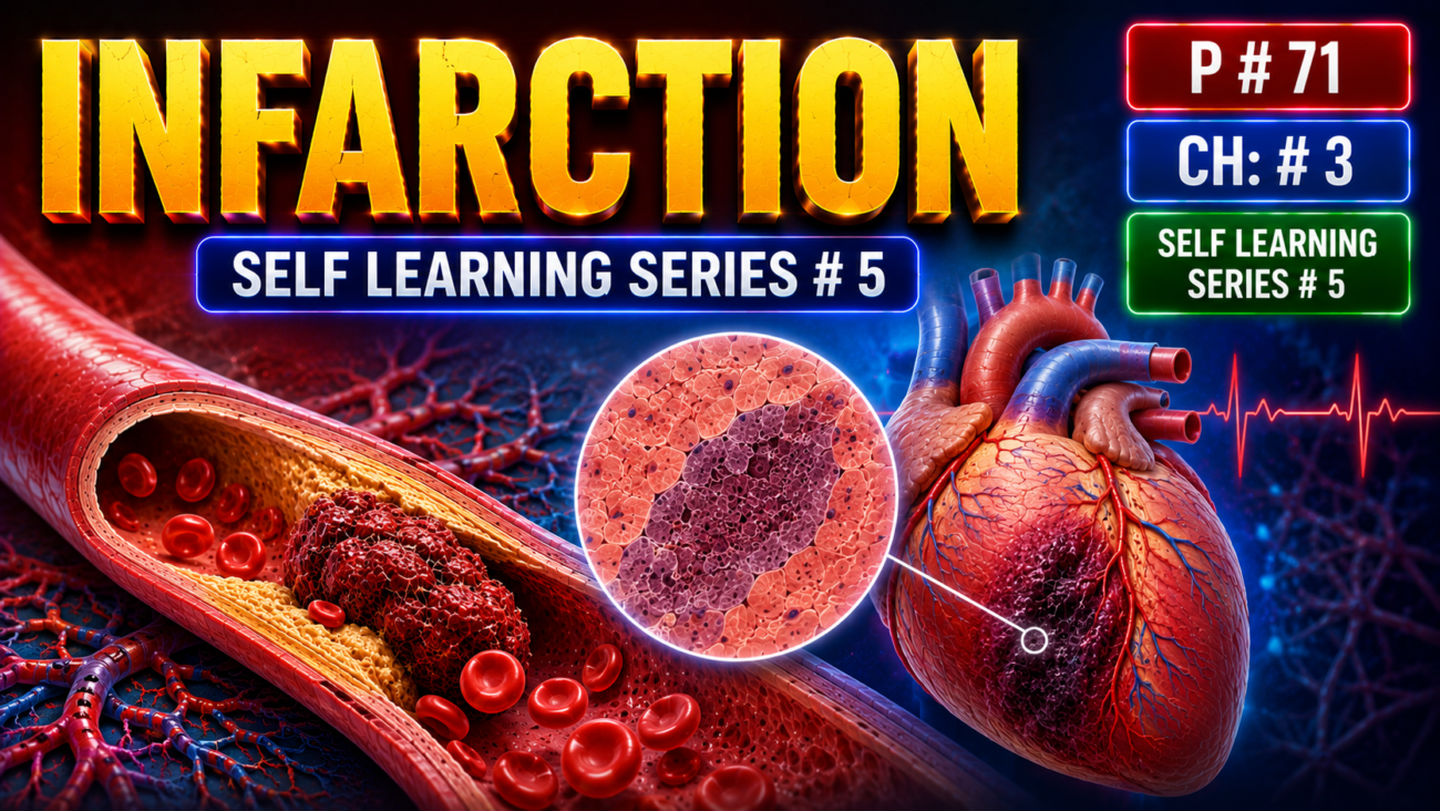
- An infarct = an area of ischemic necrosis caused by blockage of the blood supply to a tissue.
- Infarction commonly affects the heart and brain and is a major cause of illness and death.
- Other important sites include:
- lungs
- bowel
- distal extremities → ischemic necrosis may produce gangrene, especially in diabetes
- The vast majority of infarctions result from:
- arterial thrombosis, or
- arterial embolism.
- Less common causes of arterial obstruction include:
- vasospasm
- hemorrhage into an atherosclerotic plaque → plaque expands and narrows the vessel
- external compression of a vessel by:
- tumor
- dissecting aortic aneurysm
- severe edema within a confined space, such as anterior tibial compartment syndrome
- Other uncommon causes include:
- twisting of vessels → testicular torsion or bowel volvulus
- traumatic rupture of a vessel
- vascular entrapment within a hernia sac
- Venous thrombosis usually causes congestion rather than infarction.
- This is because collateral/bypass venous channels often open → restore venous outflow → allow arterial inflow to continue.
- Therefore, venous thrombosis usually causes infarction only when an organ has essentially a single venous outflow, such as:
- testis
- ovary
KEY CONCEPT
- Infarction = vascular occlusion → ischemia → tissue necrosis.
- Most infarcts are caused by arterial thrombosis or embolism.
- Arterial obstruction → infarction is common.
- Venous obstruction → usually congestion, because collateral drainage can develop.
- Venous infarction is more likely in organs with one main efferent vein.
CONCEPTUAL EXAMPLES
- Coronary artery thrombus → loss of blood supply to myocardium → myocardial infarction.
- Cerebral arterial embolus → brain ischemia → cerebral infarction.
- Testicular torsion → vessels twist → blood supply fails → testicular infarction.
- Venous thrombosis in most tissues → alternate veins drain blood → mainly congestion rather than infarction.
MORPHOLOGY
- Infarcts are classified by:
- color → reflects the amount of hemorrhage
- presence or absence of microbial infection
- Therefore, infarcts may be:
- Red (hemorrhagic) or white (anemic)
- Septic or bland
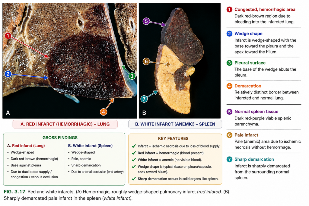
- Red infarcts occur in four main settings (Fig. 3.17A):
- Venous occlusion
- Example: testicular torsion
- Blood enters but cannot drain properly → hemorrhagic infarction.
- Tissues with dual circulation
- Examples: lung and small intestine
- Collateral arterial blood still enters the damaged area, but perfusion remains inadequate → red infarct.
- Previously congested tissues
- Slow venous outflow → blood accumulates → infarct becomes hemorrhagic.
- Reperfusion after infarction
- Blood flow returns after ischemic injury.
- Example: after angioplasty opens an arterial obstruction.
- Damaged vessels leak blood into the infarcted tissue → red infarct.
- Venous occlusion
KEY CONCEPT
- Red infarct = infarction with hemorrhage.
- Think of four classic settings:
venous blockage + dual blood supply + previous congestion + reperfusion.
CONCEPTUAL EXAMPLES
- Testicular torsion → venous drainage blocked → blood accumulates → red infarct.
- Pulmonary vessel occlusion → second blood supply still brings some blood → hemorrhagic infarct.
- Angioplasty restores flow to already damaged tissue → blood leaks into it → red reperfusion infarct.
MORPHOLOGY
- White (anemic) infarcts occur after arterial occlusion in solid organs with end-arterial circulation, especially:
- heart
- spleen
- kidney (Fig. 3.17B)
- White infarcts are usually wedge-shaped:
- apex → points toward the blocked vessel
- base → faces the outer surface of the organ
- If the base reaches a serosal surface, a fibrinous exudate may form over it.
- In an acute infarct:
- margins are initially poorly defined and slightly hemorrhagic
- with time → margins become clearer
- a narrow hyperemic rim develops because of inflammation
- Arterial infarcts in organs without dual circulation become progressively paler and more sharply defined with time.
- In contrast, organs with dual blood supply, especially the lung, usually develop hemorrhagic (red) infarcts (Fig. 3.17A).
- In hemorrhagic infarcts:
- macrophages remove extravasated RBCs
- heme iron → hemosiderin
- extensive hemorrhage may eventually leave a firm brown residue
- In most tissues, the main microscopic pattern is ischemic coagulative necrosis.
- Within a few hours → inflammation begins around the infarct.
- By about 1–2 days → the inflammatory border becomes well defined.
- Later:
- inflammation → repair
- repair begins at the preserved margins
- some surviving peripheral cells may regenerate if the supporting tissue framework remains intact
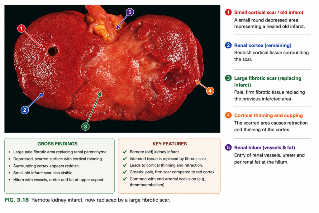
- most infarcts eventually become a scar (Fig. 3.18)
- Brain is the major exception:
- ischemic injury in the CNS → liquefactive necrosis, not coagulative necrosis.
- Septic infarcts occur when:
- infected cardiac valve vegetations embolize, or
- microbes infect already necrotic tissue.
- The infected infarct may become an abscess → stronger inflammatory response → healing by organization and fibrosis.
KEY CONCEPT
- White infarct → arterial blockage + solid end-arterial organ.
- Red infarct → commonly occurs in tissues with dual blood supply.
- Most infarcts → coagulative necrosis → inflammation → scar.
- Brain infarct → liquefactive necrosis.
- Septic infarct → infected infarct → abscess.
CONCEPTUAL EXAMPLES
- Coronary artery blocked → pale wedge-like myocardial infarct → white infarct.
- Pulmonary arterial blockage → blood still enters from another circulation → red infarct.
- Kidney infarct → coagulative necrosis → inflammation → fibrous scar.
- Brain ischemia → tissue softens and liquefies → liquefactive necrosis.
- Infected valve vegetation embolizes → infected infarct → abscess.
Factors That Influence Infarct Development
- Vascular occlusion may cause little effect or severe tissue necrosis, depending mainly on three factors.
- Anatomy of vascular supply:
- The most important factor is whether the tissue has an alternative blood supply.
- Lung → dual supply from pulmonary + bronchial arteries → pulmonary arteriole blockage usually does not cause infarction unless bronchial flow is also reduced.
- Liver → hepatic artery + portal vein → relatively resistant to infarction.
- Hand and forearm → radial + ulnar arteries → alternative blood flow reduces infarction risk.
- Kidney and spleen → end-arterial circulation → arterial blockage usually causes infarction.
- Rate of occlusion:
- Slow blockage is less likely to cause infarction because collateral vessels have time to enlarge and carry more blood.
- Small connections normally exist between the major coronary arteries.
- Slowly developing coronary obstruction → collateral flow may increase enough to prevent infarction, even if the original artery eventually becomes completely blocked.
- Tissue vulnerability to hypoxia:
- Different cells tolerate loss of blood supply for different lengths of time because their metabolic needs differ.
- Neurons → irreversible injury after only 3–4 minutes.
- Cardiac muscle cells → die after about 20–30 minutes of ischemia.
- Fibroblasts → may survive many hours of ischemia.
KEY CONCEPT
- Infarct development mainly depends on:
vascular anatomy + speed of blockage + tissue sensitivity to hypoxia. - Dual/collateral blood supply → protects against infarction.
- Slow occlusion → allows collateral circulation to develop.
- Neurons are most vulnerable, myocardium is next, and fibroblasts are much more resistant.
CONCEPTUAL EXAMPLES
- Pulmonary artery branch blocked but bronchial circulation remains intact → lung infarction may not occur.
- Renal artery blocked → no major alternative supply → kidney infarction likely.
- Coronary artery narrows slowly → collateral vessels enlarge → myocardium may remain viable.
- Brain loses blood flow → neurons can die within 3–4 minutes.
