


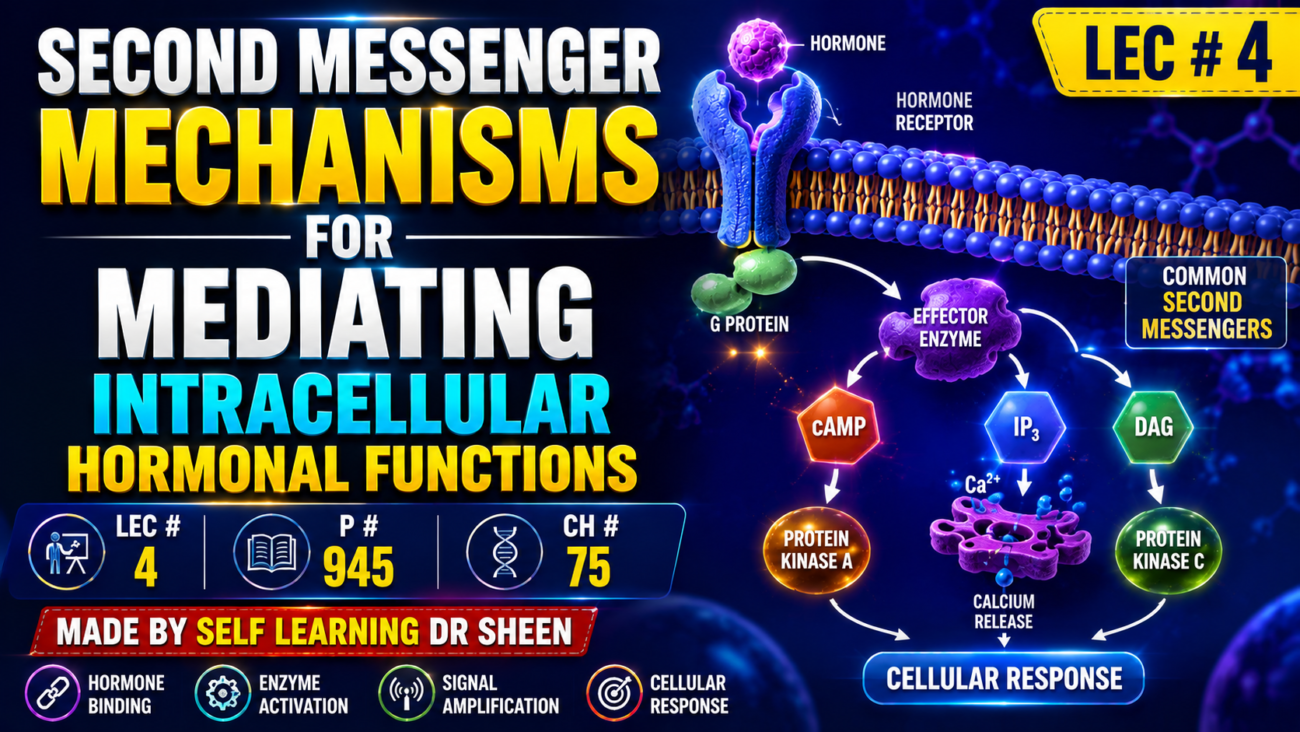
- Some hormones act inside cells by causing the formation of a second messenger, such as cAMP, inside the cell membrane.
- The cAMP then produces the hormone’s effects inside the cell.
- Therefore, the hormone’s direct action is mainly to activate one specific membrane receptor.
- The second messenger carries out the remaining intracellular effects.
- cAMP is not the only second messenger used by hormones.
- Two other important second messengers are:
- Calcium ions (Ca²⁺) and calmodulin
- Products formed from membrane phospholipid breakdown
- In some cases, one hormone can activate more than one second-messenger system in the same target tissue.
KEY CONCEPT
- Hormone → membrane receptor → second messenger → intracellular effects.
- Important second messengers include cAMP, Ca²⁺ with calmodulin, and membrane phospholipid breakdown products.
CONCEPTUAL EXAMPLES
- cAMP: Hormone activates its membrane receptor → cAMP is formed → cAMP produces intracellular effects.
- Ca²⁺/calmodulin: A hormone can use calcium ions and calmodulin as a second-messenger system.
- Multiple systems: One hormone may activate more than one second-messenger system in the same target tissue.
Adenylyl Cyclase–cAMP Second Messenger System
- Table 75.3 lists some hormones that use the adenylyl cyclase–cAMP mechanism, while Fig. 75.7 shows how this system works.
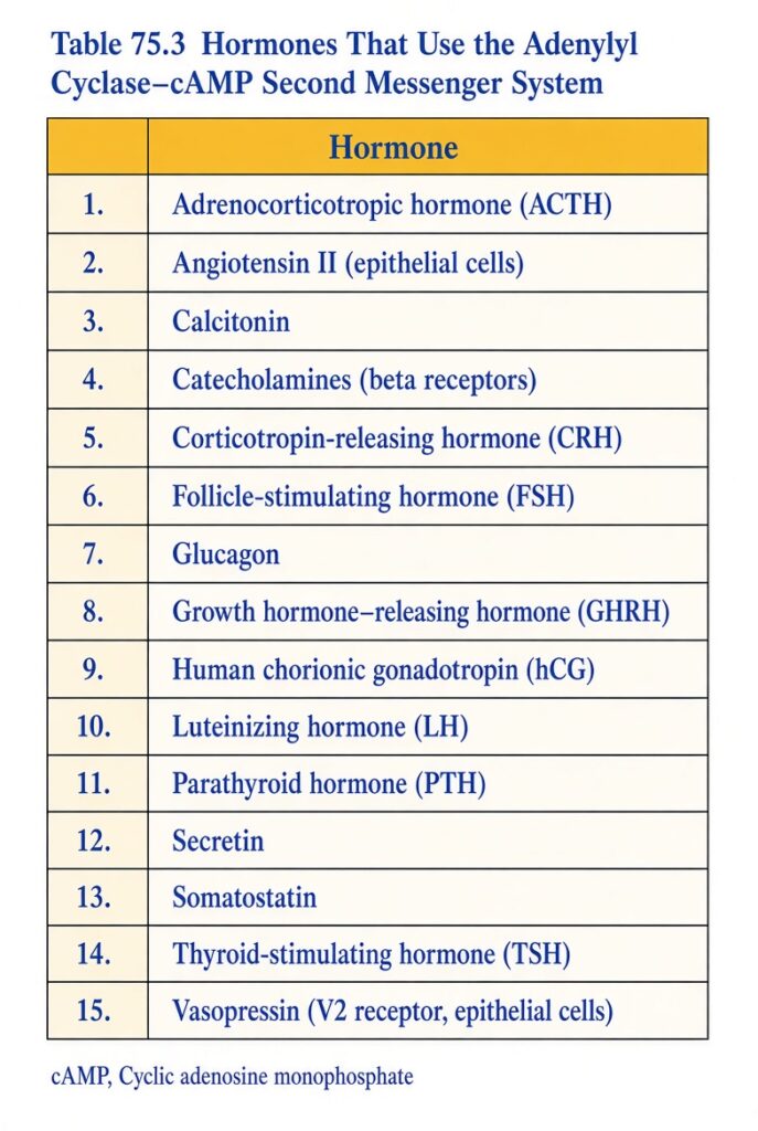
- When a hormone binds to its receptor, the receptor connects with a G protein.
- If this G protein stimulates adenylyl cyclase, it is called a Gs protein (stimulatory G protein).
- Gs activates adenylyl cyclase, a membrane-bound enzyme.
- Adenylyl cyclase converts a small amount of cytoplasmic ATP into cAMP inside the cell.
- cAMP activates cAMP-dependent protein kinase, which adds phosphate groups to specific cell proteins.
- These protein changes start biochemical reactions that produce the cell’s response to the hormone.
- Once cAMP is formed, it usually starts an enzyme cascade.
- One enzyme activates another, which activates another, and so on.
- This creates amplification, so a very small amount of hormone can produce a powerful response throughout the cell.
- If the hormone receptor is connected to an inhibitory G protein (Gi), adenylyl cyclase is inhibited.
- This reduces cAMP formation and usually produces an inhibitory effect in the cell.
- Therefore, Gs increases cAMP, whereas Gi decreases cAMP.
- Changes in cAMP can also change the phosphorylation of important proteins inside the cell.
- The final effect of cAMP depends on the intracellular machinery of the target cell.
- Different cells contain different enzymes, so the same cAMP signal can produce different effects.
- These effects may include:
- Synthesis of specific intracellular chemicals
- Muscle contraction or relaxation
- Secretion by cells
- Changes in cell permeability
- In thyroid cells, cAMP stimulates formation of thyroxine and triiodothyronine.
- In adrenocortical cells, cAMP causes secretion of adrenocortical steroid hormones.
- In some epithelial cells of the distal and collecting tubules of the kidney, cAMP increases water permeability.
KEY CONCEPT
- Hormone → receptor → G protein → adenylyl cyclase → cAMP → protein kinase → protein phosphorylation → cell response.
- Gs → ↑ adenylyl cyclase → ↑ cAMP → stimulation.
- Gi → ↓ adenylyl cyclase → ↓ cAMP → inhibition.
- Small hormone signal → enzyme cascade → large cellular response.
CONCEPTUAL EXAMPLES
- Gs example: Hormone binds receptor → Gs activates adenylyl cyclase → cAMP increases → cell is stimulated.
- Gi example: Hormone binds receptor → Gi inhibits adenylyl cyclase → cAMP decreases → cell activity is inhibited.
- Thyroid cell: cAMP → formation of thyroxine and triiodothyronine.
- Adrenocortical cell: cAMP → secretion of steroid hormones.
- Kidney epithelial cell: cAMP → increased permeability to water.
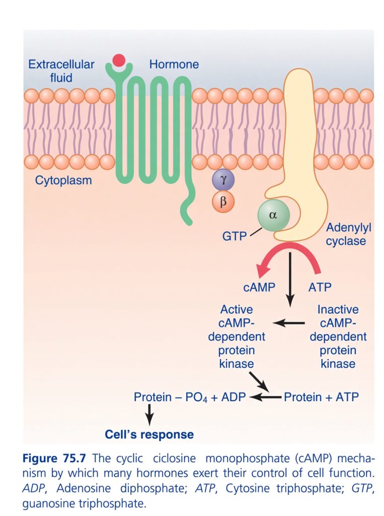
Absolutely. Let’s turn this Guyton Figure 75.7 — cAMP second-messenger system into a very simple story.
🧠 The BIG IDEA
Think of a hormone as a person knocking on the outside door of a house.
The hormone cannot enter the cell.
So it gives a message to a receptor on the cell membrane.
The receptor then activates an internal messenger system that carries the message inside the cell.
The whole pathway in ONE line:
Hormone → Receptor → G-protein → Adenylyl cyclase → cAMP → Protein kinase → Protein phosphorylation → Cell response
That is the entire figure. Now let’s understand every part.
1️⃣ Hormone = The Outside Messenger 📩
At the top of the figure:
Extracellular fluid → Hormone → Receptor
The hormone is outside the cell.
Why?
Because many hormones are water-soluble and cannot freely cross the lipid cell membrane.
So they need a receptor on the outside surface of the cell membrane.
👉 Think:
Hormone = WhatsApp message arriving at the cell’s door.
2️⃣ Receptor = The Doorbell 🔔
The green structure crossing the membrane is the hormone receptor.
When the hormone binds to it:
Hormone + receptor → receptor changes shape
This starts the signaling process.
Important:
The hormone usually does NOT go inside.
Instead, the receptor communicates the message inward.
3️⃣ The Cell Membrane = The Wall 🧱
The orange/pink structure is the cell membrane.
It separates:
Outside → Extracellular fluid
from
Inside → Cytoplasm
So:
OUTSIDE ↓Hormone ↓Receptor──────────── CELL MEMBRANE ──────────── ↓G-protein ↓Adenylyl cyclase ↓cAMP ↓Protein kinase ↓Cell responseINSIDE
4️⃣ G-Protein = The Middleman 🚶
Look near the adenylyl cyclase:
α, β, γ
These are the three components of a G-protein.
The important one here is mainly the α-subunit.
Normally:
α has GDP
When the hormone activates the receptor:
GDP is replaced by GTP
So:
Inactive G-protein → Active G-protein
Easy memory:
GDP = OFF
GTP = ON
5️⃣ What does the α-subunit do?
The activated α-subunit moves toward adenylyl cyclase.
Think:
Receptor says: “Hey G-protein, we’ve received a message!”
G-protein says:
“I’ll tell adenylyl cyclase.”
6️⃣ Adenylyl Cyclase = The Factory 🏭
This yellow structure is adenylyl cyclase.
Its job is extremely important:
It converts:
ATP → cAMP
So:
ATP → cAMP
cAMP stands for:
Cyclic adenosine monophosphate
You don’t need to memorize the entire chemical name initially.
Just remember:
Adenylyl cyclase makes cAMP.
7️⃣ cAMP = The SECOND Messenger 🏃♂️
This is the most important concept in the figure.
The hormone is the:
FIRST messenger
cAMP is the:
SECOND messenger
Why?
Because the hormone delivered the original message.
Then cAMP carries that message inside the cell.
So:
Hormone = First messenger
cAMP = Second messenger
ELI5:
Mom calls you:
“Clean your room!”
Mom = first messenger
You tell your brother:
“We need to clean!”
Your brother = second messenger
😂
8️⃣ Why does the cell need a second messenger?
Because the hormone often cannot enter the cell.
Therefore:
Hormone stays outside
but
cAMP carries the signal inside
This is why the system is called a:
Second-messenger system
9️⃣ cAMP Activates Protein Kinase
Now look at the middle-bottom part of the figure.
cAMP acts on:
cAMP-dependent protein kinase
This enzyme is commonly called:
Protein Kinase A (PKA)
So:
cAMP → activates PKA
Very important.
Remember:
cAMP = activates Protein Kinase A
🔟 What does Protein Kinase do?
This is where the cell actually starts changing its activity.
Protein kinase transfers a:
Phosphate (PO₄)
onto another protein.
This process is called:
Phosphorylation
So:
Protein + ATP → Protein–PO₄ + ADP
The protein gets a phosphate group.
1️⃣1️⃣ Why phosphorylate a protein?
Because adding phosphate can change the protein’s activity.
It may:
- activate the protein
- inhibit the protein
- change its function
- alter enzyme activity
- produce a cellular response
Therefore:
Phosphorylation → changes protein activity → cell response
1️⃣2️⃣ The final result = CELL RESPONSE 🎯
At the bottom of the figure:
Protein → Cell’s response
This is the final objective.
The cell may respond by changing things such as:
- enzyme activity
- metabolism
- ion transport
- secretion
- contraction
- other cellular functions
The exact response depends on which hormone, receptor, and intracellular proteins are involved.
🔥 NOW READ THE WHOLE FIGURE LIKE A STORY
Imagine this happening:
Step 1
A hormone arrives outside the cell.
⬇️
Step 2
Hormone binds to its receptor.
⬇️
Step 3
The receptor activates a G-protein.
⬇️
Step 4
The G-protein’s α-subunit binds GTP.
⬇️
Step 5
The α-subunit activates adenylyl cyclase.
⬇️
Step 6
Adenylyl cyclase converts:
ATP → cAMP
⬇️
Step 7
cAMP acts as the second messenger.
⬇️
Step 8
cAMP activates cAMP-dependent protein kinase (PKA).
⬇️
Step 9
PKA phosphorylates proteins.
Protein + ATP → Protein–PO₄ + ADP
⬇️
Step 10
The modified protein produces the:
CELLULAR RESPONSE
🧠 SUPER-SHORT MEMORY CHAIN
Memorize this:
H → R → G → AC → cAMP → PKA → P → Response
Where:
| Abbreviation | Meaning |
|---|---|
| H | Hormone |
| R | Receptor |
| G | G-protein |
| AC | Adenylyl cyclase |
| cAMP | Second messenger |
| PKA | Protein kinase A |
| P | Phosphorylation |
| Response | Cellular effect |
⭐ What is the role of ATP?
The figure shows ATP twice conceptually.
First:
ATP → cAMP
This is done by:
Adenylyl cyclase
Second:
ATP → ADP
This happens when protein kinase transfers phosphate to a protein.
So don’t confuse them.
Adenylyl cyclase:
ATP → cAMP
Protein kinase:
Protein + ATP → Protein–PO₄ + ADP
⚡ Why can a tiny amount of hormone cause a BIG response?
This is a very important conceptual point.
The signal can be amplified.
One hormone-receptor interaction can activate G-proteins.
Those can activate adenylyl cyclase.
Adenylyl cyclase can generate many cAMP molecules.
cAMP can activate protein kinase.
Protein kinase can modify many proteins.
Therefore:
Small outside signal → large intracellular response
Think of it like:
One person → starts a chain reaction → many people become involved.
🎯 The MOST IMPORTANT EXAM POINTS
1. Hormone
Usually remains outside the cell in this pathway.
2. Receptor
Located in the cell membrane.
3. G-protein
Acts as the link between receptor and adenylyl cyclase.
4. GTP
Activates the relevant G-protein α-subunit.
5. Adenylyl cyclase
Converts:
ATP → cAMP
6. cAMP
Is the:
SECOND MESSENGER
7. Protein kinase A
Is activated by:
cAMP
8. Protein kinase
Causes:
Phosphorylation
9. Phosphorylation
Changes protein activity.
10. Final result
Cellular response
🧩 ONE VISUAL STORY
Think of a restaurant:
Hormone = customer placing an order
↓
Receptor = waiter receiving order
↓
G-protein = waiter carrying the message
↓
Adenylyl cyclase = kitchen machine
↓
ATP → cAMP = order ticket produced
↓
cAMP = internal message
↓
Protein kinase A = chef activating workers
↓
Phosphorylation = workers get instructions
↓
Cell response = food gets prepared
🚨 Don’t make these common mistakes
❌ Hormone enters the cell
✅ Hormone binds membrane receptor.
❌ G-protein directly makes the cellular response
✅ G-protein activates the signaling pathway.
❌ cAMP is the hormone
✅ cAMP is the second messenger.
❌ Adenylyl cyclase produces ATP
✅ Adenylyl cyclase uses ATP to produce cAMP.
❌ Protein kinase produces cAMP
✅ Adenylyl cyclase produces cAMP; cAMP activates protein kinase.
❌ Phosphorylation means removing phosphate
✅ Phosphorylation means adding phosphate.
🏆 FINAL 10-SECOND REVISION
Hormone knocks → receptor hears → G-protein switches ON → adenylyl cyclase works → ATP becomes cAMP → cAMP activates PKA → PKA phosphorylates proteins → proteins change → CELL RESPONDS.
That is the entire Guyton figure.
🔑 Golden sentence:
“The hormone gives the message outside; cAMP carries the message inside; protein kinase converts the message into a cellular action.”
Cell Membrane Phospholipid Second Messenger System
- Some hormones activate transmembrane receptors, which activate phospholipase C (PLC) on the inner side of the receptor (Table 75.4).
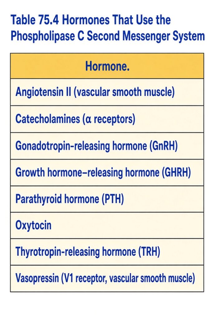
- PLC breaks down membrane phospholipids, especially phosphatidylinositol bisphosphate (PIP₂).
- PIP₂ is broken down into two second messengers:
- Inositol triphosphate (IP₃)
- Diacylglycerol (DAG)
- IP₃ releases Ca²⁺ from mitochondria and the endoplasmic reticulum.
- Ca²⁺ then acts as a second messenger, producing effects such as:
- Smooth muscle contraction
- Changes in cell secretion
- DAG activates protein kinase C (PKC).
- PKC phosphorylates many proteins, which produces the cellular response (Fig. 75.8).
- The lipid portion of DAG is arachidonic acid.
- Arachidonic acid is a precursor of prostaglandins and other local hormones.
- These substances produce multiple effects in tissues throughout the body.
KEY CONCEPT
Hormone → transmembrane receptor → PLC → PIP₂ → IP₃ + DAG
- IP₃ → Ca²⁺ release → cellular effects
- DAG → PKC → protein phosphorylation → cellular response
- Arachidonic acid → prostaglandins and other local hormones
CONCEPTUAL EXAMPLES
- Smooth muscle: PIP₂ → IP₃ → Ca²⁺ → smooth muscle contraction.
- Cell secretion: PIP₂ → IP₃ → Ca²⁺ → changes in secretion.
- Protein response: PIP₂ → DAG → PKC → protein phosphorylation → cell response.
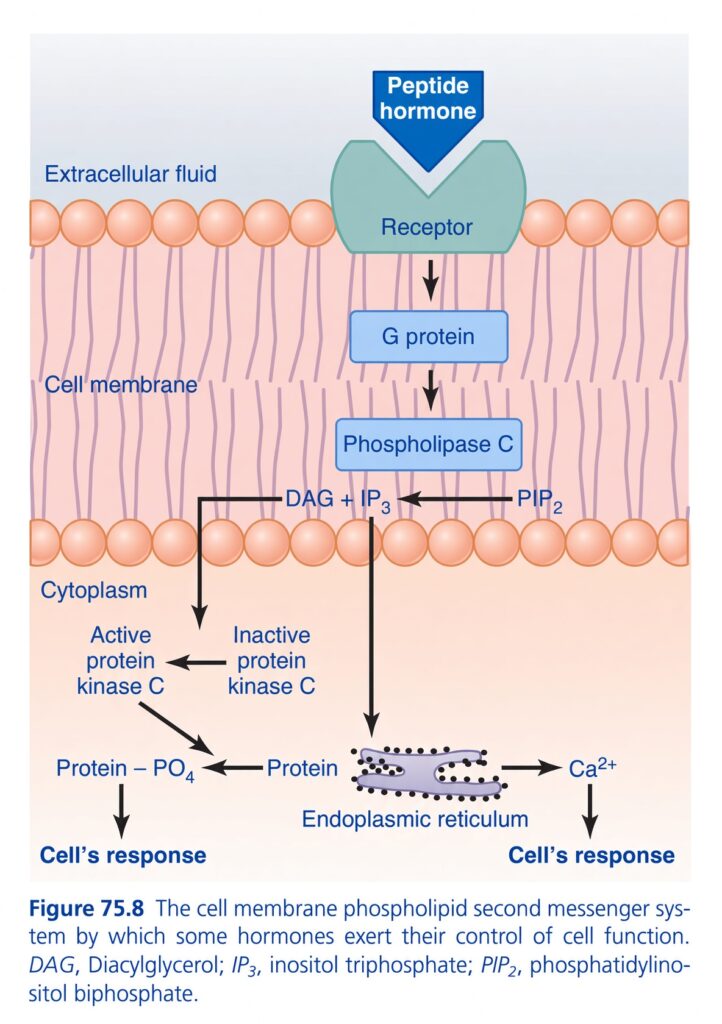
🧠 THE BIG PICTURE
This pathway is basically:
Hormone → Receptor → G-protein → Phospholipase C → PIP₂ → IP₃ + DAG → Ca²⁺ + PKC → Cell response
The most important thing is that PIP₂ gets split into TWO messengers:
🔵 IP₃ → releases Ca²⁺
🟠 DAG → activates Protein Kinase C
1️⃣ PEPTIDE HORMONE 📩
At the very top:
Peptide hormone
It is outside the cell.
Why doesn’t it simply enter the cell?
Because peptide hormones are generally water-soluble and cannot easily cross the lipid cell membrane.
So they need a receptor.
Think:
Hormone = person knocking at the door 🚪
2️⃣ RECEPTOR = THE DOORBELL 🔔
The peptide hormone binds to the:
Receptor
The receptor is embedded ( Attached ) in the cell membrane.
When the hormone binds:
Receptor becomes activated
Then the receptor sends the message toward the inside.
3️⃣ G-PROTEIN = THE MESSENGER 🚶
Immediately below the receptor is:
G protein
The receptor activates the G protein.
Think:
Receptor: “Hey G-protein, we received a hormone message!”
G-protein:
“Got it. I’ll pass the message along.”
4️⃣ PHOSPHOLIPASE C = THE SCISSORS ✂️
G-protein activates:
Phospholipase C (PLC)
This is one of the most important enzymes in the diagram.
Think:
PLC = scissors
Its job is to cut a membrane molecule.
That molecule is:
PIP₂
5️⃣ PIP₂ = THE ORIGINAL RAW MATERIAL 🧱
PIP₂ stands for:
Phosphatidylinositol bisphosphate
Don’t get stuck on the long name.
For exams, remember:
PIP₂ is the molecule that PLC cuts.
PLC cuts:
PIP₂ → IP₃ + DAG
🔥 This is the central reaction of the whole figure.
6️⃣ TWO NEW MESSENGERS ARE CREATED
Once PLC cuts PIP₂, you get:
IP₃
and
DAG
Now the pathway splits into two branches.
🔵 BRANCH 1: IP₃ → Ca²⁺
Follow the IP₃ arrow downward.
IP₃ travels toward the:
Endoplasmic reticulum (ER)
The ER acts like a:
Calcium storage warehouse 🏪
It stores lots of Ca²⁺.
IP₃ basically says:
“Open the calcium warehouse!”
So the ER releases:
Ca²⁺
7️⃣ Ca²⁺ = ANOTHER SECOND MESSENGER ⚡
The calcium released into the cytoplasm now acts as a signaling molecule.
So:
IP₃ → Ca²⁺ release
Then:
Ca²⁺ → cellular response
The response can include things such as:
- smooth muscle contraction
- secretion
- changes in enzyme activity
- other cellular functions
Easy memory:
IP₃ = Calcium releaser
🟠 BRANCH 2: DAG → Protein Kinase C
Now go back to the other product:
DAG
DAG activates:
Protein Kinase C (PKC)
In the figure you can see:
Inactive protein kinase C
⬇️
Active protein kinase C
So:
DAG → activates PKC
8️⃣ PKC = THE PHOSPHORYLATION MACHINE ⚙️
Once PKC becomes active, it acts on proteins.
It adds a phosphate group:
Protein + ATP → Protein–PO₄ + ADP
This is:
PHOSPHORYLATION
The phosphorylated protein changes its activity.
That produces:
Cell’s response
🎯 NOW FOLLOW THE ENTIRE FIGURE
Let’s read it from top to bottom:
PEPTIDE HORMONE ↓ RECEPTOR ↓ G-PROTEIN ↓PHOSPHOLIPASE C ↓ PIP₂ ↓ ┌─────┴─────┐ ↓ ↓IP₃ DAG ↓ ↓ER PKC ↓ ↓Ca²⁺ Phosphorylation ↓ ↓CELL CELLRESPONSE RESPONSE
That’s the entire figure! 🎯
🧠 WHY TWO PATHWAYS?
Because one hormone signal can produce multiple effects simultaneously.
IP₃ branch:
IP₃ → Ca²⁺ → response
DAG branch:
DAG → PKC → phosphorylation → response
So one hormone can activate different intracellular mechanisms.
🔥 THE MOST IMPORTANT DIFFERENCE
Don’t mix up these two:
| Molecule | Main job |
|---|---|
| IP₃ | Releases Ca²⁺ from ER |
| DAG | Activates Protein Kinase C |
| Ca²⁺ | Produces cellular effects |
| PKC | Phosphorylates proteins |
🔑 One-line memory:
“IP₃ opens the Calcium door; DAG turns PKC ON.”
🧩 WHAT IS THE ER DOING?
The purple structure at the bottom is:
Endoplasmic reticulum
Here, think of ER as:
Calcium storage tank 🛢️
Normally:
Ca²⁺ stored inside ER
IP₃ arrives:
IP₃ → opens Ca²⁺ release channels
Then:
Ca²⁺ moves into cytoplasm
Then Ca²⁺ helps create the cellular response.
🧠 WHY IS Ca²⁺ CALLED A SECOND MESSENGER?
Because the hormone started the message.
The hormone itself doesn’t have to enter the cell.
Instead:
Hormone → receptor → signaling pathway → Ca²⁺
Ca²⁺ then carries the message inside the cell.
Therefore:
First messenger:
Hormone
Second messengers:
IP₃, DAG
And:
Ca²⁺ can also function as a second messenger.
⚡ COMPARE WITH THE PREVIOUS cAMP FIGURE
You just learned the cAMP system.
Now put them side-by-side:
cAMP SYSTEM
Hormone
↓
Receptor
↓
G-protein
↓
Adenylyl cyclase
↓
ATP → cAMP
↓
PKA
↓
Phosphorylation
↓
Cell response
PHOSPHOLIPID SYSTEM
Hormone
↓
Receptor
↓
G-protein
↓
PLC
↓
PIP₂
↓
IP₃ + DAG
Then:
IP₃ → Ca²⁺ → response
and
DAG → PKC → phosphorylation → response
🚨 EXAM TRAPS
❌ PLC converts ATP into cAMP
No.
✅ PLC breaks down PIP₂.
❌ IP₃ activates PKC
No.
✅ DAG activates PKC.
❌ DAG releases Ca²⁺ from ER
No.
✅ IP₃ causes Ca²⁺ release.
❌ PKC releases Ca²⁺
No.
✅ PKC phosphorylates proteins.
❌ PIP₂ is the final messenger
No.
✅ PIP₂ is the substrate that gets broken down to produce IP₃ and DAG.
🏆 10-SECOND GUYTON REVISION
If you’re in an exam and see this pathway, immediately think:
“PLC CUTS PIP₂.”
Then:
PIP₂ → IP₃ + DAG
And:
IP₃ → Ca²⁺
DAG → PKC
PKC → Phosphorylation
Ca²⁺ + phosphorylation → Cell response
🧠 ULTIMATE MEMORY FORMULA
H → R → G → PLC → PIP₂ → IP₃ + DAG
Then:
🔵 IP₃ → ER → Ca²⁺ → Response
🟠 DAG → PKC → Protein phosphorylation → Response
⭐ Golden sentence:
“Hormone activates receptor, receptor activates G-protein, G-protein activates PLC, PLC cuts PIP₂ into IP₃ and DAG; IP₃ releases Ca²⁺, while DAG activates PKC.”
