


LEARNING OBJECTIVES
After studying this topic, students will be able to:
- Describe the physiological anatomy of a synapse and presynaptic terminal.
- Explain neurotransmitter release and the role of Ca²⁺ and SNARE proteins.
- Differentiate ionotropic and metabotropic receptor actions.
- Explain how cation and anion channels cause excitation or inhibition.
- Describe how second messenger systems produce prolonged neuronal effects.
INTRODUCTION
A synapse is the functional site where one neuron communicates with another neuron through presynaptic terminals, neurotransmitters, and postsynaptic receptors. Neurotransmitters released from presynaptic terminals may excite or inhibit the postsynaptic neuron, depending on the neurotransmitter and receptor involved.
Synaptic transmission may produce a rapid effect through ion channels or a prolonged effect through second messenger systems. Understanding these mechanisms explains how neurons receive, process, and respond to incoming signals and how prolonged neuronal changes can occur in functions such as memory.
- Fig. 46.6 shows a typical anterior motor neuron in the anterior horn of the spinal cord.
- The anterior motor neuron has three major parts:
- Soma → the main body of the neuron.
- Single axon → extends from the soma into a peripheral nerve that leaves the spinal cord.
- Dendrites → many branching projections coming from the soma.
- The dendrites can extend up to 1 millimeter into the surrounding areas of the spinal cord.
- About 10,000 to 200,000 small synaptic knobs, called presynaptic terminals, may be present on the surfaces of the dendrites and soma.
- About:
- 80%–95% of presynaptic terminals are present on the dendrites.
- 5%–20% are present on the soma.
- These presynaptic terminals are the ends of nerve fibers coming from many other neurons.
- Many presynaptic terminals are excitatory:
- They release a neurotransmitter.
- This neurotransmitter excites the postsynaptic neuron.
- Other presynaptic terminals are inhibitory:
- They release a neurotransmitter.
- This neurotransmitter inhibits the postsynaptic neuron.
- Neurons in other parts of the spinal cord and brain can differ from the anterior motor neuron in:
- Size of the cell body
- Length, size, and number of dendrites
- Dendrites may range from almost zero length to many centimeters
- Length and size of the axon
- Number of presynaptic terminals
- The number of presynaptic terminals may range from only a few to as many as 200,000.
- Because of these differences, neurons in different parts of the nervous system respond differently to incoming synaptic signals.
- Therefore, different neurons can perform many different functions.
Easiest Example
Presynaptic terminal → releases neurotransmitter → postsynaptic neuron → excitation or inhibition
Excitatory terminal → neurotransmitter released → postsynaptic neuron excited
Inhibitory terminal → neurotransmitter released → postsynaptic neuron inhibited
KEY CONCEPT
Anterior motor neuron receives thousands of presynaptic terminals → terminals may be excitatory or inhibitory → released neurotransmitters excite or inhibit the postsynaptic neuron → differences in neuronal structure and synaptic inputs allow different neurons to perform different functions.
PRESYNAPTIC TERMINALS
- Electron microscopic studies show that presynaptic terminals have different anatomical shapes.
- Most presynaptic terminals look like small round or oval knobs.
- Therefore, they are also called:
- Terminal knobs
- Boutons
- End-feet
- Synaptic knobs
- Fig. 46.5A shows the basic structure of a chemical synapse.
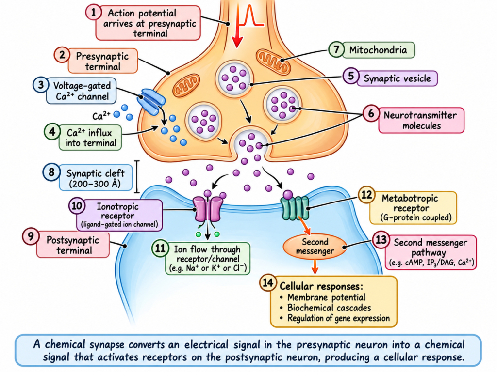
- It shows a single presynaptic terminal present on the membrane surface of a postsynaptic neuron.
- The presynaptic terminal is separated from the postsynaptic neuronal soma by a synaptic cleft.
- The synaptic cleft is usually 200–300 Å wide.
- The presynaptic terminal contains two important internal structures:
- Transmitter vesicles
- Mitochondria
- These structures are important for the excitatory or inhibitory function of the synapse.
- Transmitter vesicles contain the neurotransmitter.
- The neurotransmitter is released into the synaptic cleft and may excite or inhibit the postsynaptic neuron.
- If the postsynaptic neuronal membrane contains excitatory receptors:
- Neurotransmitter binds to them.
- → The postsynaptic neuron is excited.
- If the postsynaptic neuronal membrane contains inhibitory receptors:
- Neurotransmitter binds to them.
- → The postsynaptic neuron is inhibited.
- Mitochondria provide adenosine triphosphate (ATP).
- ATP provides the energy needed to synthesize new neurotransmitters.
- When an action potential reaches the presynaptic terminal:
- The presynaptic membrane becomes depolarized.
- → A small number of vesicles empty their neurotransmitter into the synaptic cleft.
- → The released neurotransmitter binds to a receptor on the postsynaptic neuronal membrane.
- → This immediately changes the permeability of the postsynaptic membrane.
- → The postsynaptic neuron is excited or inhibited.
- Whether the postsynaptic neuron is excited or inhibited depends on:
- The neurotransmitter released
- The characteristics of the neuronal receptor
KEY CONCEPT
Action potential reaches presynaptic terminal → membrane depolarizes → transmitter vesicles release neurotransmitter → neurotransmitter binds to postsynaptic receptors → membrane permeability changes → postsynaptic neuron is excited or inhibited.
Easiest Example
Excitatory receptor → neurotransmitter binds → postsynaptic neuron excited
Inhibitory receptor → neurotransmitter binds → postsynaptic neuron inhibited
TRANSMITTER RELEASE FROM PRESYNAPTIC TERMINALS AND ROLE OF CALCIUM IONS
- The presynaptic terminal membrane contains active zones.
- At the active zones:
- Some vesicles containing small-molecule neurotransmitters are already docked.
- These vesicles are ready for immediate release when an action potential arrives.
- Other vesicles containing small-molecule neurotransmitters remain in a reserve pool outside the active zone.
- Vesicles containing neuropeptide transmitters do not dock at the active zones.
- These neuropeptide vesicles are:
- Located outside the active zone
- Away from the terminal membrane
- Slower to release than small-molecule transmitter vesicles
- Docking of synaptic vesicles at the active zone occurs through interactions between SNARE proteins.
- SNARE means soluble N-ethylmaleimide-sensitive factor attachment protein receptor.
- SNARE proteins are present on:
- The vesicle surface
- The presynaptic terminal membrane
- The exact interactions responsible for:
- Vesicle docking
- Fusion with the presynaptic terminal membrane
- Release of neurotransmitters
are not fully understood.
- Fig. 46.7 shows a simplified model of this process.
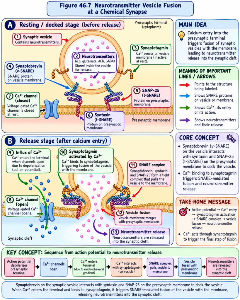
- A vesicle SNARE (v-SNARE) called synaptobrevin binds to two target membrane SNARE proteins (t-SNAREs):
- Syntaxin
- SNAP-25
- Interaction of these three proteins:
- Synaptobrevin + Syntaxin + SNAP-25
- → causes the vesicle to dock at the active zone.
- The active zones of the presynaptic terminal membrane contain many voltage-gated calcium channels.
- When an action potential depolarizes the presynaptic membrane:
- Voltage-gated calcium channels open.
- → Calcium ions enter the presynaptic terminal.
- This is shown in Figs. 46.5A and 46.7.
- The amount of neurotransmitter released into the synaptic cleft is directly related to the number of calcium ions entering the terminal (Video 46.2).
- Calcium ions entering the presynaptic terminal bind to a vesicle-bound calcium-sensing protein called synaptotagmin.
- Synaptotagmin then interacts with the membrane-bound SNARE proteins.
- This interaction forms a molecular structure called the trans-SNARE complex.
- The trans-SNARE complex causes:
- Fusion of the vesicular membrane with the presynaptic terminal membrane.
- This is shown in Fig. 46.7.
- As the vesicular membrane merges with the terminal cell membrane:
- The contents of the vesicle are released into the synaptic cleft.
- → The released neurotransmitter can then interact with postsynaptic receptors.
- This process is called exocytosis.
- Exocytosis of synaptic vesicles and release of neurotransmitters occurs after each single action potential.
- In vesicles containing acetylcholine:
- Each vesicle contains about 2,000–10,000 acetylcholine molecules.
- The presynaptic terminal contains enough vesicles to transmit:
- From a few hundred
- To more than 10,000 action potentials
KEY CONCEPT
Action potential arrives → presynaptic membrane depolarizes → voltage-gated Ca²⁺ channels open → Ca²⁺ enters → Ca²⁺ binds synaptotagmin → SNARE proteins interact → trans-SNARE complex forms → vesicle fuses with terminal membrane → neurotransmitter is released by exocytosis → neurotransmitter interacts with postsynaptic receptors.
Easiest Example
More Ca²⁺ enters the presynaptic terminal → more neurotransmitter is released into the synaptic cleft.
TRANSMITTER ACTIONS ON POSTSYNAPTIC NEURONS—FUNCTION OF RECEPTOR PROTEINS
- The membrane of the postsynaptic neuron contains large numbers of receptor proteins, as shown in Fig. 46.5A.
- Each receptor protein has two important components:
- Binding component
- Projects outward from the membrane into the synaptic cleft.
- Binds the neurotransmitter coming from the presynaptic terminal.
- Intracellular component
- Passes through the postsynaptic membrane.
- Extends into the inside of the postsynaptic neuron.
- Binding component
- Activation of the receptor controls the opening of ion channels in the postsynaptic cell in two ways:
- 1. Directly through ion channels
- The receptor directly opens ion channels.
- → Specific types of ions pass through the membrane.
- 2. Through a second messenger
- The receptor activates a second messenger.
- The second messenger is not an ion channel.
- It is a molecule that extends into the cell cytoplasm.
- → It activates one or more substances inside the postsynaptic neuron.
- → These second messengers increase or decrease specific cellular functions.
- 1. Directly through ion channels
- Neurotransmitter receptors that directly control ion channels are called ionotropic receptors.
- Neurotransmitter receptors that act through second messenger systems are called metabotropic receptors.
KEY CONCEPT
Neurotransmitter binds to postsynaptic receptor → receptor acts either directly on an ion channel or through a second messenger → postsynaptic cell function changes.
Easiest Example
Ionotropic receptor → directly controls ion channel
Metabotropic receptor → activates second messenger → changes cellular functions
CATION AND ANION CHANNELS
- Ion channels in the postsynaptic neuronal membrane are usually of two types:
- 1. Cation channels
- Usually allow sodium ions (Na⁺) to pass when opened.
- Sometimes also allow potassium ions (K⁺) and/or calcium ions (Ca²⁺) to pass.
- 2. Anion channels
- Mainly allow chloride ions (Cl⁻) to pass.
- May also allow very small amounts of other anions to pass.
- 1. Cation channels
- As discussed in Chapter 4, these ion channels are highly selective for one or more specific ions.
- Channel selectivity depends on:
- Diameter
- Shape
- Electrical charges along the inside surface
- Chemical bonds along the inside surface
- Cation channels that carry sodium ions are lined with negative charges.
- These negative charges:
- Attract positively charged sodium ions into the channel.
- This occurs when the channel becomes wide enough for the hydrated sodium ion to pass.
- The same negative charges:
- Repel chloride ions and other anions.
- → Prevent their passage through the channel.
- In anion channels:
- When the channel becomes wide enough, chloride ions enter the channel.
- → Chloride ions pass through to the opposite side.
- However:
- Sodium
- Potassium
- Calcium
are blocked mainly because their hydrated ions are too large to pass.
- When cation channels open:
- Positively charged sodium ions enter the neuron.
- → Positive electrical charge enters.
- → The neuron is excited.
- Therefore, a neurotransmitter that opens cation channels is called an excitatory transmitter.
- When anion channels open:
- Negative electrical charges enter the neuron.
- → The neuron is inhibited.
- Therefore, neurotransmitters that open anion channels are called inhibitory transmitters.
- When a neurotransmitter activates an ion channel:
- The channel usually opens within a fraction of a millisecond.
- When the neurotransmitter is no longer present:
- The channel closes equally rapidly.
- Therefore, opening and closing of ion channels provides very rapid control of postsynaptic neurons.
KEY CONCEPT
Cation channel opens → Na⁺ enters → positive charge enters → neuron excited → excitatory effect
Anion channel opens → negative charge enters → neuron inhibited → inhibitory effect
Easiest Example
Na⁺ entry → excitation
Cl⁻ entry → inhibition
“SECOND MESSENGER” SYSTEM IN THE POSTSYNAPTIC NEURON
- Many functions of the nervous system, such as memory, need long-lasting changes in neurons.
- These changes may continue for:
- Seconds
- To months
- They can continue even after the original neurotransmitter is no longer present.
- Ion channels are not suitable for producing these prolonged changes.
- This is because ion channels close within milliseconds after the neurotransmitter is no longer present.
- In many cases, prolonged excitation or inhibition of the postsynaptic neuron occurs through a second messenger chemical system inside the neuron.
- The second messenger then produces the prolonged effect.
- There are several types of second messenger systems.
- One of the most common systems uses proteins called G proteins.
- Fig. 46.8 shows a membrane receptor G protein.
- The inactive G protein complex is free in the cytosol.
- It contains:
- Guanosine diphosphate (GDP)
- Alpha (α) component
- Beta (β) component
- Gamma (γ) component
- The α component is the activator part of the G protein.
- The β and γ components are attached to the α component.
- As long as the G protein complex is bound to GDP:
- → The G protein remains inactive.
- After a nerve impulse, a neurotransmitter activates the receptor.
- The receptor then undergoes a conformational change.
- This change exposes a binding site for the G protein complex.
- The G protein complex then binds to the part of the receptor that extends into the inside of the cell.
- This allows the α subunit to:
- Release GDP
- Bind guanosine triphosphate (GTP) at the same time
- Separate from the β and γ subunits
- The separated α-GTP complex can then move freely through the cytoplasm.
- It can perform one or more functions depending on the specific characteristics of the neuron.
- Fig. 46.8 shows the following four possible changes:
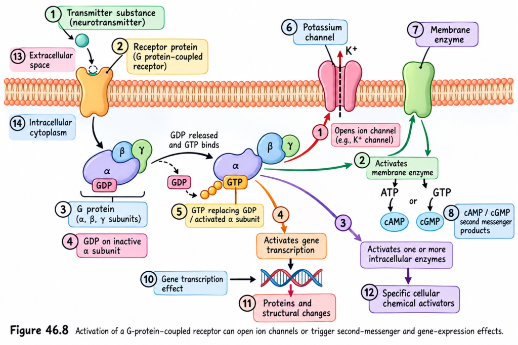
1. Opening Specific Ion Channels
- The G protein can open specific ion channels in the postsynaptic cell membrane.
- The upper right part of Fig. 46.8 shows a potassium channel opened in response to the G protein.
- This channel often remains open for a prolonged time.
- This is different from directly activated ion channels, which close rapidly and do not use a second messenger system.
2. Activation of cAMP or cGMP
- The G protein can activate:
- Cyclic adenosine monophosphate (cAMP)
- Cyclic guanosine monophosphate (cGMP)
- cAMP or cGMP can activate highly specific metabolic machinery inside the neuron.
- This can produce many different chemical effects.
- These effects may include long-term changes in the structure of the cell.
- These structural changes can alter the long-term excitability of the neuron.
3. Activation of Intracellular Enzymes
- The G protein can directly activate one or more intracellular enzymes.
- These enzymes can then produce many specific chemical functions inside the cell.
4. Activation of Gene Transcription
- The G protein system can activate gene transcription.
- This is one of the most important effects of second messenger systems.
- Gene transcription can cause formation of new proteins inside the neuron.
- These new proteins can change:
- The neuron’s metabolic machinery
- The neuron’s structure
- Structural changes occur in appropriately activated neurons, especially during long-term memory processes.
- The G protein becomes inactive when the GTP attached to the α subunit is hydrolyzed to GDP.
- This causes the α subunit to:
- Leave its target protein
- → Inactivate the second messenger system
- The α subunit then combines again with:
- β subunit
- γ subunit
- The complete G protein complex returns to its inactive state.
- Activation of second messenger systems, including G protein systems and other types, is very important for changing the long-term response characteristics of neuronal pathways.
- This subject is discussed again in Chapter 58 in relation to memory functions of the nervous system.
KEY CONCEPT
Neurotransmitter activates receptor → G protein binds receptor → α subunit releases GDP and binds GTP → α-GTP separates → activates ion channels, cAMP/cGMP, enzymes, or gene transcription → prolonged neuronal effects occur.
GTP → hydrolyzed to GDP → α subunit returns to β and γ → G protein becomes inactive again.
Easiest Example
Direct ion channel system → effect lasts for milliseconds
Second messenger system → produces prolonged neuronal changes → may last from seconds to months
CLINICAL IMPORTANCE
- Synaptic excitation and inhibition: Presynaptic terminals release neurotransmitters that can either excite or inhibit the postsynaptic neuron, depending on the neurotransmitter and receptor involved.
- Role of calcium in neurotransmitter release: Entry of Ca²⁺ into the presynaptic terminal triggers vesicle fusion and neurotransmitter release; the amount released is directly related to the amount of Ca²⁺ entering the terminal.
- Role of SNARE proteins: Synaptobrevin, syntaxin, and SNAP-25 help dock synaptic vesicles and participate in their fusion with the presynaptic membrane, allowing exocytosis of neurotransmitter.
- Fast neuronal responses: Ionotropic receptors directly control ion channels and therefore produce very rapid changes in the postsynaptic neuron.
- Excitatory effect: Opening of cation channels, especially allowing Na⁺ entry, brings positive charge into the neuron and causes excitation.
- Inhibitory effect: Opening of anion channels, mainly allowing Cl⁻ movement, produces an inhibitory effect on the neuron.
- Rapid versus prolonged effects: Directly controlled ion channels act for a very short time, whereas second messenger systems can produce neuronal effects lasting from seconds to months.
- Importance in memory: Long-lasting neuronal changes required for functions such as memory are produced through second messenger systems rather than short-lived ion-channel effects.
- Long-term neuronal changes: G-protein second messenger systems can affect ion channels, cAMP/cGMP, intracellular enzymes, and gene transcription, producing prolonged changes in neuronal function.
- Gene transcription and long-term memory: Activation of gene transcription can produce new proteins that alter neuronal metabolism and structure, especially during long-term memory processes.
HIGH-YIELD POINTS
- An anterior motor neuron has three major parts: soma, single axon, and dendrites.
- About 10,000–200,000 presynaptic terminals may be present on the dendrites and soma.
- About 80%–95% of presynaptic terminals are on dendrites, while 5%–20% are on the soma.
- Presynaptic terminals may be excitatory or inhibitory.
- Different neuronal structures and synaptic inputs allow different neurons to perform different functions.
- Presynaptic terminals are also called terminal knobs, boutons, end-feet, or synaptic knobs.
- The synaptic cleft is about 200–300 Å wide.
- Presynaptic terminals contain transmitter vesicles and mitochondria.
- Transmitter vesicles contain neurotransmitter, while mitochondria provide ATP for neurotransmitter synthesis.
- An action potential depolarizes the presynaptic membrane → neurotransmitter is released → it binds postsynaptic receptors → membrane permeability changes → the neuron is excited or inhibited.
- Small-molecule transmitter vesicles may be docked at active zones and ready for immediate release.
- Vesicle docking involves SNARE proteins.
- Synaptobrevin + syntaxin + SNAP-25 help dock the vesicle at the active zone.
- An action potential opens voltage-gated Ca²⁺ channels in the presynaptic membrane.
- The amount of neurotransmitter released is directly related to the amount of Ca²⁺ entering the terminal.
- Ca²⁺ binds synaptotagmin, which interacts with SNARE proteins and promotes vesicle fusion.
- Neurotransmitter release by vesicle fusion is called exocytosis.
- Postsynaptic receptors have a binding component and an intracellular component.
- Ionotropic receptors directly control ion channels.
- Metabotropic receptors act through second messenger systems.
- Postsynaptic ion channels are mainly cation channels and anion channels.
- Opening cation channels, especially allowing Na⁺ entry, causes neuronal excitation.
- Opening anion channels, mainly involving Cl⁻, causes neuronal inhibition.
- Direct ion-channel actions are very rapid and occur within fractions of a millisecond.
- Second messenger systems are important for prolonged neuronal changes, including memory-related changes.
- These prolonged effects may last from seconds to months.
- One common second messenger system uses G proteins.
- An inactive G protein contains GDP + α + β + γ subunits.
- After receptor activation, the α subunit releases GDP, binds GTP, and separates from β and γ.
- The α-GTP complex can activate:
- Specific ion channels
- cAMP or cGMP
- Intracellular enzymes
- Gene transcription
- Gene transcription can produce new proteins and cause long-term changes in neuronal metabolism and structure, especially during long-term memory.
- Hydrolysis of GTP to GDP inactivates the G protein system.
Quickest exam line:
Action potential → Ca²⁺ entry → synaptotagmin + SNARE interaction → vesicle fusion → exocytosis → neurotransmitter binds postsynaptic receptor → excitation/inhibition.
EXCITATORY OR INHIBITORY RECEPTORS IN THE POSTSYNAPTIC MEMBRANE
- When postsynaptic receptors are activated, they can cause:
- Excitation of the postsynaptic neuron, or
- Inhibition of the postsynaptic neuron.
- Having both excitatory and inhibitory receptors gives an additional level of control to nervous system function.
- These receptors allow nervous activity to be either:
- Excited, or
- Restrained/Inhibited.
- Different receptors use different molecular and membrane mechanisms to produce excitation or inhibition.
Excitation
- Opening of sodium (Na⁺) channels
- Sodium channels open.
- Large numbers of positive charges enter the postsynaptic cell.
- The inside of the cell becomes less negative and more positive.
- This causes depolarization of the cell membrane.
- The membrane potential moves toward the threshold level for excitation.
- This is the most widely used mechanism for excitation.
- Decreased conduction through chloride (Cl⁻) or potassium (K⁺) channels
- Movement through chloride channels, potassium channels, or both is reduced.
- Less negatively charged chloride (Cl⁻) enters the neuron.
- Or less positively charged potassium (K⁺) leaves the neuron.
- In either case, the inside of the neuron becomes less negative than normal.
- This produces an excitatory effect.
- Changes in internal metabolism
- Changes can occur in the internal metabolism of the postsynaptic neuron.
- These changes can increase cell activity.
- They may also:
- Increase the number of excitatory membrane receptors, or
- Decrease the number of inhibitory membrane receptors.
- These changes promote excitation.
Inhibition
- Opening of chloride (Cl⁻) channels
- Chloride channels in the postsynaptic membrane open.
- Negatively charged chloride ions (Cl⁻) rapidly move from outside to inside the neuron.
- More negative charges enter the neuron.
- The inside of the neuron becomes more negative.
- This produces inhibition.
- Increased potassium (K⁺) movement out of the neuron
- Potassium conductance increases.
- Positively charged potassium ions (K⁺) move out of the neuron.
- Positive charges are lost from inside the neuron.
- The inside becomes more negative.
- This produces inhibition.
- Activation of receptor enzymes
- Receptor enzymes may:
- Inhibit cellular metabolic functions.
- Increase the number of inhibitory synaptic receptors.
- Decrease the number of excitatory receptors.
- These changes promote inhibition.
- Receptor enzymes may:
Easiest Example
- Na⁺ enters the neuron → inside becomes more positive → excitation.
- Cl⁻ enters the neuron → inside becomes more negative → inhibition.
- K⁺ leaves the neuron → inside becomes more negative → inhibition.
Reference
Guyton and Hall Textbook of Medical Physiology, 15th Edition
Chapter 46 — Basic Functions of Synapses, and Neurotransmitters
