


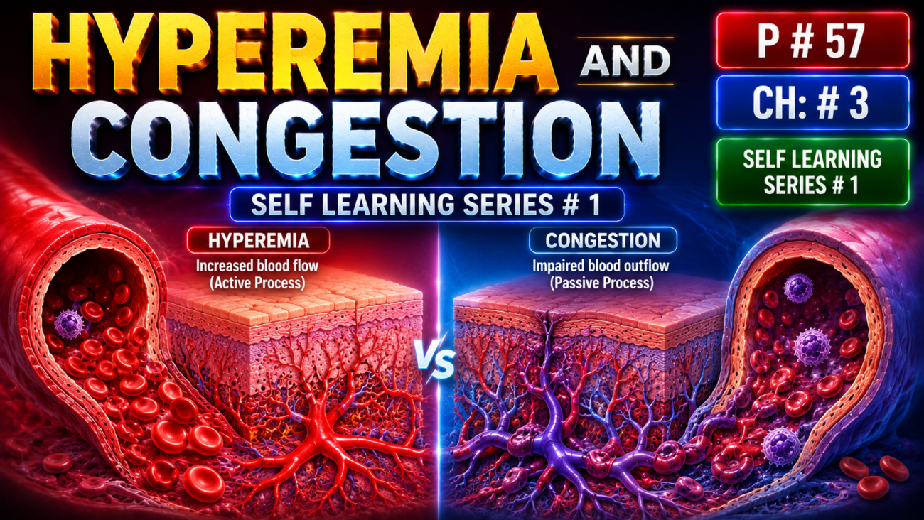
- Both mean increased blood volume within a tissue, but their mechanisms are different.
- Hyperemia is an active process:
- arterioles dilate → blood inflow increases.
- Seen in inflammation and exercising skeletal muscle.
- Tissue becomes red because it contains more oxygenated blood.
- Congestion is a passive process:
- venous blood cannot leave the tissue properly.
- Occurs systemically in cardiac failure or locally with venous obstruction.
- Tissue becomes blue-red (cyanotic) because deoxygenated hemoglobin accumulates.
- Long-standing congestion → poor tissue perfusion + persistent hypoxia → parenchymal cell death → fibrosis.
- Increased intravascular pressure may also → edema or capillary rupture → focal hemorrhage.
KEY CONCEPT
- Hyperemia = active ↑ arterial inflow → oxygenated blood → red tissue.
- Congestion = passive ↓ venous outflow → deoxygenated blood → blue-red tissue.
- Vessel injury → hemostasis; too little clotting causes hemorrhage, while inappropriate clotting causes thrombosis/embolism → ischemia and infarction.
CONCEPTUAL EXAMPLES
- Exercising muscle → arterioles dilate → more blood enters → hyperemia.
- Cardiac failure → venous blood backs up → congestion.
- Fluid enters lung tissue/alveoli → pulmonary edema → hypoxia.
- Clot blocks a coronary artery → ischemia → myocardial infarction.
MORPHOLOGY
- Cut surfaces of hyperemic or congested tissues are usually wet and may ooze blood.
- Acute pulmonary congestion:
- Alveolar capillaries become engorged with blood.
- Alveolar septa may develop edema.
- Intra-alveolar hemorrhage may occur.
- Chronic pulmonary congestion:
- Alveolar septa become thickened and fibrotic.
- Alveoli contain many macrophages filled with hemosiderin.
- These macrophages are called “heart failure cells” and come from phagocytosed red blood cells (Fig. 3.1).
- Acute hepatic congestion:
- Central vein and sinusoids become distended with blood.
- Centrally located hepatocytes may undergo necrosis because of greater hypoxia.
- Periportal hepatocytes are closer to hepatic arterioles → receive relatively more oxygen → suffer less hypoxia.
- These periportal hepatocytes may develop fatty change.
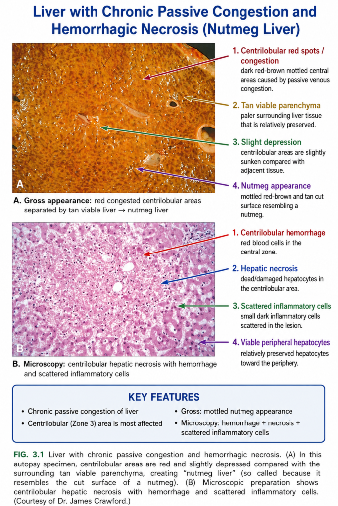
- Chronic passive liver congestion:
- Central parts of hepatic lobules become congested, red-brown, and slightly depressed because of necrosis and cell loss.
- Surrounding periportal hepatocytes appear tan and may show fatty change.
- This characteristic appearance is called “nutmeg liver” (Fig. 3.1A, B).
KEY CONCEPT
- Acute lung congestion → blood-filled capillaries + edema + hemorrhage.
- Chronic lung congestion → fibrosis + hemosiderin-laden heart failure cells.
- Acute liver congestion → central hepatocyte necrosis + periportal fatty change.
- Chronic liver congestion → characteristic nutmeg liver.
CONCEPTUAL EXAMPLES
- Long-standing blood backup in lungs → RBCs leak into alveoli → macrophages eat RBCs → hemosiderin accumulates → heart failure cells.
- Long-standing venous backup in liver → central areas receive less oxygen and are damaged most → red-brown congested centers surrounded by paler fatty areas → nutmeg liver.
EDEMA
- About 60% of lean body weight is water.
- 2/3 of body water is intracellular.
- Most remaining water is interstitial fluid.
- Only about 5% of body water is in plasma.
- Edema = abnormal accumulation of interstitial fluid in tissues.
- Fluid may also collect in body cavities as an effusion:
- Pleural cavity → hydrothorax
- Pericardial cavity → hydropericardium
- Peritoneal cavity → hydroperitoneum / ascites
- Anasarca = severe generalized edema with marked subcutaneous swelling and fluid accumulation in body cavities.
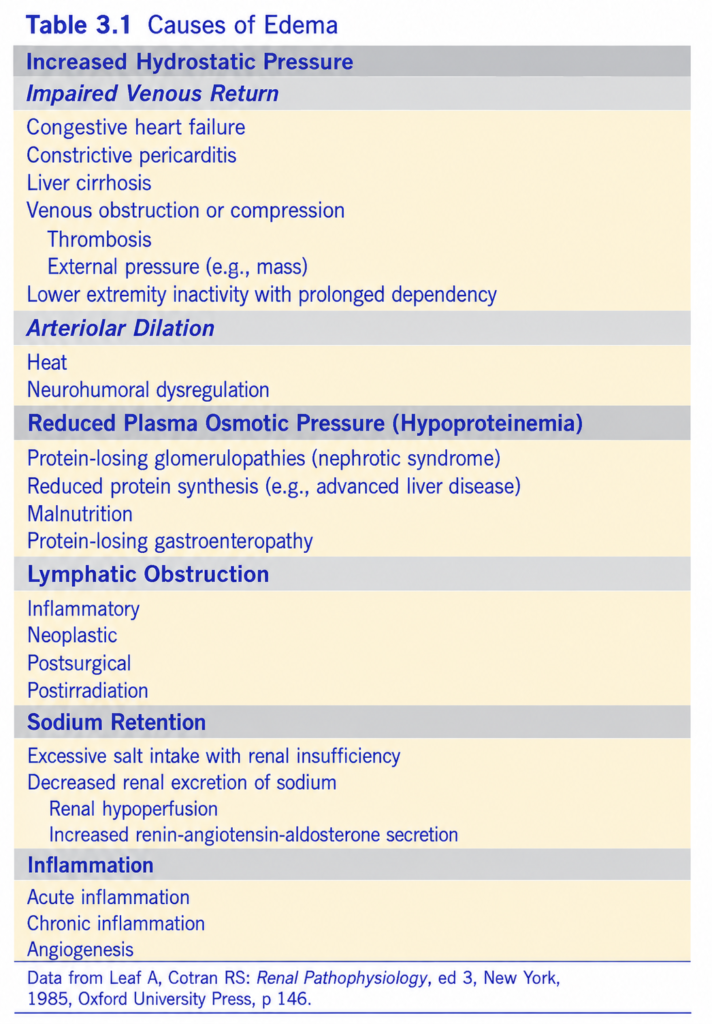
- Major causes of edema are listed in Table 3.1.
- In inflammation, edema occurs mainly because of increased vascular permeability.
- Movement of fluid between blood vessels and tissues mainly depends on two opposite forces:
- Vascular hydrostatic pressure → pushes fluid out of vessels.
- Plasma colloid osmotic pressure → pulls fluid back into vessels.
- Normally:
- At the arteriolar end, hydrostatic pressure causes fluid to move out.
- At the venular end, osmotic pressure causes fluid to move back in.
- The small amount of extra interstitial fluid is removed by lymphatics → thoracic duct → bloodstream.
- Therefore, tissues normally remain relatively dry.
- ↑ Hydrostatic pressure or ↓ colloid osmotic pressure → more water enters the interstitium (Fig. 3.2).
- If fluid accumulation exceeds lymphatic drainage capacity → edema develops.
- Edema caused by:
- High hydrostatic pressure or low colloid osmotic pressure → usually protein-poor transudate.
- Increased vascular permeability in inflammation → protein-rich exudate.
KEY CONCEPT
- Edema = excess interstitial fluid.
- ↑ Hydrostatic pressure → pushes fluid out.
- ↓ Plasma colloid osmotic pressure → less fluid pulled back in.
- If lymphatics cannot remove the excess fluid → edema.
- Transudate = protein-poor; exudate = protein-rich.
CONCEPTUAL EXAMPLES
- More pressure inside vessels → more fluid pushed into tissues → edema.
- Less plasma protein effect → less water returns to vessels → edema.
- Inflammation → vessels become more permeable → protein-rich fluid escapes → exudate.
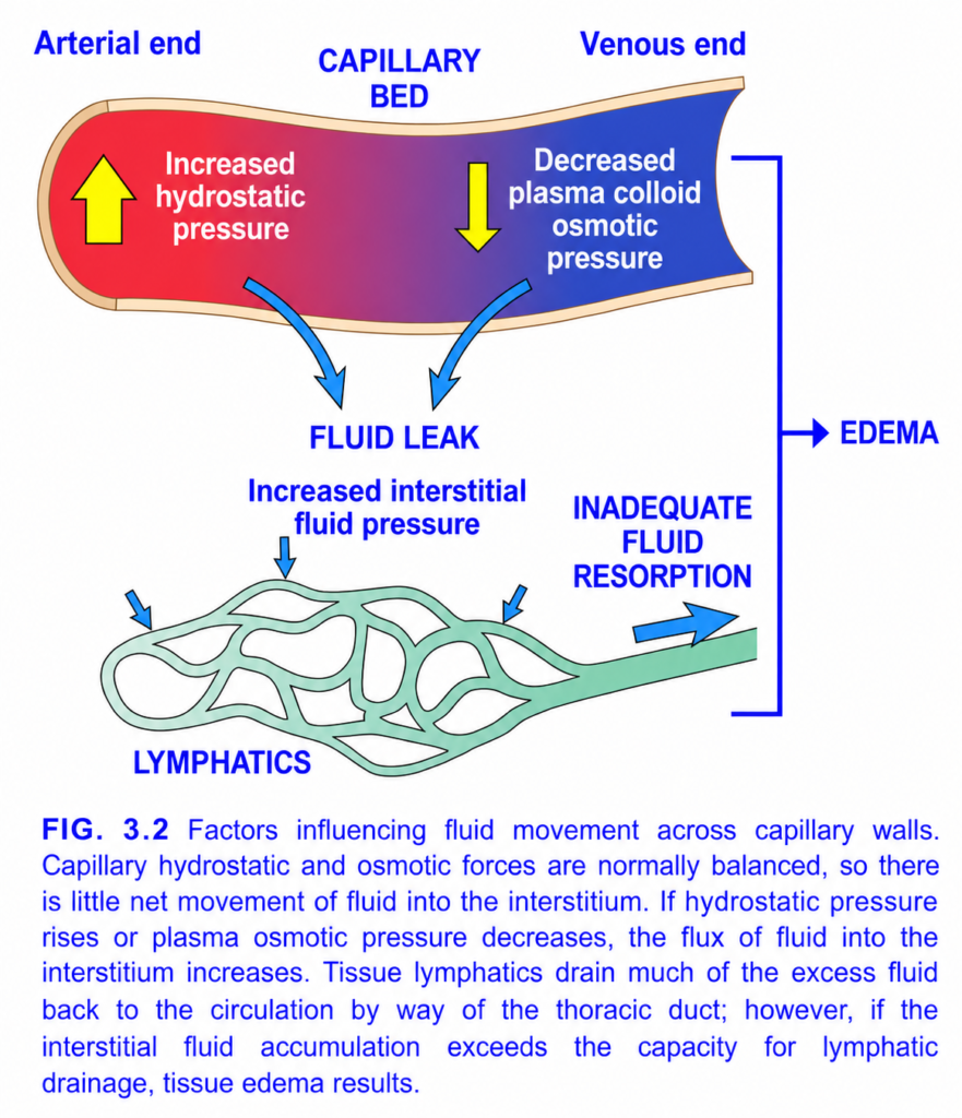
FIG. 3.2 — Why Fluid Leaves Capillaries and Causes Edema
🧠 Whole figure in one idea
Too much fluid leaves capillary OR too little returns → interstitial fluid accumulates → EDEMA
1️⃣ 🔴 Arterial end — ↑ Hydrostatic pressure
🔴 Red side = arterial end of capillary
🟨 Up yellow arrow = increased hydrostatic pressure
Hydrostatic pressure = pressure of blood pushing outward on the capillary wall.
So:
↑ Capillary hydrostatic pressure
→ pushes more water OUT of capillary
⬇️
💧 FLUID LEAK
→ edema
Easy memory:
Hydrostatic = PUSHES fluid OUT.
2️⃣ 🔵 Venous end — ↓ Plasma colloid osmotic pressure
🔵 Blue side = venous end
Normally, blood proteins—especially albumin—pull water back into the capillary.
🟨 Down yellow arrow = decreased plasma colloid osmotic pressure
↓ Albumin / plasma proteins
→ ↓ pulling force
→ less fluid comes back into blood
→ fluid stays outside
⬇️ Blue arrow
💧 FLUID LEAK / accumulation
Easy memory:
Oncotic pressure = PULLS fluid IN.
So:
↓ oncotic pressure → ↓ pull inward → edema
3️⃣ 🔵 Blue curved arrows = Fluid moving OUT
These arrows show excessive fluid entering the:
Interstitial space
= space between tissue cells and blood vessels.
More fluid outside vessels
→ ↑ interstitial fluid pressure
4️⃣ 🟢 Green network = LYMPHATICS
Think of lymphatics as the body’s:
“Drainage pipes”
Normally:
Extra interstitial fluid → lymphatics → returned to blood
So lymphatics protect against edema.
5️⃣ 🔵 Small arrows into lymphatics
They show:
Excess tissue fluid → enters lymphatic vessels
As tissue fluid rises, lymphatic drainage normally increases.
6️⃣ ➡️ Large blue arrow — Inadequate fluid resorption
If lymphatics:
- are blocked, OR
- cannot handle all the excess fluid,
then fluid cannot be removed fast enough.
Fluid production > lymphatic drainage
⬇️
💧 EDEMA
🎨 Color map
- 🔴 Red capillary side = arterial end
- 🔵 Blue capillary side = venous end
- 🟨 Yellow arrows = change in pressure
- 🔵 Blue downward arrows = fluid leaving capillary
- 🟢 Green vessels = lymphatic drainage
- ➡️ Blue bracket/arrow = all mechanisms can end in edema
⭐ Three mechanisms shown
1. ↑ Hydrostatic pressure
→ more fluid PUSHED OUT
2. ↓ Plasma colloid osmotic pressure
→ less fluid PULLED IN
3. ↓ Lymphatic drainage
→ fluid cannot be removed
All lead to:
↑ Interstitial fluid → EDEMA
🎯 2-line exam recall
Hydrostatic pressure PUSHES fluid OUT; plasma oncotic pressure PULLS fluid IN.
↑ Hydrostatic pressure + ↓ plasma oncotic pressure + lymphatic obstruction → EDEMA.
Increased Hydrostatic Pressure
- ↑ Hydrostatic pressure mainly occurs when venous return is impaired.
- Deep venous thrombosis (DVT) → venous blockage in one leg → edema mainly distal to the obstruction.
- Congestive heart failure → systemic ↑ venous pressure → often generalized edema.
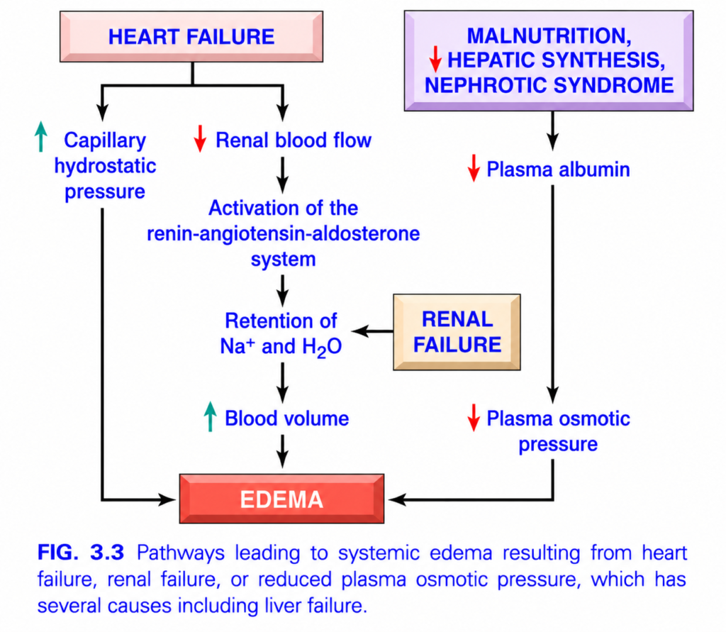
- The mechanisms of generalized edema in cardiac, renal, and hepatic failure are shown in Fig. 3.3.
- In congestive heart failure:
- ↓ Cardiac output → blood pools in veins → ↑ capillary hydrostatic pressure → edema.
- ↓ Cardiac output → ↓ renal perfusion → activates renin–angiotensin–aldosterone system (RAAS).
- RAAS activation → Na⁺ + water retention = secondary hyperaldosteronism.
- With a normal heart:
- Na⁺ + water retention → ↑ cardiac filling → ↑ cardiac output → improved renal perfusion.
- With a failing heart:
- Heart cannot adequately increase output despite increased filling.
- Therefore:
↓ cardiac output → renal hypoperfusion → Na⁺/water retention → ↑ venous pressure → more edema → further fluid retention. - This forms a vicious cycle.
- The cycle improves if:
- cardiac output is restored, or
- renal fluid retention is reduced by salt restriction, diuretics, or aldosterone antagonists.
- Secondary hyperaldosteronism can also occur in noncardiac generalized edema.
Reduced Plasma Osmotic Pressure
- Low plasma albumin is an important cause of edema due to ↓ colloid osmotic pressure.
- Albumin forms almost half of plasma protein and is the major contributor to plasma colloid osmotic pressure.
- Albumin falls mainly because of:
- ↑ urinary loss, or
- ↓ hepatic synthesis.
- Nephrotic syndrome:
- Glomerular damage → albumin and other plasma proteins leak into urine → albuminuria.
- Severe liver disease, such as cirrhosis, and protein malnutrition:
- ↓ albumin synthesis.
- Regardless of the cause:
↓ albumin → ↓ plasma osmotic pressure → edema → ↓ intravascular volume → renal hypoperfusion → secondary hyperaldosteronism. - Kidney then retains more salt and water, but edema worsens because the main problem—low plasma protein—still remains.
Lymphatic Obstruction
- Lymphatic obstruction → reduced removal of interstitial fluid → lymphedema.
- It usually results from a localized inflammatory or neoplastic obstruction.
- Filariasis:
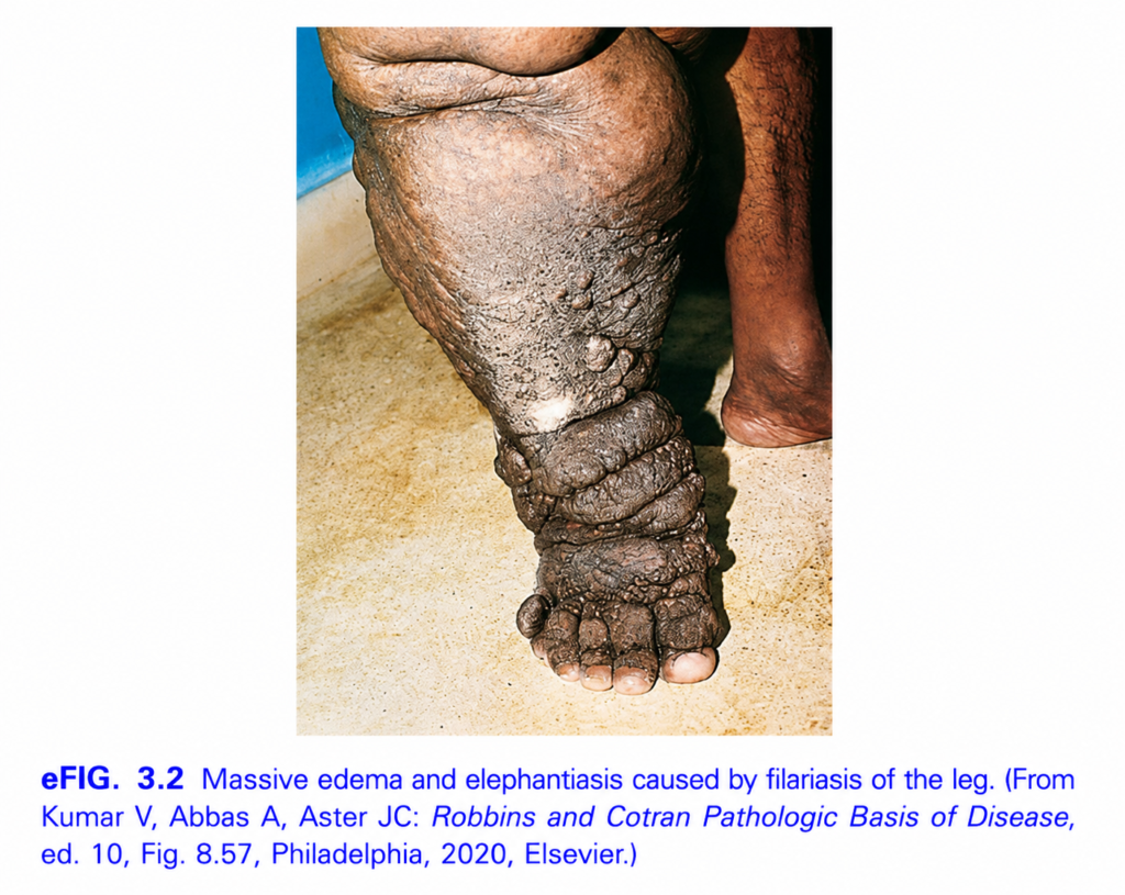
- Fibrosis of inguinal lymphatics and lymph nodes → massive edema of lower limbs and external genitalia → elephantiasis (eFig. 3.2).
- Breast cancer:
- Tumor may block superficial lymphatics → edema of overlying skin.
- Fine pitting gives an orange-peel appearance = peau d’orange.
- Treatment of breast cancer can also cause lymphedema:
- Axillary lymph-node removal and/or irradiation → impaired lymph drainage → severe arm lymphedema.
Sodium and Water Retention
- Excess Na⁺ and water retention causes edema by two mechanisms:
- ↑ intravascular volume → ↑ hydrostatic pressure.
- Dilution of plasma proteins → ↓ plasma osmotic pressure.
- It occurs in diseases with impaired renal function, including:
- poststreptococcal glomerulonephritis
- acute renal failure
KEY CONCEPT
- ↑ Hydrostatic pressure → pushes more fluid out of vessels.
- ↓ Albumin → decreases osmotic pull back into vessels.
- Lymphatic obstruction → prevents removal of interstitial fluid.
- Na⁺ + water retention → ↑ hydrostatic pressure + ↓ plasma osmotic pressure.
- In heart failure:
↓ cardiac output → ↓ renal perfusion → RAAS → Na⁺/water retention → ↑ venous pressure → worsening edema.
CONCEPTUAL EXAMPLES
- DVT → blocked venous return → localized leg edema.
- Heart failure → venous backup + renal salt/water retention → generalized edema.
- Nephrotic syndrome → albumin lost in urine → ↓ osmotic pressure → edema.
- Cirrhosis → ↓ albumin synthesis → edema.
- Filariasis → lymphatic blockage → elephantiasis.
- Breast cancer/axillary node removal → impaired lymph drainage → peau d’orange or arm lymphedema.
MORPHOLOGY
- Edema is easiest to recognize by gross examination.
- Microscopically, edema causes subtle clearing and separation of extracellular matrix (ECM) elements.
- Any tissue may develop edema, but it is most common in the subcutaneous tissues, lungs, and brain.
- Subcutaneous edema is greatest in body parts located furthest below the heart, because hydrostatic pressure is highest there.
- Standing → edema is greatest in the legs.
- Lying down → edema is greatest over the sacrum.
- This is called dependent edema.
- Pressing edematous skin with a finger pushes away interstitial fluid → leaves a temporary finger-shaped depression = pitting edema.
- Edema caused by renal dysfunction or nephrotic syndrome often appears first in loose connective tissue.
- Example: eyelids → periorbital edema.
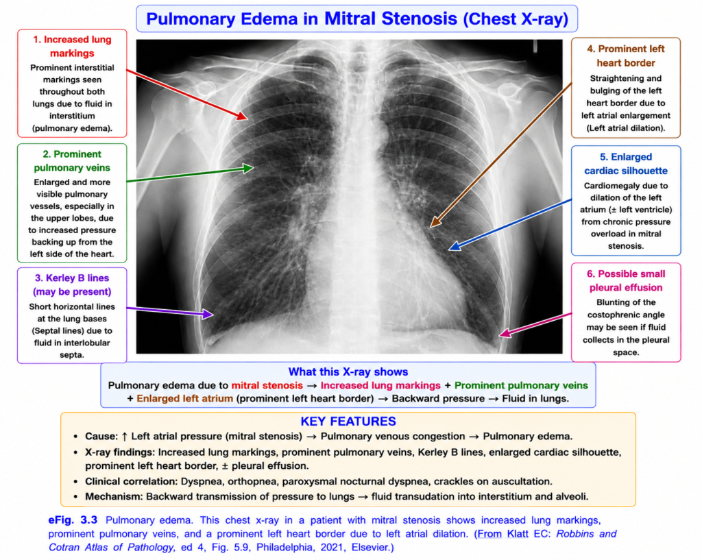
- In pulmonary edema (eFig. 3.3):
- Lungs may become 2–3 times their normal weight.
- On cutting, they release frothy fluid, which may be blood-tinged.
- The froth contains air + edema fluid + extravasated red blood cells.
- Brain edema may be:
- Localized → for example, around an abscess or tumor.
- Generalized → depending on the extent of injury or disease.
- In generalized brain edema:
- Brain gyri swell.
- Sulci become narrow.
- Swollen gyri become flattened against the skull.
KEY CONCEPT
- Dependent edema → gravity-dependent areas: legs when standing, sacrum when lying.
- Pitting edema → finger pressure leaves a depression.
- Renal edema → often first appears around the eyes.
- Pulmonary edema → heavy lungs + frothy fluid.
- Brain edema → swollen gyri + narrowed sulci.
CONCEPTUAL EXAMPLES
- Standing for a long time → fluid collects more in the legs → dependent edema.
- Press swollen skin → temporary dent remains → pitting edema.
- Nephrotic syndrome → swelling around the eyelids → periorbital edema.
- Lung edema → lungs become heavy and release frothy fluid.
- Generalized brain swelling → gyri enlarge and sulci become narrow.
MORPHOLOGY
- Edema is easiest to recognize by gross examination.
- Microscopically, edema causes subtle clearing and separation of extracellular matrix (ECM) elements.
- Any tissue may develop edema, but it is most common in the subcutaneous tissues, lungs, and brain.
- Subcutaneous edema is greatest in body parts located furthest below the heart, because hydrostatic pressure is highest there.
- Standing → edema is greatest in the legs.
- Lying down → edema is greatest over the sacrum.
- This is called dependent edema.
- Pressing edematous skin with a finger pushes away interstitial fluid → leaves a temporary finger-shaped depression = pitting edema.
- Edema caused by renal dysfunction or nephrotic syndrome often appears first in loose connective tissue.
- Example: eyelids → periorbital edema.
- In pulmonary edema (eFig. 3.3):
- Lungs may become 2–3 times their normal weight.
- On cutting, they release frothy fluid, which may be blood-tinged.
- The froth contains air + edema fluid + extravasated red blood cells.
- Brain edema may be:
- Localized → for example, around an abscess or tumor.
- Generalized → depending on the extent of injury or disease.
- In generalized brain edema:
- Brain gyri swell.
- Sulci become narrow.
- Swollen gyri become flattened against the skull.
KEY CONCEPT
- Dependent edema → gravity-dependent areas: legs when standing, sacrum when lying.
- Pitting edema → finger pressure leaves a depression.
- Renal edema → often first appears around the eyes.
- Pulmonary edema → heavy lungs + frothy fluid.
- Brain edema → swollen gyri + narrowed sulci.
CONCEPTUAL EXAMPLES
- Standing for a long time → fluid collects more in the legs → dependent edema.
- Press swollen skin → temporary dent remains → pitting edema.
- Nephrotic syndrome → swelling around the eyelids → periorbital edema.
- Lung edema → lungs become heavy and release frothy fluid.
- Generalized brain swelling → gyri enlarge and sulci become narrow.
Clinical Features
- Effects of edema can range from mild discomfort to rapidly fatal complications.
- Subcutaneous edema is important because it may indicate underlying cardiac or renal disease.
- If severe, it may also:
- impair healing of skin wounds
- reduce clearance of skin infections
- Pulmonary edema is a common clinical problem.
- It occurs most often with left ventricular failure, but can also occur with:
- renal failure
- acute lung injury
- inflammatory lung disorders
- infectious lung disorders
- Pulmonary edema can become fatal because fluid interferes with normal ventilation.
- Fluid inside alveoli also provides a favorable environment for secondary infection.
- Brain edema is life threatening.
- Severe brain swelling may cause the brain to herniate through the foramen magnum.
- Increased intracranial pressure may reduce the blood supply to the brain stem.
- Brain-stem injury may damage the medullary centers controlling respiration and other vital functions → death.
KEY CONCEPT
- Subcutaneous edema → may signal heart or kidney disease.
- Pulmonary edema → impairs ventilation and increases infection risk.
- Brain edema → may cause herniation, brain-stem ischemia, and death.
CONCEPTUAL EXAMPLES
- Heart or kidney disease → visible swelling of subcutaneous tissues.
- Left ventricular failure → fluid accumulates in lungs → breathing becomes difficult.
- Severe brain swelling → increased intracranial pressure → brain-stem compression → failure of vital functions.
