


- Hemorrhage = escape of blood from blood vessels into surrounding tissues or outside the body.
- It occurs after blood-vessel damage and may become worse when blood clotting is defective.
- Causes include:
- chronic congestion with capillary bleeding
- trauma
- atherosclerosis
- inflammatory erosion of vessel walls
- neoplastic erosion of vessel walls
- Bleeding may be massive when a large artery or vein is affected.
- Hemorrhagic diatheses = disorders with an increased tendency to bleed, sometimes even after minor injury.
- They may result from inherited or acquired defects involving:
- vessel walls
- platelets
- coagulation factors
- All three normally work together for proper hemostasis.
- Hemorrhage may occur as:
- external bleeding, or
- accumulation of blood inside tissue = hematoma.
- Hematomas may range from:
- minor → bruise
- severe → massive internal hematoma that can be fatal.
- Large bleeding into body cavities is named according to location:
- Hemothorax → blood in pleural cavity
- Hemopericardium → blood in pericardial cavity
- Hemoperitoneum → blood in peritoneal cavity
- Hemarthrosis → blood in a joint
- Large hemorrhages may occasionally cause jaundice because macrophages break down large amounts of red cells and hemoglobin.
- Petechiae:
- tiny hemorrhages, about 1–2 mm
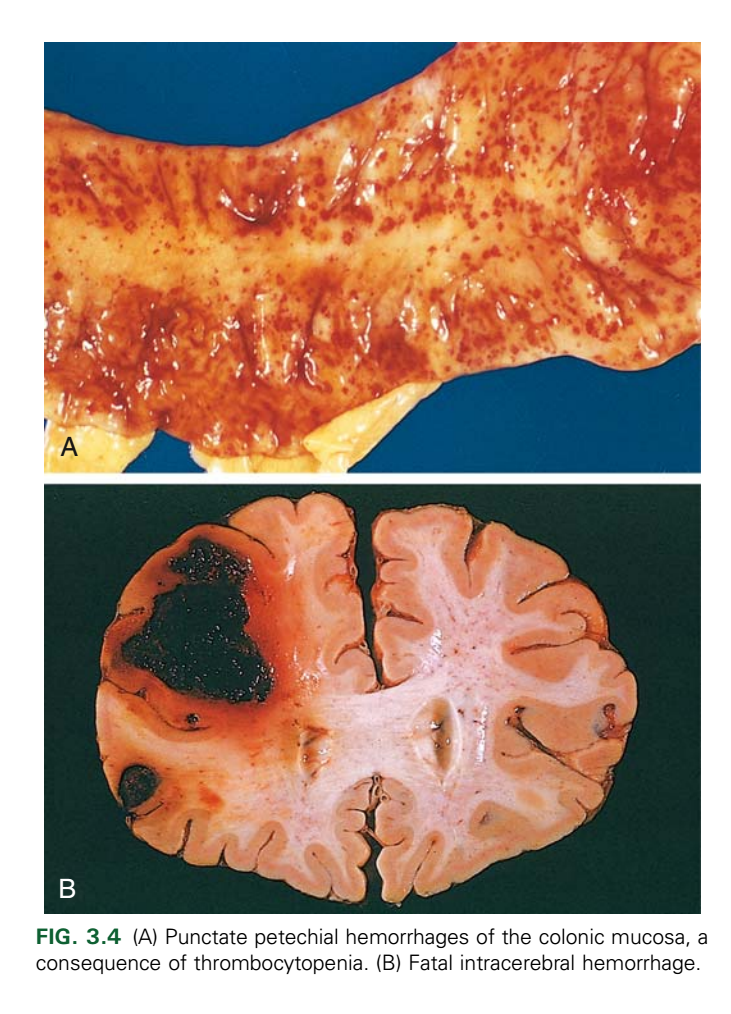
- occur in skin, mucous membranes, or serosal surfaces (Fig. 3.4A)
- Causes:
- thrombocytopenia
- defective platelet function
- loss of vascular wall support, as in vitamin C deficiency
- Purpura:
- slightly larger hemorrhages, about 3–5 mm
- Causes include those of petechiae plus:
- trauma
- vasculitis
- increased vascular fragility
- Ecchymoses:
- larger subcutaneous hematomas, about 1–2 cm
- commonly called bruises
- Macrophages remove and break down extravasated red cells.
- Color changes occur as:
hemoglobin (red-blue) → bilirubin (blue-green) → hemosiderin (golden-brown).
- Severity of hemorrhage depends on:
- amount of blood lost
- speed of blood loss
- site of bleeding
- health of the patient
- Rapid loss of up to about 20% of blood volume may be tolerated by a healthy adult.
- The same loss may cause cardiovascular decompensation in patients with underlying heart or lung disease.
- Greater blood loss may cause hemorrhagic (hypovolemic) shock.
- Location is extremely important:
- a small subcutaneous bleed may be harmless
- a similar bleed in the brain may be fatal (Fig. 3.4B).
- Chronic or repeated external blood loss, such as from peptic ulcer or menstrual bleeding:
→ loss of hemoglobin iron → iron deficiency anemia. - Internal bleeding, such as a hematoma, usually does not cause iron deficiency because iron from red cells is efficiently recycled.
KEY CONCEPT
- Hemorrhage = blood escapes from vessels.
- Petechiae = 1–2 mm, Purpura = 3–5 mm, Ecchymoses = 1–2 cm.
- Clinical danger depends mainly on volume + rate + location + patient condition.
- Large rapid blood loss → hypovolemic shock.
- Chronic external bleeding → iron deficiency anemia, while internal blood is usually recycled.
CONCEPTUAL EXAMPLES
- Low platelets → tiny skin spots → petechiae.
- Larger small hemorrhages → purpura.
- Trauma under the skin → ecchymosis/bruise with changing colors.
- Massive rapid bleeding → ↓ circulating blood volume → hypovolemic shock.
- Chronic peptic-ulcer bleeding → repeated iron loss → iron deficiency anemia.
HEMOSTASIS AND THROMBOSIS
- Hemostasis = normal formation of a blood clot after traumatic vascular injury to stop bleeding.
- Thrombosis = pathologic formation of a thrombus inside a vessel damaged by disease.
- Thus:
Normal vessel injury → hemostasis
Disease-related vessel injury → thrombosis
Hemostasis
- Hemostasis is a carefully controlled process involving:
- platelets
- clotting factors
- endothelium
- It occurs at the site of vascular injury and forms a clot that prevents or limits bleeding.
- The overall sequence is shown in Fig. 3.5.
- Immediately after injury:
Arteriolar vasoconstriction → ↓ blood flow to injured area. - Vasoconstriction is produced by:
- neurogenic reflexes
- local release of endothelin, a strong vasoconstrictor from endothelium.
- This effect is temporary; permanent control requires platelets + coagulation factors.
- Primary hemostasis = platelet plug formation:
- Endothelial injury → exposes subendothelial collagen.
- Collagen binds von Willebrand factor (vWF).
- vWF promotes platelet adhesion and activation.
- Activated platelets change from small discs into flat, spiky cells → ↑ surface area.
- Platelets release granules → recruit more platelets.
- Platelets aggregate → form the primary hemostatic plug.
- Secondary hemostasis = fibrin deposition:
- Vascular injury exposes tissue factor.
- Tissue factor is normally present on subendothelial cells such as smooth muscle cells and fibroblasts.
- Tissue factor binds and activates factor VII.
- This starts the coagulation cascade → thrombin generation.
- Thrombin converts fibrinogen → insoluble fibrin.
- Fibrin forms a mesh around the platelet plug.
- Thrombin also activates more platelets → more aggregation.
- Result → platelet plug becomes stronger and consolidated.
- Clot stabilization:
- Fibrin is cross-linked by factor XIII.
- Platelet aggregates contract.
- Together → form a solid, permanent plug that prevents further bleeding.
- Counterregulatory mechanisms limit clotting to the injury site and later promote clot removal and tissue repair.
- The endothelium controls whether a clot forms, grows, or dissolves.
- Healthy endothelial cells:
- inhibit platelet aggregation
- inhibit coagulation
- promote fibrinolysis
- After endothelial injury or activation → balance shifts toward clot formation.
- Endothelium may be activated by:
- microbial pathogens
- abnormal hemodynamic forces
- proinflammatory mediators
- These changes can increase the risk of thrombosis.
KEY CONCEPT
- Hemostasis occurs in a clear sequence:
Vessel injury → vasoconstriction → platelet adhesion/activation → primary platelet plug → tissue factor + factor VII → thrombin → fibrin → factor XIII cross-linking → stable clot
- Primary hemostasis = platelets.
- Secondary hemostasis = fibrin formation.
- Factor XIII = stabilizes fibrin.
- Healthy endothelium is mainly anticoagulant, while injured/activated endothelium favors clotting.
CONCEPTUAL EXAMPLES
- Small vessel cut → vessel constricts first → reduces blood loss.
- Exposed collagen + vWF → platelets stick → temporary platelet plug.
- Tissue factor activates coagulation → thrombin forms fibrin → stronger clot.
- Factor XIII cross-links fibrin → stable permanent plug.
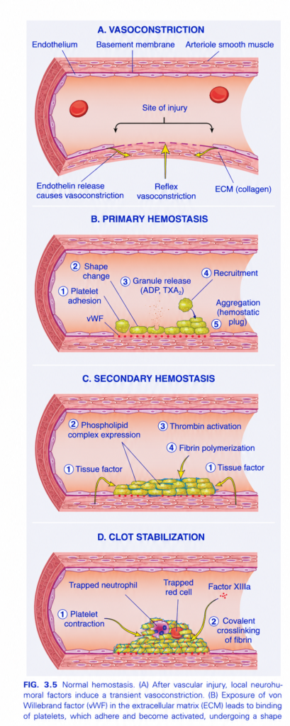
FIG. 3.5 — NORMAL HEMOSTASIS
Easiest concept: Blood vessel injury → stop bleeding → make stable clot
Whole figure in one line
Vasoconstriction → platelet plug → fibrin clot → clot stabilization
A. VASOCONSTRICTION
🔴 Red vessel lumen = blood inside arteriole
🩷 Pink inner lining = endothelium
🟤 Outer muscular layer = arteriole smooth muscle
🟣 Thin supporting layer = basement membrane
🧵 ECM collagen becomes exposed at injury.
At the injury:
Vessel injury
→ reflex contraction + endothelin release
→ smooth muscle contracts
→ vessel becomes narrower
→ blood flow decreases
🟨 Yellow arrows = contraction toward the injured area.
Memory:
First response = SQUEEZE the vessel.
B. PRIMARY HEMOSTASIS = PLATELET PLUG
🟢/yellow cells = platelets
① Platelet adhesion
Injury exposes vWF + collagen.
vWF = von Willebrand factor
vWF acts like glue:
Damaged vessel → vWF exposed → platelets stick
② Shape change
Platelets become activated and change from smooth discs into sticky irregular cells.
③ Granule release
Activated platelets release:
- ADP
- TXA₂ = thromboxane A₂
These signals call and activate more platelets.
④ Recruitment
More platelets arrive.
⑤ Aggregation
Platelets stick to each other.
➡️ Result = temporary platelet plug
Memory:
Adhere → Activate → Release → Recruit → Aggregate
C. SECONDARY HEMOSTASIS = FIBRIN
Now the platelet plug must become stronger.
① Tissue factor
Damaged tissue exposes/releases tissue factor.
→ starts the coagulation cascade.
② Phospholipid complex
Activated platelet surfaces provide a platform for clotting reactions.
③ Thrombin activation
Coagulation cascade produces thrombin.
Thrombin is the key enzyme that converts:
Fibrinogen → fibrin
④ Fibrin polymerization
🟦/green mesh around platelets = fibrin strands
Fibrin forms a net around the platelet plug.
➡️ Temporary plug becomes a strong clot.
Memory:
Tissue factor → Thrombin → Fibrin
D. CLOT STABILIZATION
Now the clot is tightened and reinforced.
① Platelet contraction
Platelets contract.
→ clot becomes smaller, tighter, and stronger.
② Factor XIIIa
Factor XIIIa makes covalent cross-links between fibrin strands.
Think:
Loose fibrin net → XIIIa → locked strong fibrin net
🔴 Trapped red cell = RBC caught in clot
🟣 Trapped neutrophil = inflammatory cell caught in clot
🎨 COLOR / STRUCTURE MAP
- 🔴 Red discs = red blood cells
- 🟢/yellow cells = platelets
- 🟦/green mesh = fibrin
- 🩷 vessel lining = endothelium
- 🟤 vessel wall = smooth muscle
- 🟨 arrows = direction of each hemostatic step
🧠 Fastest exam recall
1. Vasoconstriction
Endothelin → vessel narrows
2. Primary hemostasis
vWF → platelet adhesion → platelet plug
3. Secondary hemostasis
Tissue factor → thrombin → fibrin
4. Stabilization
Factor XIIIa → cross-links fibrin
⭐ Ultimate memory:
Squeeze → Stick → Strengthen → Stabilize
Platelets
- Platelets are essential for hemostasis because they:
- form the primary platelet plug that initially seals vessel injury.
- provide a surface where activated coagulation factors gather and work.
- Platelets are disc-shaped, anucleate cell fragments released from bone-marrow megakaryocytes into blood.
- Their function depends on:
- surface glycoprotein receptors
- a contractile cytoskeleton
- α-granules
- dense (δ) granules
- α-Granules contain:
- P-selectin
- fibrinogen
- factor V
- vWF
- fibronectin
- platelet factor 4
- PDGF
- TGF-β
- Dense granules contain:
- ADP
- ATP
- polyphosphate
- Ca²⁺
- serotonin
- epinephrine
- After vascular injury, platelets contact exposed collagen + vWF and begin forming the platelet plug (Fig. 3.5B).
- Platelet adhesion:
- vWF acts as a bridge:
- exposed collagen ← vWF → platelet GpIb
- Thus, platelets stick to the injured vessel wall (Fig. 3.6).
- vWF deficiency → von Willebrand disease.
- GpIb deficiency → Bernard-Soulier syndrome.
- Both can cause bleeding.
- After adhesion, platelets rapidly change from smooth discs into spiky “sea-urchin” shapes → greatly ↑ surface area.
- Platelet activation also changes GpIIb/IIIa, increasing its ability to bind fibrinogen.
- Negatively charged phospholipids, especially phosphatidylserine, move to the platelet surface.
- They bind Ca²⁺.
- This surface helps assemble coagulation factor complexes.
- Platelets then release their granules.
- Shape change + granule release = platelet activation.
- Important activators include:
- thrombin
- ADP
- Thrombin activates platelets through protease-activated receptors (PARs).
- ADP released from dense granules activates more platelets:
platelet activation → ADP release → more platelet activation
= recruitment. - Activated platelets also produce thromboxane A₂ (TxA₂) → strongly promotes platelet aggregation.
- Aspirin inhibits cyclooxygenase → ↓ TxA₂ formation → ↓ platelet aggregation → mild bleeding tendency.
- Platelet growth factors such as PDGF contribute to repair of the damaged vessel wall.
- Platelet aggregation occurs after activation:
- activated GpIIb/IIIa binds fibrinogen.
- fibrinogen bridges neighboring platelets.
- platelets therefore stick to one another → aggregation.
- GpIIb/IIIa deficiency → Glanzmann thrombasthenia → bleeding disorder.
- Early platelet aggregation is reversible.
- Thrombin then strengthens the plug by:
- further platelet activation
- further aggregation
- irreversible platelet contraction
- Platelet contraction depends on the cytoskeleton and tightens the platelet mass.
- At the same time:
thrombin → fibrinogen → insoluble fibrin. - Thrombin also activates factor XIIIa → cross-links fibrin → firmly locks platelets in place.
- This produces the definitive secondary hemostatic plug.
- Red cells and leukocytes may become trapped in the plug.
- Leukocytes can also adhere to P-selectin on activated platelets.
KEY CONCEPT
Vessel injury → collagen exposed → vWF binds GpIb → platelet adhesion → platelet activation → ADP + TxA₂ recruit more platelets → GpIIb/IIIa binds fibrinogen → platelet aggregation → thrombin makes fibrin → factor XIIIa cross-links fibrin → stable clot
- GpIb = adhesion
- GpIIb/IIIa = aggregation
- vWF = bridge to collagen
- ADP + TxA₂ = recruit/aggregate platelets
- Thrombin + fibrin = stabilize the plug
CONCEPTUAL EXAMPLES
- vWF deficiency → platelets cannot adhere properly → bleeding.
- GpIb deficiency → defective adhesion → Bernard-Soulier syndrome.
- GpIIb/IIIa deficiency → defective aggregation → Glanzmann thrombasthenia.
- Aspirin → ↓ TxA₂ → ↓ platelet aggregation.
- Thrombin + factor XIIIa → fibrin becomes strong and cross-linked → stable hemostatic plug.
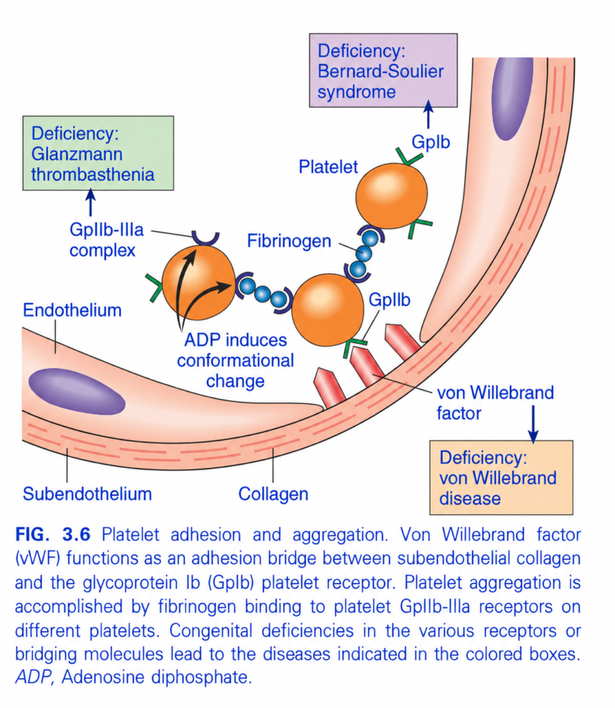
FIG. 3.6 — Platelet Adhesion & Aggregation
Easiest concept: Platelets first STICK to the injured vessel, then STICK to each other
Whole figure in one line
Collagen exposed → vWF binds collagen → platelet GpIb binds vWF → platelet activated by ADP → GpIIb/IIIa activated → fibrinogen bridges platelets → platelet aggregation
1️⃣ Vessel injury exposes subendothelial collagen
🩷 Pink vessel wall = endothelium/subendothelium
🔴 Red rod-like structures = exposed collagen
Normally collagen is hidden beneath intact endothelium.
Injury → endothelium breaks → collagen becomes exposed
2️⃣ von Willebrand factor (vWF) attaches to collagen
vWF works like a bridge/glue.
Think:
Collagen — vWF — Platelet
So vWF connects the injured vessel wall to the platelet.
🟦 Label pointing to the bridge = von Willebrand factor
❌ If vWF is deficient:
➡️ von Willebrand disease
3️⃣ Platelet GpIb binds vWF
🟠 Orange circles = platelets
🟢 receptor on platelet = GpIb
GpIb is the platelet receptor that grabs vWF.
Therefore:
Collagen → vWF → GpIb → platelet adhesion
❌ GpIb deficiency:
➡️ Bernard-Soulier syndrome
🧠 Memory:
Bernard = “B” = GpIb
4️⃣ Platelet activation → ADP released
Once the platelet sticks, it becomes activated.
ADP acts on platelets and causes a:
Conformational change
= change in receptor shape so it can work properly.
The black arrows in the platelet show this activation/change in shape.
5️⃣ ADP activates GpIIb/IIIa
🟣 U-shaped receptor = GpIIb/IIIa
Before activation, it does not bind fibrinogen efficiently.
ADP → changes GpIIb/IIIa shape → receptor becomes active
6️⃣ Fibrinogen bridges two platelets
🔵 Blue bead-like chain = fibrinogen
Fibrinogen binds:
GpIIb/IIIa on platelet 1
↔️ fibrinogen
↔️ GpIIb/IIIa on platelet 2
This joins platelets together.
This is called:
Platelet aggregation
❌ If GpIIb/IIIa is deficient
Platelets can stick to the vessel, but they cannot aggregate properly.
➡️ Glanzmann thrombasthenia
🧠 Memory:
Glanzmann = GpIIb/IIIa problem
🎨 Color/structure map
- 🟠 Orange spheres = platelets
- 🟢 Green receptor = GpIb
- 🟣 Purple receptor = GpIIb/IIIa
- 🔵 Blue beads = fibrinogen
- 🔴 Red structures in wall = collagen
- 🩷 Pink wall = endothelium/subendothelium
⭐ Most important distinction
ADHESION
Collagen → vWF → GpIb → platelet sticks to vessel
AGGREGATION
GpIIb/IIIa → fibrinogen → platelet sticks to platelet
🧠 Fast exam memory
vWF + GpIb = ADHESION
Fibrinogen + GpIIb/IIIa = AGGREGATION
Diseases:
- vWF deficiency → von Willebrand disease
- GpIb deficiency → Bernard-Soulier syndrome
- GpIIb/IIIa deficiency → Glanzmann thrombasthenia
⭐ One-line recall
GpIb grabs vWF; GpIIb/IIIa grabs fibrinogen.
Coagulation Factors
- Coagulation factors work in an amplifying enzyme cascade → finally produce an insoluble fibrin clot.
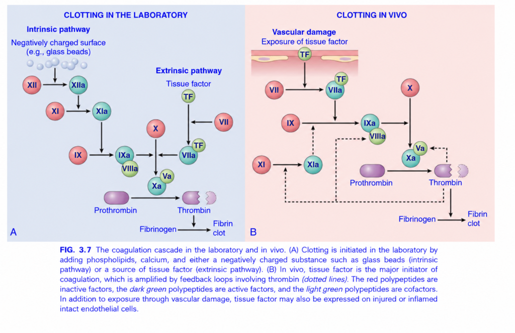
- The importance of individual factors differs between laboratory testing and clotting inside the body (Fig. 3.7), but the basic principles are similar.
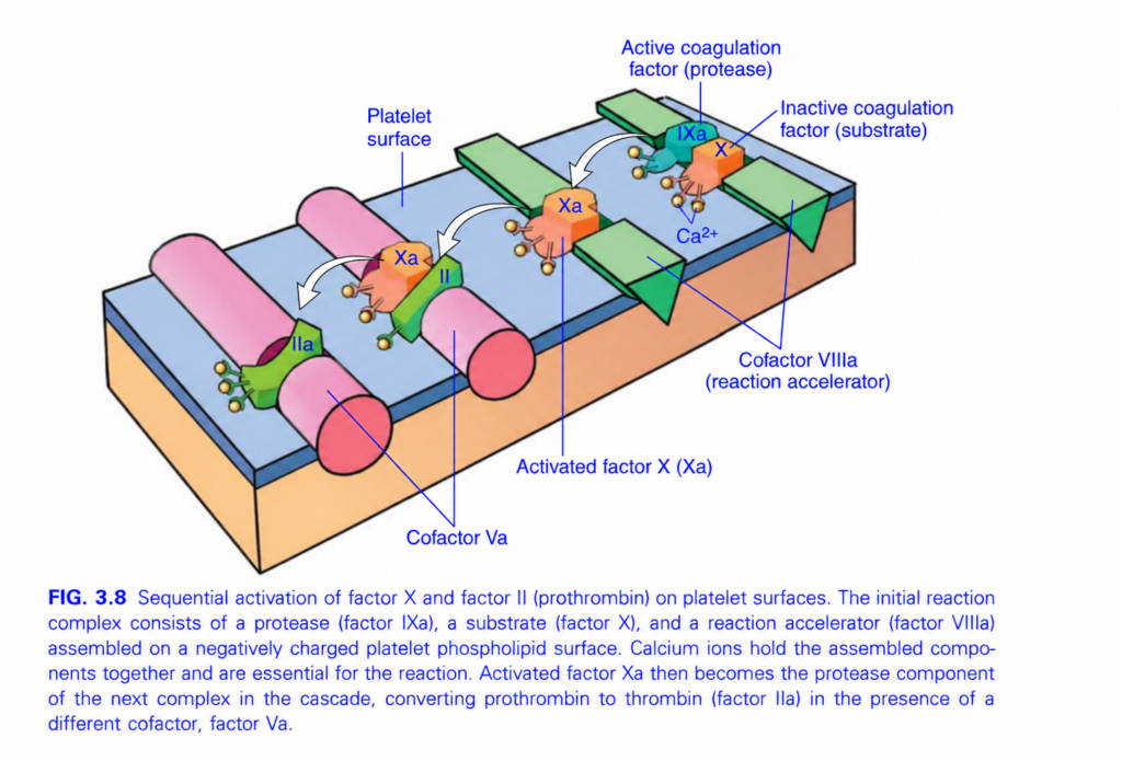
- The coagulation cascade can be imagined as a “dance”, where activation passes from one coagulation factor to the next (Fig. 3.8).
- Each reaction needs:
- Enzyme = activated coagulation factor
- Substrate = inactive coagulation factor waiting to be activated
- Cofactor = accelerates the reaction
- These components assemble on the negatively charged phospholipid surface of activated platelets.
- Calcium (Ca²⁺) is also required for assembly of coagulation-factor complexes.
- Ca²⁺ binds to specially modified glutamic acid residues in factors II, VII, IX, and X.
- Formation of these modified residues requires vitamin K.
- Warfarin interferes with vitamin K metabolism → reduces the normal function of these vitamin K–dependent factors.
- In laboratory testing, coagulation is divided into extrinsic and intrinsic pathways.
- Prothrombin time (PT) evaluates the extrinsic pathway:
- factors X, VII, V, II (prothrombin), and fibrinogen
- Tissue factor + phospholipids + Ca²⁺ are added to plasma.
- The time required to form a fibrin clot is measured.
- Partial thromboplastin time (PTT) evaluates the intrinsic pathway:
- factors XII, XI, X, IX, VIII, V, II, and fibrinogen
- Negatively charged particles + phospholipids + Ca²⁺ are added.
- Factor XII is activated, and the time to fibrin-clot formation is measured.
- PT and PTT are very useful clinically, but they do not exactly reproduce coagulation inside blood vessels.
- Factor deficiencies show this difference:
- Deficiency of V, VII, VIII, IX, or X → moderate to severe bleeding.
- Prothrombin deficiency → incompatible with life.
- Factor XI deficiency → usually mild bleeding.
- Factor XII deficiency → no bleeding disorder.
- The normal physiologic role of factor XII is uncertain.
- Excess factor XII activity can predispose to angioedema because factor XII can promote bradykinin generation by cleaving high-molecular-weight kininogen.
- In the body:
- Factor VIIa + tissue factor is considered the major activator of factor IX.
- Factor IXa + factor VIIIa is the major activator of factor X.
- Thrombin can also activate factor XI → provides positive feedback and amplifies coagulation.
- Thrombin is the most important coagulation factor because it controls several parts of hemostasis and links clotting with inflammation and repair.
- Thrombin converts:
soluble fibrinogen → fibrin monomers → insoluble fibrin clot. - Thrombin further increases fibrin formation by activating:
- factor V
- factor VIII
- factor XI
- Thrombin activates factor XIII → factor XIII cross-links fibrin → makes the clot more stable.
- Thrombin also activates platelets through PARs → platelet activation + aggregation + contraction.
- PARs are also present on inflammatory cells, endothelial cells, and other cells (Fig. 3.9).
- Thrombin activation of these receptors contributes to tissue repair.
- Thrombin can also have an anticoagulant effect.
- When thrombin encounters normal endothelium, it can change from procoagulant to anticoagulant activity → helps prevent the clot from spreading beyond the injured area.
KEY CONCEPT
- Coagulation cascade → thrombin → fibrin → stable clot.
- PT = extrinsic pathway.
- PTT = intrinsic pathway.
- Vitamin K is required for factors II, VII, IX, X.
- Warfarin interferes with vitamin K.
- Thrombin is central:
fibrin formation + factor XIII activation + platelet activation + cascade amplification. - Normal endothelium can change thrombin toward an anticoagulant role, limiting clot extension.
CONCEPTUAL EXAMPLES
- Vessel injury → tissue factor + VIIa → activates IX → IXa + VIIIa → activates X → more coagulation.
- Thrombin → fibrinogen becomes fibrin → clot forms.
- Thrombin → factor XIII activation → fibrin is cross-linked → clot becomes stronger.
- Warfarin → interferes with vitamin K → factors II, VII, IX, X function less effectively.
- Factor XII deficiency → abnormal laboratory pathway but no clinical bleeding disorder.
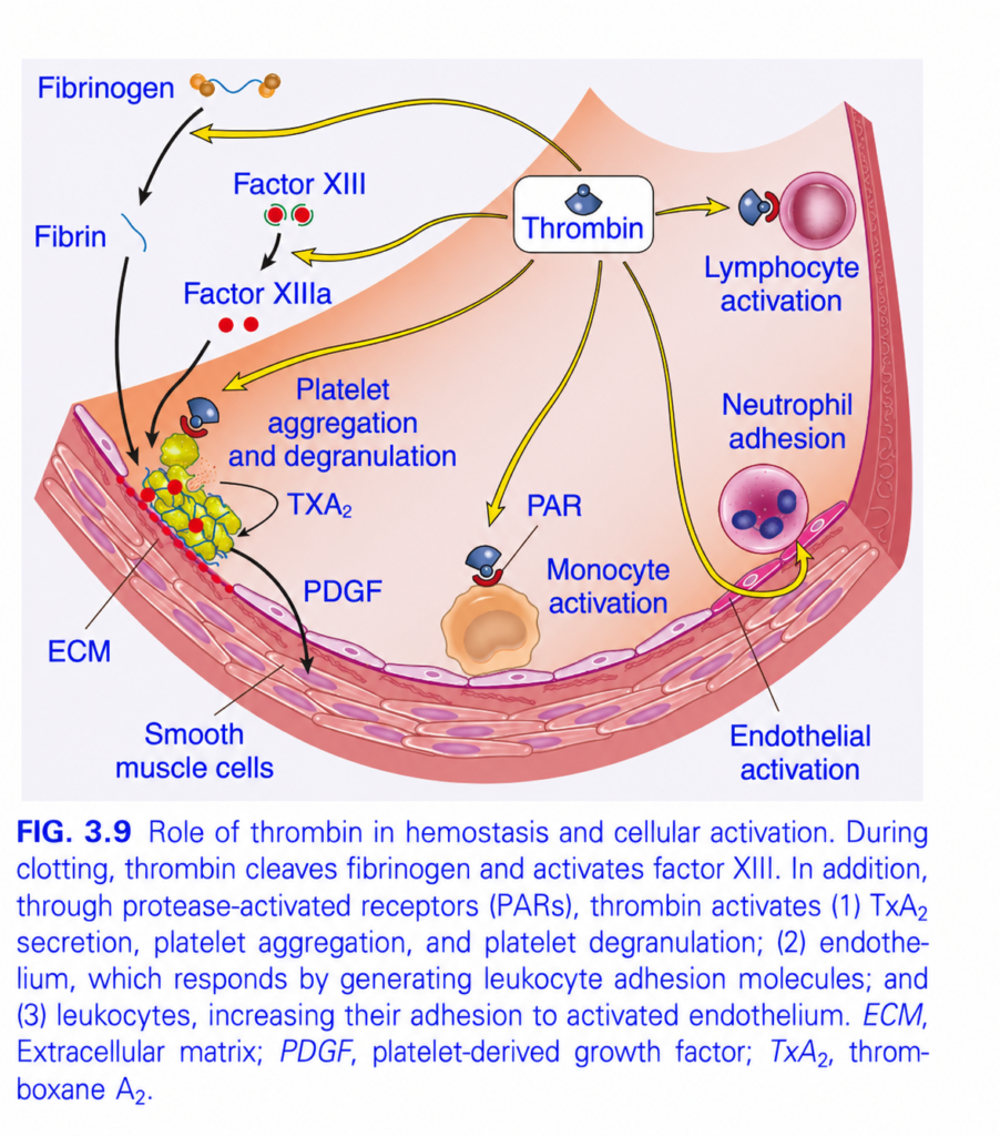
FIG. 3.9 — THROMBIN: The Central Player in Hemostasis
🧠 Easiest idea: Thrombin makes the clot AND activates cells around the clot
Whole figure in one flow
Thrombin → fibrin clot + stronger platelets + activated endothelium/leukocytes + tissue repair
1️⃣ Thrombin → Fibrinogen → Fibrin
🔵 Fibrinogen = soluble protein floating in blood.
⬇️ Thrombin cuts it
🔵 Fibrin = insoluble threads.
Fibrinogen → Fibrin → fibrin mesh → clot
Memory:
Thrombin = fibrin maker
2️⃣ Thrombin → Factor XIII → Factor XIIIa
🟢 Factor XIII = inactive form
🔴 Factor XIIIa = active form
Thrombin activates:
Factor XIII → XIIIa
Then XIIIa cross-links fibrin strands.
➡️ Loose fibrin → strong, stable fibrin clot
Exam:
Factor XIIIa = fibrin cross-linker
3️⃣ Thrombin → 🟡 Platelet activation
🟡/green clump = activated platelets.
Thrombin activates platelets through PAR
(protease-activated receptors).
This causes:
Platelet activation
→ aggregation
→ degranulation
→ more TXA₂
TXA₂
Thromboxane A₂
→ recruits/activates more platelets
→ promotes vasoconstriction
➡️ Clot becomes bigger and stronger.
4️⃣ Platelets release PDGF
PDGF = platelet-derived growth factor
⬇️ Black arrow toward vessel wall
PDGF stimulates:
Smooth muscle cells / fibroblast-like repair cells
→ proliferation
→ production of ECM
ECM
= extracellular matrix, the supporting material used in tissue repair.
So:
Platelet → PDGF → repair of damaged vessel wall
5️⃣ Thrombin → Endothelial activation
🩷 Thin cells lining the vessel = endothelial cells.
Thrombin activates them.
Activated endothelium produces more leukocyte adhesion molecules.
⬇️
White cells can now stick to the vessel wall.
6️⃣ 🟣 Neutrophil adhesion
The purple cell attached to the vessel wall = neutrophil.
Thrombin → endothelial activation → adhesion molecules ↑ → neutrophil sticks
➡️ Links clotting with inflammation.
7️⃣ 🟠 Monocyte activation
Thrombin binds PAR on monocytes.
Thrombin → PAR → monocyte activation
➡️ Monocytes participate in inflammation and tissue repair.
8️⃣ 🩷 Lymphocyte activation
Yellow arrow from thrombin toward the lymphocyte:
Thrombin → lymphocyte activation
Again, this shows that thrombin is not only a clotting enzyme—it also promotes cellular/inflammatory responses.
🎨 COLOR / ARROW MAP
- 🔵 Blue curved strand = fibrin/fibrinogen
- 🟢 Factor XIII = inactive
- 🔴 Factor XIIIa / red dots = activated cross-linking factor
- 🟡 Green-yellow clump = platelets
- 🩷 vessel lining = endothelium
- 🟠 cell = monocyte
- 🟣 round leukocytes = neutrophil / lymphocyte
- 🟨 Yellow arrows = effects produced by thrombin
- ⚫ Black arrows = downstream clot/repair effects
⭐ Fastest exam recall
THROMBIN does 4 major things:
1. Fibrinogen → Fibrin
2. Factor XIII → XIIIa → cross-links fibrin
3. Activates platelets → aggregation + degranulation + TXA₂
4. Activates endothelium + leukocytes → inflammation
🧠 One-line memory
THROMBIN = “Clot + Platelets + Inflammation + Repair.”
Factors That Limit Coagulation
- Once coagulation begins, it must stay limited to the injured vessel site; otherwise, harmful widespread clotting may occur.
- Several mechanisms prevent the clot from spreading:
- Flowing blood dilutes and washes away activated coagulation factors.
- These activated factors are then rapidly removed by the liver.
- Coagulation also requires negatively charged phospholipids, mainly supplied by activated platelets.
- Activated platelets are concentrated at the injury site, so coagulation is limited mainly to that area.
- The most important control mechanisms come from the intact endothelium surrounding the injured site.
- Activation of coagulation also activates the fibrinolytic system → limits clot size and later helps dissolve the clot (Fig. 3.10).
- Fibrinolysis = breakdown of fibrin within a clot.
- The main enzyme responsible is plasmin.
- Plasmin → breaks down fibrin.
- It also interferes with further fibrin polymerization.
- Breakdown of fibrin produces fibrin split products.
- An important fibrin breakdown product is D-dimer.
- Increased D-dimer levels can be useful clinical markers of several thrombotic conditions.
- Plasmin is formed from an inactive circulating precursor:
plasminogen → plasmin. - The most important activator of plasminogen is tissue plasminogen activator (t-PA).
- t-PA is produced mainly by endothelial cells.
- t-PA works most effectively when it is bound to fibrin.
- Therefore, its fibrinolytic action is mainly concentrated at the site of the clot.
- Once plasmin is formed, it must also be controlled.
- α₂-plasmin inhibitor binds free plasmin → rapidly inhibits it → prevents excessive fibrin breakdown.
KEY CONCEPT
- Clotting is limited by:
blood flow washing away factors + liver removal + localized platelet phospholipids + intact endothelium. - Clot removal occurs through:
t-PA → plasminogen → plasmin → fibrin breakdown → fibrin split products/D-dimers
- t-PA starts fibrinolysis.
- Plasmin breaks fibrin.
- D-dimer indicates fibrin breakdown.
- α₂-plasmin inhibitor stops free plasmin.
CONCEPTUAL EXAMPLES
- Vessel injury → clot forms only at the damaged area because activated factors are washed away from surrounding normal vessels.
- After the clot has done its job → t-PA activates plasmin → fibrin is broken down → clot gradually dissolves.
- Fibrin breakdown → D-dimers appear in blood.
- Plasmin escapes away from the clot → α₂-plasmin inhibitor quickly neutralizes it.
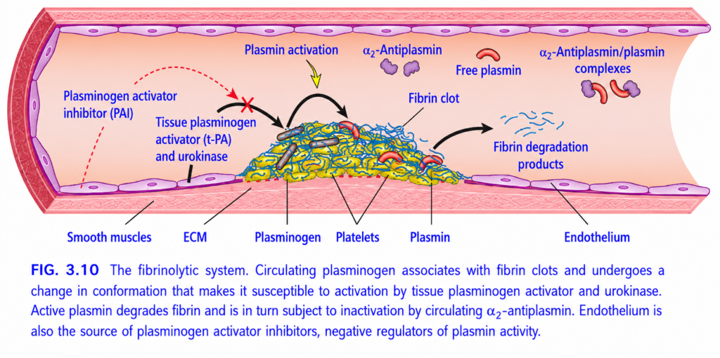
FIG. 3.10 — FIBRINOLYTIC SYSTEM
🧠 Easiest concept: After a clot has done its job, plasmin cuts the fibrin clot and removes it.
Whole figure in one flow
Plasminogen → t-PA/urokinase → PLASMIN → fibrin breakdown → fibrin degradation products
1️⃣ 🟡/🔵 Central mass = Fibrin clot
The clot contains:
- 🟡 Platelets
- 🔵 tangled strands = fibrin
- Gray structures = trapped cellular material
- Plasminogen becomes attached to the fibrin clot.
Think:
Plasminogen = inactive clot-dissolving enzyme
2️⃣ Endothelium releases t-PA and urokinase
🩷 Flat cells lining the vessel = endothelium.
They produce:
t-PA = tissue plasminogen activator
and urokinase
These convert:
Plasminogen → PLASMIN
⬇️
🔴 Red curved structures = active plasmin
🧠 Memory:
t-PA turns plasminogen into plasmin.
3️⃣ Plasmin cuts fibrin
⚫ Black arrow:
Plasmin → attacks fibrin clot
The fibrin mesh is chopped into small pieces:
🔵 Small blue fragments = fibrin degradation products
So:
Fibrin clot → Plasmin → Fibrin degradation products
➡️ The clot gradually dissolves.
⭐ This process = FIBRINOLYSIS
4️⃣ 🟣 α₂-Antiplasmin stops free plasmin
Plasmin must mainly act at the clot, not everywhere in the blood.
🟣 Purple molecules = α₂-antiplasmin
It binds any free plasmin:
Free plasmin + α₂-antiplasmin
→ inactive α₂-antiplasmin/plasmin complex
➡️ Prevents excessive fibrin breakdown elsewhere.
Memory:
Antiplasmin = anti-plasmin → STOP plasmin
5️⃣ PAI = Plasminogen Activator Inhibitor
🔴 Red dotted arrows = inhibition.
PAI blocks:
t-PA / plasminogen activators
Therefore:
PAI ↓ plasmin formation → ↓ fibrinolysis
So PAI protects against too much clot dissolution.
🎨 ARROW / COLOR MAP
- 🔵 tangled fibers = fibrin
- 🟡 cells in clot = platelets
- 🔴 curved pieces = plasmin
- 🟣 blobs = α₂-antiplasmin
- ⚫ Black arrows = activation/breakdown pathway
- 🟨 Yellow arrows = important regulatory actions
- 🔴 Dotted arrow + ❌ = PAI inhibition
- 🩷 flat vessel cells = endothelium
⭐ Two opposite forces
🟢 Clot removal
t-PA → plasmin → fibrin breakdown
🛑 Clot removal controlled
PAI blocks t-PA
α₂-antiplasmin blocks free plasmin🧠 Fastest exam recall
t-PA activates plasminogen → plasmin.
Plasmin destroys fibrin → fibrin degradation products (including D-dimer from cross-linked fibrin).
🔥 One-line memory:
t-PA STARTS plasmin; plasmin CUTS fibrin; α₂-antiplasmin STOPS plasmin; PAI STOPS t-PA.
Endothelium
- The endothelium controls the balance between clot formation and clot breakdown.
- This balance determines whether a clot forms, grows, stays limited, or dissolves (Fig. 3.11).
- Normal endothelial cells are mainly antithrombotic.
- They:
- inhibit platelets
- inhibit coagulation factors
- promote fibrinolysis
- Together, these actions prevent thrombosis and keep clotting limited to the site of vascular injury.
- When endothelium is injured or activated by proinflammatory factors, it loses many of these antithrombotic properties.
- Antithrombotic actions of normal endothelium can be grouped into three major effects:
- Platelet inhibitory effects:
- Intact endothelium acts as a physical barrier → prevents platelets from contacting subendothelial vWF and collagen.
- It releases prostacyclin (PGI₂) and nitric oxide (NO) → inhibit platelet activation and aggregation.
- Adenosine diphosphatase breaks down ADP → decreases platelet activation.
- PGI₂ and NO also cause vasodilation → helps wash away coagulation factors.
- Endothelial cells also bind thrombin → reduce thrombin-induced platelet activation.
- Anticoagulant effects:
- Normal endothelium separates circulating coagulation factors from tissue factor in the vessel wall.
- Important anticoagulant molecules include:
- thrombomodulin
- endothelial protein C receptor
- heparin-like molecules
- tissue factor pathway inhibitor (TFPI)
- Thrombomodulin pathway:
- Thrombomodulin binds thrombin.
- Once bound, thrombin loses much of its normal procoagulant activity.
- Instead, thrombin activates protein C.
- Protein C is vitamin K–dependent and works with protein S.
- Activated protein C + protein S → inhibit factors Va and VIIIa.
- Result → less coagulation.
- Heparin-like molecules:
- Bind and activate antithrombin III.
- Antithrombin III inhibits:
- thrombin
- factor IXa
- factor Xa
- factor XIa
- factor XIIa
- The clinical effect of heparin depends on increasing antithrombin activity.
- TFPI:
- Works with protein S.
- Inhibits the tissue factor–factor VIIa complex.
- Result → reduces activation of the coagulation pathway.
- Fibrinolytic effects:
- Normal endothelial cells produce t-PA.
- t-PA promotes fibrinolysis by helping generate plasmin.
KEY CONCEPT
- Normal endothelium is strongly antithrombotic.
- It protects against clotting by:
↓ platelet activation + ↓ coagulation + ↑ fibrinolysis
- Important pathways:
- PGI₂ + NO + ADP breakdown → inhibit platelets
- Thrombomodulin → protein C + protein S → inhibit Va and VIIIa
- Heparin-like molecules → antithrombin III → inhibit thrombin and IXa, Xa, XIa, XIIa
- TFPI → inhibits tissue factor–VIIa
- t-PA → promotes fibrinolysis
CONCEPTUAL EXAMPLES
- Healthy endothelium → platelets cannot contact collagen → no unnecessary platelet plug.
- Thrombomodulin binds thrombin → thrombin switches from clot-promoting to protein C activation.
- Protein C + protein S → shut down Va and VIIIa → coagulation slows.
- Heparin-like molecules → activate antithrombin III → major clotting enzymes are inhibited.
- Endothelium releases t-PA → fibrin clot is gradually broken down.
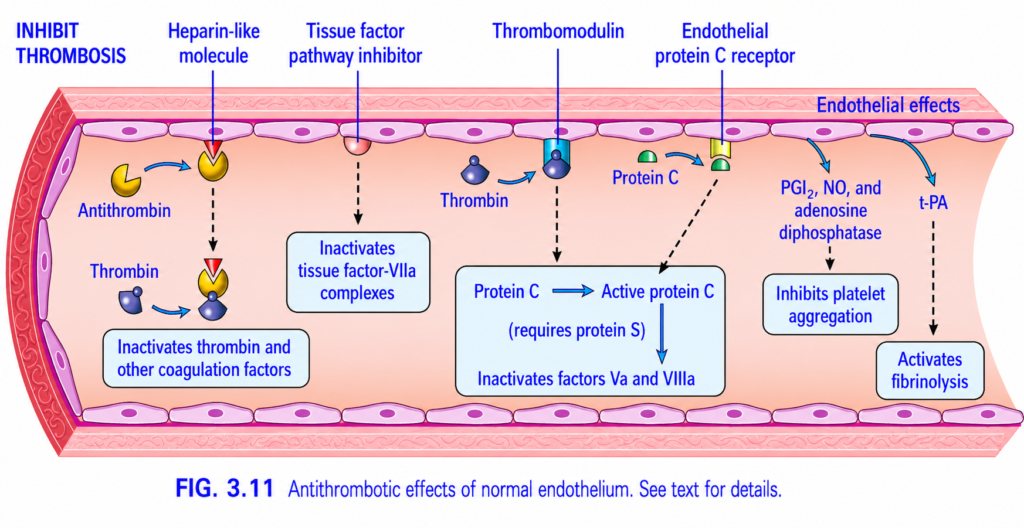
FIG. 3.11 — ANTITHROMBOTIC EFFECTS OF NORMAL ENDOTHELIUM
🧠 Easiest idea: Healthy endothelium prevents unwanted clotting in 3 ways
Whole figure in one line
Healthy endothelium → blocks coagulation + blocks platelets + promotes fibrinolysis → prevents thrombosis
1️⃣ LEFT — Heparin-like molecules + Antithrombin
🟡 Antithrombin = natural anticoagulant in blood.
🔻 Heparin-like molecule on endothelium greatly increases antithrombin activity.
Then:
Heparin-like molecule → activates antithrombin → inhibits thrombin + other coagulation factors
Result:
❌ Less fibrin formation
❌ Less clotting
Memory:
Heparin helps Antithrombin STOP thrombin.
2️⃣ Tissue Factor Pathway Inhibitor (TFPI)
TFPI is made by normal endothelium.
⬇️ Black dotted arrow
It inactivates the Tissue factor–VIIa complex.
So:
TFPI → blocks tissue factor pathway → ↓ coagulation
Memory:
TFPI = Tissue Factor Pathway OFF
3️⃣ CENTER — Thrombomodulin + Protein C
🔵 Thrombin binds to thrombomodulin on the endothelial surface.
Normally thrombin promotes clotting, but when it binds thrombomodulin:
It changes job:
Thrombin + thrombomodulin → activates Protein C
The endothelial protein C receptor also helps this process.
Active Protein C needs Protein S
Protein C → Active Protein C
Then:
Active Protein C + Protein S
→ inactivates Factor Va and Factor VIIIa
Result:
↓ thrombin generation
↓ coagulation
↓ thrombosis
Exam memory:
Protein C/S cut off 5 and 8
➡️ Va + VIIIa inhibited
4️⃣ RIGHT — PGI₂, NO, and ADPase
Normal endothelium produces:
PGI₂ = Prostacyclin
NO = Nitric oxide
Adenosine diphosphatase
These all reduce platelet activity.
⬇️
Inhibit platelet aggregation
Simple concept:
They tell platelets:
“Do NOT stick together.”
5️⃣ t-PA → Fibrinolysis
Normal endothelium releases:
t-PA = tissue plasminogen activator
t-PA converts:
Plasminogen → plasmin
Plasmin breaks down fibrin.
So:
t-PA → fibrinolysis → clot breakdown
🎨 COLOR / ARROW MAP
- 🩷 Flat purple-pink cells = endothelium
- 🟡 Yellow molecules = antithrombin
- 🔵 Dark blue structure = thrombin
- 🟢 Green structures = Protein C system
- ⬛ Black dotted arrows = downstream inhibitory effects
- 🔵 Blue arrows = endothelial protective actions
⭐ THREE BIG ANTITHROMBOTIC ACTIONS
1. Anticoagulant
- Heparin-like molecules → antithrombin
- TFPI
- Protein C + Protein S
2. Antiplatelet
- PGI₂
- NO
- ADPase
3. Fibrinolytic
- t-PA → plasmin → fibrin breakdown
🧠 Fastest exam recall
Healthy endothelium = anti-clot surface
Antithrombin stops thrombin
Protein C/S stop Va & VIIIa
PGI₂ + NO stop platelets
t-PA breaks the clot
⭐ One-line memory
Normal endothelium = STOP coagulation + STOP platelets + DISSOLVE fibrin.
