


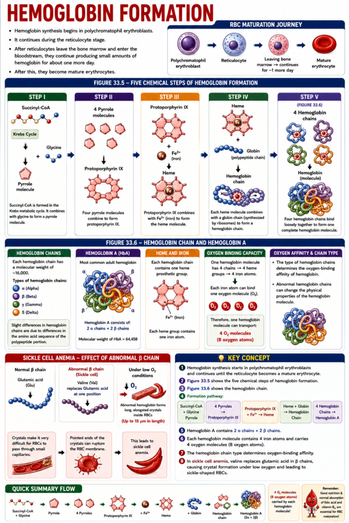
- Hemoglobin synthesis begins in polychromatophil erythroblasts.
- It continues during the reticulocyte stage of RBC development.
- After reticulocytes leave the bone marrow and enter the bloodstream, they continue producing small amounts of hemoglobin for about one more day.
- After this, they become mature erythrocytes.
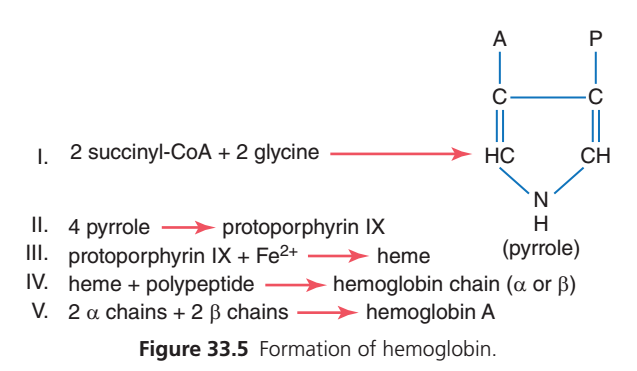
- Figure 33.5 shows the basic chemical steps of hemoglobin formation.
- Step I
- Succinyl-CoA is formed in the Krebs metabolic cycle.
- Succinyl-CoA combines with glycine.
- This forms a pyrrole molecule.
- Step II
- Four pyrrole molecules combine.
- This forms protoporphyrin IX.
- Step III
- Protoporphyrin IX combines with Fe²⁺ (iron).
- This forms the heme molecule.
- Step IV
- Each heme molecule combines with a long polypeptide chain (globin).
- The globin is synthesized by ribosomes.
- This forms a hemoglobin chain.
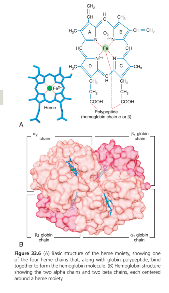
- Figure 33.6
- Step V
- Four hemoglobin chains bind loosely together.
- This forms one complete hemoglobin molecule.
- Each hemoglobin chain has a molecular weight of about 16,000.
- Four chains together form one hemoglobin molecule.
- There are slight differences in hemoglobin chains because of differences in the amino acid sequence of the polypeptide portion.
- The different hemoglobin chains are:
- Alpha (α)
- Beta (β)
- Gamma (γ)
- Delta (δ)
- The most common adult hemoglobin is Hemoglobin A (HbA).
- Hemoglobin A consists of:
- Two alpha (α) chains
- Two beta (β) chains
- Hemoglobin A has a molecular weight of 64,458.
- Each hemoglobin chain contains one heme prosthetic group.
- Each heme group contains one iron atom.
- Since one hemoglobin molecule has four chains, it contains four iron atoms.
- Each iron atom can bind one oxygen molecule (O₂).
- Therefore, one hemoglobin molecule can transport:
- Four oxygen molecules (O₂)
- Eight oxygen atoms
- The type of hemoglobin chains determines the oxygen-binding affinity of hemoglobin.
- Abnormal hemoglobin chains can change the physical properties of the hemoglobin molecule.
- In sickle cell anemia:
- Valine replaces glutamic acid at one position in each of the two beta chains.
- When this abnormal hemoglobin is exposed to low oxygen:
- It forms long, elongated crystals inside RBCs.
- These crystals may reach 15 micrometers in length.
- These crystals make it very difficult for RBCs to pass through small capillaries.
- The pointed ends of the crystals can rupture the RBC membrane.
- This leads to sickle cell anemia.
KEY CONCEPT
- Hemoglobin synthesis starts in polychromatophil erythroblasts and continues until the reticulocyte becomes a mature erythrocyte.
- Figure 33.5 shows the five chemical steps of hemoglobin formation.
- Figure 33.6 shows the hemoglobin chain.
- Formation pathway:
- Succinyl-CoA + Glycine → Pyrrole
- 4 Pyrroles → Protoporphyrin IX
- Protoporphyrin IX + Fe²⁺ → Heme
- Heme + Globin → Hemoglobin Chain
- Four Hemoglobin Chains → Hemoglobin A
- Hemoglobin A contains 2 α chains + 2 β chains.
- Each hemoglobin molecule contains 4 iron atoms and carries 4 oxygen molecules (8 oxygen atoms).
- The hemoglobin chain type determines oxygen-binding affinity.
- In sickle cell anemia, valine replaces glutamic acid in the β chains, causing crystal formation under low oxygen and leading to sickle-shaped RBCs.
Hemoglobin Combines Reversibly With Oxygen
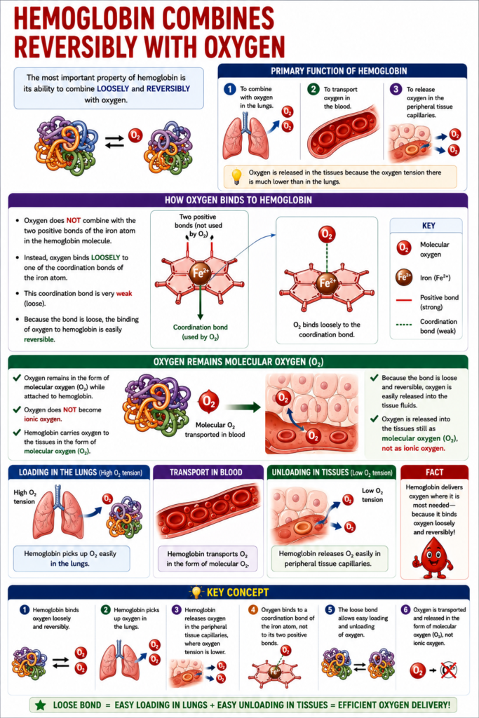
- The most important property of hemoglobin is its ability to combine loosely and reversibly with oxygen.
- The primary function of hemoglobin is:
- To combine with oxygen in the lungs.
- To transport oxygen in the blood.
- To release oxygen in the peripheral tissue capillaries.
- Oxygen is released in the tissues because the oxygen tension there is much lower than in the lungs.
- Oxygen does not combine with the two positive bonds of the iron atom in the hemoglobin molecule.
- Instead, oxygen binds loosely to one of the coordination bonds of the iron atom.
- This coordination bond is very weak (loose).
- Because the bond is loose, the binding of oxygen to hemoglobin is easily reversible.
- Oxygen remains in the form of molecular oxygen (O₂) while attached to hemoglobin.
- Oxygen does not become ionic oxygen.
- Hemoglobin carries oxygen to the tissues in the form of molecular oxygen (O₂).
- Because the bond is loose and reversible, oxygen is easily released into the tissue fluids.
- Oxygen is released into the tissues still as molecular oxygen (O₂), not as ionic oxygen.
KEY CONCEPT
- Hemoglobin binds oxygen loosely and reversibly.
- Hemoglobin picks up oxygen in the lungs.
- Hemoglobin releases oxygen in the peripheral tissue capillaries, where oxygen tension is lower.
- Oxygen binds to a coordination bond of the iron atom, not to its two positive bonds.
- The loose bond allows easy loading and unloading of oxygen.
- Oxygen is transported and released in the form of molecular oxygen (O₂), not ionic oxygen.
MADE BY SELF LEARNING CEO AND FOUNDER DR SHEEN

This material is very useful
thanks too much