


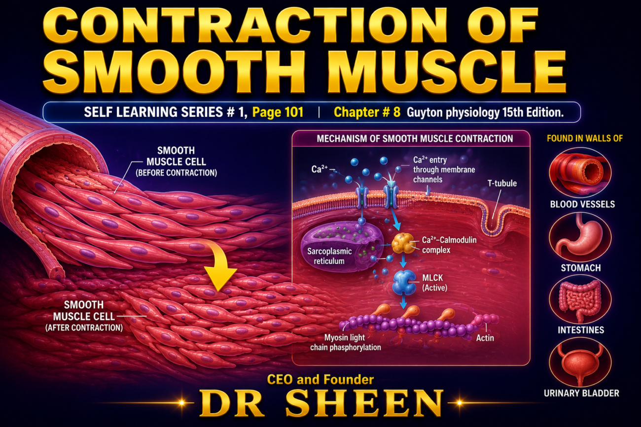
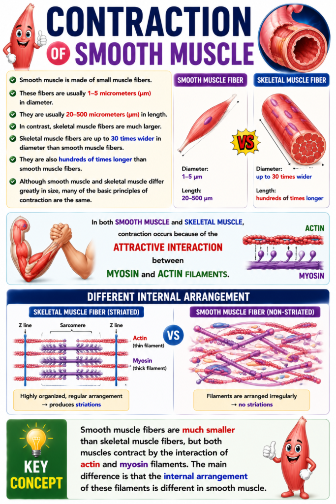
- Smooth muscle is made of small fibers.
- These fibers are usually 1 to 5 micrometers in diameter.
- They are only 20 to 500 micrometers in length.
- Skeletal muscle fibers are much larger.
- They can be up to 30 times greater in diameter.
- They can be hundreds of times longer.
- Despite these size differences, many of the same contraction principles apply to both smooth and skeletal muscle.
- The same attractive forces between myosin and actin filaments cause contraction in smooth muscle as in skeletal muscle.
- The internal physical arrangement of smooth muscle fibers is different.
KEY CONCEPT
- Smooth muscle fibers are much smaller than skeletal muscle fibers, but both muscles contract by the interaction of actin and myosin filaments. The main difference is that the internal arrangement of these filaments is different in smooth muscle.
YPES OF SMOOTH MUSCLE
- Smooth muscle of each organ is different from that of most other organs in several ways.
- These differences include: (1) physical dimensions; (2) organization into bundles or sheets; (3) response to different stimuli; (4) characteristics of innervation; and (5) function.
- For simplicity, smooth muscle can generally be divided into two major types.
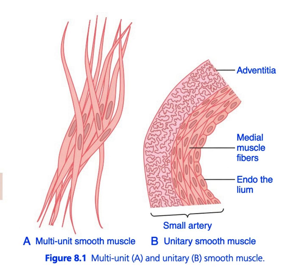
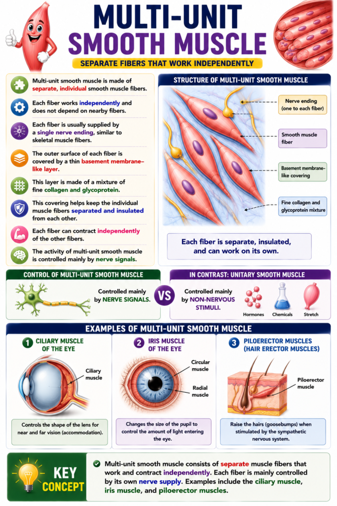
- These two types are multi-unit smooth muscle and unitary (or single-unit) smooth muscle, shown in Fig. 8.1.
KEY CONCEPT Smooth muscle varies by organ but is mainly divided into multi-unit and unitary (single-unit) types.
MULTI-UNIT SMOOTH MUSCLE
- Multi-unit smooth muscle is made of separate, individual smooth muscle fibers.
- Each fiber works independently and does not depend on nearby fibers.
- Each fiber is usually supplied by a single nerve ending, similar to skeletal muscle fibers.
- The outer surface of each fiber is covered by a thin basement membrane–like layer.
- This layer is made of a mixture of fine collagen and glycoprotein.
- This covering helps keep the individual muscle fibers separated and insulated from each other.
- Each fiber can contract independently of the other fibers.
- The activity of multi-unit smooth muscle is controlled mainly by nerve signals.
- In contrast, unitary smooth muscle is controlled mainly by non-nervous stimuli.
- Examples of multi-unit smooth muscle include:
- Ciliary muscle of the eye
- Iris muscle of the eye
- Piloerector muscles, which raise the hairs when stimulated by the sympathetic nervous system.
KEY CONCEPT
- Multi-unit smooth muscle consists of separate muscle fibers that work and contract independently. Each fiber is mainly controlled by its own nerve supply. Examples include the ciliary muscle, iris muscle, and piloerector muscles.
UNITARY SMOOTH MUSCLE
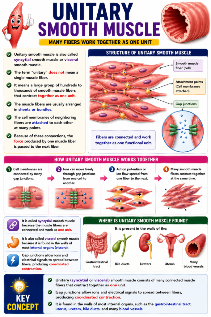
- Unitary smooth muscle is also called syncytial smooth muscle or visceral smooth muscle.
- The term “unitary” does not mean a single muscle fiber.
- It means a large group of hundreds to thousands of smooth muscle fibers that contract together as one unit.
- The muscle fibers are usually arranged in sheets or bundles.
- The cell membranes of neighboring fibers are attached to each other at many points.
- Because of these connections, the force produced by one muscle fiber is passed to the next fiber.
- The cell membranes are also connected by many gap junctions.
- Ions can move freely through these gap junctions from one muscle cell to another.
- Therefore, action potentials or ion flow can spread from one fiber to the next.
- This allows many smooth muscle fibers to contract together at the same time.
- It is called syncytial smooth muscle because the muscle fibers are connected and work as one functional unit.
- It is also called visceral smooth muscle because it is found in the walls of most internal organs (viscera).
- It is present in the walls of the:
- Gastrointestinal tract
- Bile ducts
- Ureters
- Uterus
- Many blood vessels
KEY CONCEPT
- Unitary (syncytial or visceral) smooth muscle consists of many connected muscle fibers that contract together as one unit. Gap junctions allow ions and electrical signals to spread between fibers, producing coordinated contraction. It is found in the walls of most internal organs, such as the gastrointestinal tract, uterus, ureters, bile ducts, and many blood vessels.
CONTRACTILE MECHANISM IN SMOOTH MUSCLE
Chemical Basis for Smooth Muscle Contraction
- Smooth muscle contains both actin and myosin filaments.
- These actin and myosin filaments have chemical properties similar to those in skeletal muscle.
- However, smooth muscle does not contain the troponin complex, which is required for skeletal muscle contraction.
- Therefore, the mechanism that controls contraction in smooth muscle is different from skeletal muscle.
- Chemical studies show that actin and myosin filaments in smooth muscle interact in almost the same way as they do in skeletal muscle.
- Contraction is activated by Ca²⁺ (calcium ions).
- During contraction, ATP is broken down into ADP.
- The energy released from ATP breakdown is used for muscle contraction.
- However, there are major differences between smooth muscle and skeletal muscle.
- These differences include:
- Physical organization of the muscle fibers
- Excitation–contraction coupling
- Control of contraction by Ca²⁺
- Duration of contraction
- Amount of energy required for contraction
KEY CONCEPT
- Smooth muscle contains actin and myosin filaments like skeletal muscle, but it does not have the troponin complex. Contraction is activated by Ca²⁺, and ATP is converted into ADP to provide energy. Smooth muscle differs from skeletal muscle in its structure, excitation–contraction coupling, calcium control, duration of contraction, and energy requirement.
PHYSICAL BASIS FOR SMOOTH MUSCLE CONTRACTION
- Smooth muscle does not have the same striated arrangement of actin and myosin filaments as skeletal muscle.
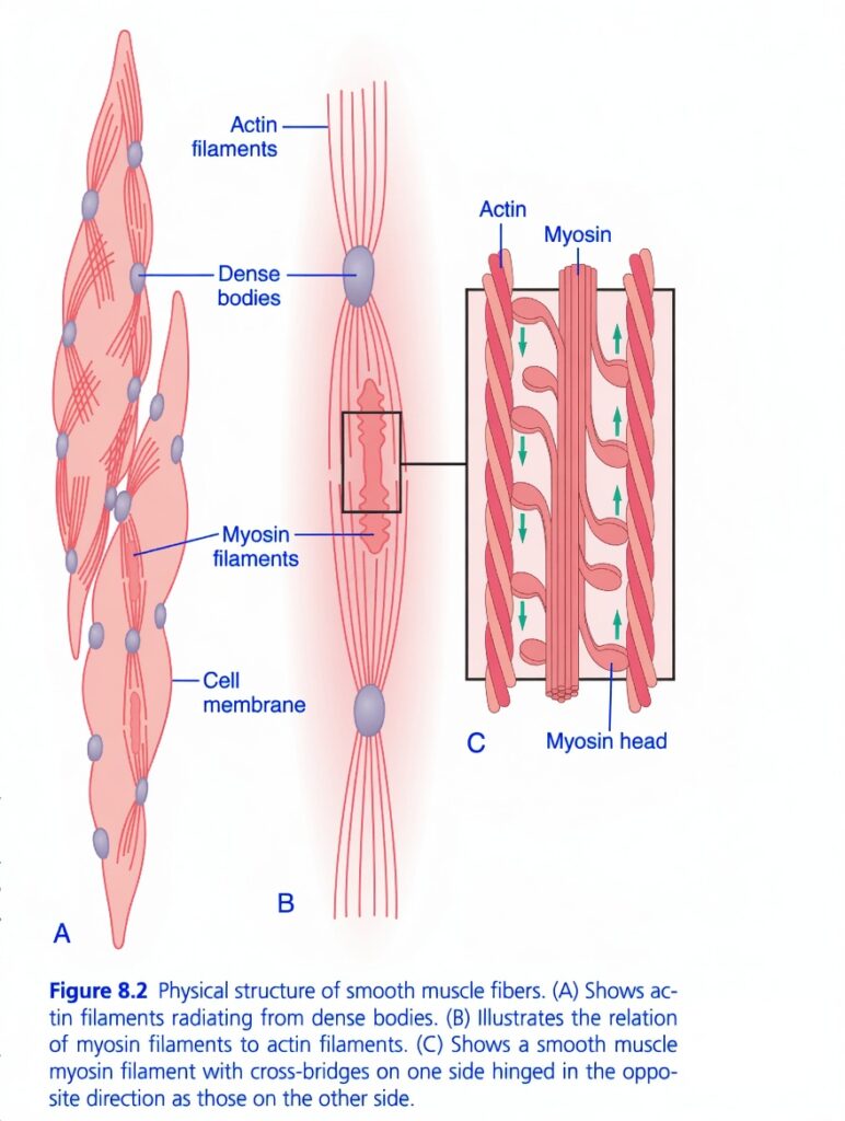
- Instead, electron microscopy shows that many actin filaments are attached to dense bodies.
- Some dense bodies are attached to the cell membrane, while others are present inside the cell.
- Some membrane dense bodies of adjacent smooth muscle cells are connected by intercellular protein bridges.
- These protein bridges help transmit the force of contraction from one cell to the next.
- Myosin filaments are located between the actin filaments.
- Myosin filaments are more than twice as thick as actin filaments.
- Electron microscopy usually shows 5–10 times more actin filaments than myosin filaments.
- Figure 8.2B shows a proposed contractile unit of a smooth muscle cell.
- In this unit, many actin filaments extend from two dense bodies.
- The ends of the actin filaments overlap with a myosin filament located between the two dense bodies.
- This contractile unit is similar to that of skeletal muscle, but its arrangement is not regular.
- The dense bodies in smooth muscle perform the same function as the Z discs in skeletal muscle.
- Another difference is that most myosin filaments have side-polar cross-bridges.
- The cross-bridges on one side face in the opposite direction to those on the other side.
- This arrangement allows one myosin filament to pull one actin filament in one direction while pulling another actin filament in the opposite direction at the same time.
- Because of this organization, smooth muscle cells can shorten by about 80% of their original length.
- In contrast, skeletal muscle fibers usually shorten by less than 30% of their original length.
Figure: Figure 8.2A, Figure 8.2B, Figure 8.2C
KEY CONCEPT
- Smooth muscle has a non-striated arrangement of actin and myosin filaments. Actin filaments are attached to dense bodies, which function like Z discs in skeletal muscle. Side-polar myosin filaments allow pulling in opposite directions, enabling smooth muscle to shorten by about 80% of its length, much more than skeletal muscle.
COMPARISON OF SMOOTH MUSCLE CONTRACTION AND SKELETAL MUSCLE CONTRACTION
- Most skeletal muscles contract and relax quickly.
- In contrast, most smooth muscles produce prolonged (tonic) contractions.
- Smooth muscle contraction may last for hours or even days.
- Therefore, the physical and chemical properties of smooth muscle contraction are different from those of skeletal muscle contraction.
Slow Cycling of the Myosin Cross-Bridges
- In smooth muscle, the myosin cross-bridges attach to actin, detach, and reattach much more slowly than in skeletal muscle.
- The cycling rate of myosin cross-bridges is only about 1/10 to 1/300 of that in skeletal muscle.
- However, the cross-bridges remain attached to the actin filaments for a much longer time.
- This longer attachment increases the force of smooth muscle contraction.
- One possible reason for the slow cycling is that the myosin cross-bridge heads have much lower ATPase activity than those in skeletal muscle.
- Therefore, ATP is broken down more slowly.
- As a result, the movements of the myosin cross-bridge heads become slower, leading to slower cross-bridge cycling.
KEY CONCEPT
- Smooth muscle contracts much more slowly and for a much longer time than skeletal muscle. Its myosin cross-bridges cycle 1/10 to 1/300 as fast because they have lower ATPase activity. The cross-bridges stay attached to actin longer, producing strong and sustained (tonic) contractions.
COMPARISON OF SMOOTH MUSCLE CONTRACTION AND SKELETAL MUSCLE CONTRACTION
Low Energy Requirement to Sustain Smooth Muscle Contraction
- Smooth muscle requires only about 1/10 to 1/300 of the energy needed by skeletal muscle to maintain the same force of contraction.
- This low energy requirement is mainly due to the slow attachment and detachment of the myosin cross-bridges.
- Each cross-bridge cycle uses only one ATP molecule, no matter how long the cycle lasts.
- Therefore, slow cross-bridge cycling reduces overall energy use.
- This energy-saving property is important because many internal organs maintain tonic smooth muscle contraction for long periods.
- Examples include:
- Intestines
- Urinary bladder
- Gallbladder
- Other viscera
Slowness of Onset of Contraction and Relaxation of the Total Smooth Muscle Tissue
- Smooth muscle usually begins to contract about 50–100 milliseconds after stimulation.
- It reaches maximum contraction about 0.5 second later.
- The contractile force then decreases over the next 1–2 seconds.
- Therefore, the total contraction lasts about 1–3 seconds.
- This is about 30 times longer than the contraction of an average skeletal muscle fiber.
- Depending on the type of smooth muscle, contraction may last as little as 0.2 second or as long as 30 seconds.
- The slow onset and prolonged contraction are mainly due to the slow attachment and detachment of myosin cross-bridges with actin filaments.
- In addition, contraction begins more slowly in response to Ca²⁺ than it does in skeletal muscle.
KEY CONCEPT
- Smooth muscle uses only 1/10 to 1/300 of the energy required by skeletal muscle to maintain the same tension because its cross-bridges cycle slowly. It begins contracting 50–100 ms after stimulation, reaches maximum contraction in about 0.5 second, and usually completes contraction in 1–3 seconds, making its contraction much slower and longer-lasting than skeletal muscle.
Maximum Force of Contraction Is Often Greater in Smooth Muscle Than in Skeletal Muscle
- Smooth muscle has fewer myosin filaments than skeletal muscle.
- The myosin cross-bridges in smooth muscle cycle more slowly.
- Despite these differences, smooth muscle can produce a greater maximum force of contraction than skeletal muscle.
- Smooth muscle can generate about 4–6 kg/cm² of cross-sectional area.
- Skeletal muscle can generate about 3–4 kg/cm² of cross-sectional area.
- The greater force in smooth muscle is due to the longer attachment of myosin cross-bridges to actin filaments.
Latch Mechanism Facilitates Prolonged Holding of Contractions of Smooth Muscle
- After smooth muscle reaches full contraction, the level of stimulation can be greatly reduced.
- Even with less stimulation, the muscle continues to maintain its full force of contraction.
- Very little energy is needed to maintain this contraction.
- Sometimes, smooth muscle uses as little as 1/300 of the energy needed for a similar sustained contraction in skeletal muscle.
- This energy-saving process is called the latch mechanism.
- The latch mechanism allows smooth muscle to maintain tonic contraction for many hours.
- It does this while using very little energy.
- Only a small amount of continuous stimulation from nerves or hormones is needed to maintain the contraction.
KEY CONCEPT
- Smooth muscle can generate a greater maximum force (4–6 kg/cm²) than skeletal muscle (3–4 kg/cm²) because its myosin cross-bridges remain attached to actin for a longer time. The latch mechanism allows smooth muscle to maintain prolonged tonic contraction for hours with very little energy and minimal continuous stimulation.
Stress-Relaxation of Smooth Muscle
- An important property of smooth muscle, especially visceral unitary smooth muscle, is stress-relaxation.
- This type of smooth muscle can return almost to its original force of contraction after it is stretched or shortened.
- This adjustment occurs within seconds or minutes.
- For example, when the urinary bladder suddenly fills with more fluid, the bladder wall stretches.
- This stretch immediately causes a large increase in bladder pressure.
- After about 15–60 seconds, the bladder pressure falls back almost to its original level, even though the bladder remains stretched.
- If the bladder volume increases again, the same response occurs again.
- Conversely, when the bladder volume suddenly decreases, the pressure first falls sharply.
- After a few seconds or minutes, the pressure rises again to near its original level.
- The decrease in pressure after stretching is called stress-relaxation.
- The increase in pressure after shortening is called reverse stress-relaxation.
- These responses allow hollow organs to maintain nearly constant internal pressure despite large and long-lasting changes in volume.
KEY CONCEPT
- Visceral unitary smooth muscle shows stress-relaxation and reverse stress-relaxation. After stretching or shortening, it gradually returns close to its original force of contraction within seconds or minutes. This helps hollow organs, such as the urinary bladder, maintain nearly constant internal pressure despite large changes in volume.
Made by Self Learning CEO and Founder Dr sheen.
