


Learning Objectives
After studying this topic, students will be able to:
- Explain the photochemistry of color vision in cones.
- Describe light and dark adaptation.
- Compare the roles of photochemical, pupillary, and neural adaptation.
Introduction
The photochemicals in cones have almost the same chemical composition as rhodopsin in rods.
The main difference is in the protein part of the visual pigment.
In cones, these proteins are called photopsins, whereas rods contain scotopsin.
The retinal portion is exactly the same in both rods and cones.
Different cone pigments make cones selectively sensitive to blue, green, or red light.
The sensitivity of the retina also automatically changes according to whether the eye is exposed to bright light or darkness.
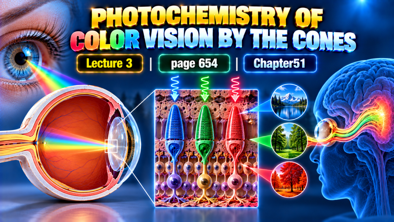
Photochemistry of Color Vision by the Cones
The photochemicals of cones are almost chemically identical to the rhodopsin found in rods.
The main difference is their protein portion.
The protein portion in cones is called:
Photopsin
The protein portion in rods is:
Scotopsin
The retinal part of the visual pigment is exactly the same in:
- Rods
- Cones
Therefore, the color-sensitive pigments of cones consist of:
Retinal + photopsin
Three Types of Cone Pigments
Each cone contains only one of three types of color pigments.
Therefore, different cones are selectively sensitive to different colors:
- Blue
- Green
- Red
The three pigments are called:
- Blue-sensitive pigment
- Green-sensitive pigment
- Red-sensitive pigment
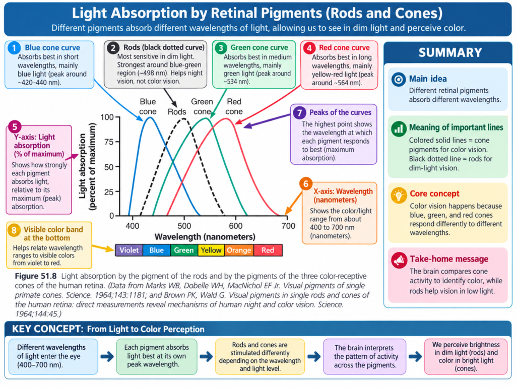
Their peak light absorption occurs at different wavelengths, as shown in Fig. 51.8.
Blue-sensitive pigment → 445 nm
Green-sensitive pigment → 535 nm
Red-sensitive pigment → 570 nm
These wavelengths are also the wavelengths at which each type of cone has its greatest light sensitivity.
This difference in sensitivity begins to explain how the retina distinguishes different colors.
For comparison, the peak absorption of rhodopsin in rods is:
505 nm
Key Concept
Each cone contains only one type of color-sensitive pigment:
Blue cone → peak at 445 nm
Green cone → peak at 535 nm
Red cone → peak at 570 nm
Rod rhodopsin → peak at 505 nm
Example
Light near 445 nm produces the greatest response in the blue-sensitive pigment, whereas light near 570 nm produces the greatest response in the red-sensitive pigment.
AUTOMATIC REGULATION OF RETINAL SENSITIVITY—LIGHT AND DARK ADAPTATION
Light Adaptation
When a person remains in bright light for several hours, large amounts of the photochemicals in rods and cones are broken down into:
Retinal + opsins
In addition, much of the retinal is converted into:
Vitamin A
Because of these changes, the amount of photosensitive chemicals remaining in the rods and cones becomes much smaller.
Therefore, the sensitivity of the eye to light also decreases.
This process is called:
Light adaptation
Key Concept
Bright light for a long time
↓
Photochemicals decrease
↓
More retinal is converted to vitamin A
↓
Photosensitive pigments decrease
↓
Retinal sensitivity decreases
↓
Light adaptation
Dark Adaptation
When a person remains in darkness for a long time, the opposite changes occur.
Retinal and opsins are converted back into:
Light-sensitive pigments
Vitamin A is also converted back into:
Retinal
This increases the amount of light-sensitive pigment.
The final amount that can be formed depends on the amount of opsin available to combine with retinal.
This process is called:
Dark adaptation
Key Concept
Darkness
↓
Retinal + opsins form light-sensitive pigments
↓
Vitamin A is converted back into retinal
↓
More photosensitive pigment forms
↓
Sensitivity to light increases
↓
Dark adaptation
Course of Dark Adaptation
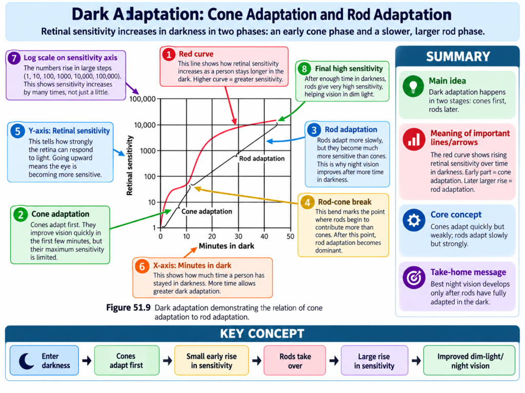
The course of dark adaptation after several hours of bright-light exposure is shown in Fig. 51.9.
When a person first enters complete darkness, retinal sensitivity is very low.
Within approximately:
1 minute
the sensitivity increases about:
10-fold
This means the retina can now respond to light with only:
One-tenth of the previously required intensity
After approximately:
20 minutes
sensitivity increases about:
6000-fold
After approximately:
40 minutes
sensitivity increases about:
25,000-fold
Easy Concept of Fold Increase
If sensitivity becomes 10 times greater, the retina needs only about one-tenth as much light to respond.
As dark adaptation continues, progressively smaller amounts of light can stimulate the retina.
Dark Adaptation Curve
The curve showing these changes is called the:
Dark adaptation curve
The curve has an inflection, meaning its pattern changes during adaptation.
The early part of the curve is caused mainly by:
Cone adaptation
This is because the chemical events of vision occur about:
4 times faster in cones than in rods
However, cones cannot achieve nearly as great an increase in sensitivity as rods.
Therefore, cones adapt rapidly but stop adapting after only a few minutes.
Rods adapt more slowly.
However, rods continue to adapt for:
- Many minutes
- Even hours
Their sensitivity increases tremendously during this period.
Additional Sensitivity of Rods
Rod sensitivity is increased further because signals from:
100 or more rods
can converge onto:
One ganglion cell
The signals from these rods summate.
This increases their sensitivity.
Key Concept
Cones
- Adapt rapidly
- Adapt about 4 times faster
- Stop adapting after a few minutes
- Show a smaller increase in sensitivity
Rods
- Adapt more slowly
- Continue adapting much longer
- Show a much greater increase in sensitivity
Other Mechanisms of Light and Dark Adaptation
Changes in the amount of rhodopsin and color photochemicals are not the only mechanisms for adaptation.
The eye also uses:
- Changes in pupillary size
- Neural adaptation
Change in Pupillary Size
Changing pupil size alters how much light can enter the eye.
This mechanism can produce approximately:
30-fold adaptation
It occurs within:
A fraction of a second
The change occurs because the pupil changes the amount of light allowed through the pupillary opening.
Key Concept
Pupil changes size → amount of entering light changes → rapid adaptation
This effect occurs much faster than full photochemical adaptation.
Neural Adaptation
Neural adaptation involves neurons in successive stages of the visual pathway in:
- Retina
- Brain
When light intensity first increases, signals transmitted by the following cells are strong:
- Bipolar cells
- Horizontal cells
- Amacrine cells
- Ganglion cells
However, most of these signals rapidly decrease at different stages of neural transmission.
The amount of neural adaptation is only:
A fewfold
This is much smaller than the many-thousandfold adaptation produced by changes in the photochemical system.
However, neural adaptation occurs:
Within a fraction of a second
Photochemical adaptation requires:
Many minutes to hours
for complete adaptation.
Key Concept
Neural adaptation = small but very fast
Photochemical adaptation = very large but much slower
Importance of Light and Dark Adaptation in Vision
Between maximum dark adaptation and maximum light adaptation, the sensitivity of the eye can change approximately:
500,000 to 1 million times
The sensitivity automatically adjusts according to the level of illumination.
For the retina to register an image properly, it must detect both:
- Light areas
- Dark areas
Therefore, retinal sensitivity must be adjusted so that receptors respond to the lighter areas but not excessively to the darker areas.
Example: Leaving a Movie Theater
When a person leaves a dark movie theater and suddenly enters bright sunlight, even the dark areas of the visual image initially appear extremely bright.
As a result, the entire image seems:
Bleached
There is very little contrast between different parts of the image.
Poor vision continues until the retina adapts enough that the darker areas no longer stimulate the receptors excessively.
Key Concept
Dark-adapted eye + sudden bright light
↓
Too much retinal sensitivity
↓
Even dark areas strongly stimulate receptors
↓
Image appears bleached
↓
Light adaptation occurs
↓
Normal contrast improves
Example: Entering Darkness
When a person first enters darkness, retinal sensitivity is initially very low.
Even the lighter areas of the image may not be able to excite the retina.
As dark adaptation develops, retinal sensitivity increases.
The lighter areas then begin to register.
Key Concept
Bright-adapted eye + sudden darkness
↓
Sensitivity initially too low
↓
Light areas may not stimulate retina sufficiently
↓
Dark adaptation develops
↓
Sensitivity rises
↓
Light areas become visibleExtreme Range of Illumination
The intensity of sunlight is approximately:
10 billion times greater than starlight
Despite this enormous difference, the eye can function:
In bright sunlight after light adaptation
and
In starlight after dark adaptation
Key Concepts With Examples
Color Vision
Cone pigment = retinal + photopsin
Each cone contains one color-sensitive pigment.
Blue → 445 nm
Green → 535 nm
Red → 570 nm
Example: Different peak sensitivities allow the retina to differentiate colors.
Light Adaptation
Bright light → fewer photosensitive pigments → lower retinal sensitivity
Example: After leaving a dark movie theater for bright sunlight, vision initially appears bleached until light adaptation occurs.
Dark Adaptation
Darkness → more photosensitive pigments → greater retinal sensitivity
Example: On first entering darkness, objects are difficult to see, but visibility improves as dark adaptation progresses.
Speed of Adaptation
Pupil and neural mechanisms → fraction of a second
Photochemical mechanism → minutes to hours
Clinical Note or Importance
Proper light and dark adaptation is essential for maintaining visual contrast under very different levels of illumination.
After sudden movement from darkness into bright light, the image may temporarily appear bleached because retinal sensitivity is too high.
After sudden movement from bright light into darkness, the retina may initially be too insensitive to detect even lighter areas.
Adaptation gradually adjusts retinal sensitivity to the new level of illumination.
High-Yield Points
- Cone pigments consist of retinal + photopsins.
- The retinal component is the same in rods and cones.
- Each cone contains only one of three color pigments.
- Blue-sensitive pigment peaks at 445 nm.
- Green-sensitive pigment peaks at 535 nm.
- Red-sensitive pigment peaks at 570 nm.
- Rod rhodopsin peaks at 505 nm.
- Bright light decreases photosensitive pigments and causes light adaptation.
- Darkness increases photosensitive pigments and causes dark adaptation.
- Dark sensitivity increases about 10-fold in 1 minute.
- It increases about 6000-fold in 20 minutes.
- It increases about 25,000-fold in 40 minutes.
- Cones adapt about 4 times faster than rods.
- Rods ultimately achieve much greater sensitivity in darkness.
- Signals from 100 or more rods can converge on one ganglion cell.
- Pupillary adaptation can produce approximately a 30-fold change within a fraction of a second.
- Neural adaptation occurs within a fraction of a second.
- Photochemical adaptation may require minutes to hours.
- Total retinal sensitivity can change about 500,000 to 1 million times.
- Sunlight is approximately 10 billion times more intense than starlight.
Common Student Mistakes
- Thinking cone pigments use a different retinal molecule from rods.
Remember:
Retinal is exactly the same in rods and cones.
The protein portions differ:
Cones → photopsins
Rods → scotopsin
- Mixing up the peak wavelengths.
Remember:
Blue = 445 nm
Green = 535 nm
Red = 570 nm
Rod rhodopsin = 505 nm
- Thinking light adaptation increases retinal sensitivity.
Remember:
Light adaptation → sensitivity decreases
Dark adaptation → sensitivity increases
- Thinking cones adapt more slowly than rods.
Remember:
Cones adapt about 4 times faster, but rods eventually become much more sensitive.
- Confusing speed with degree of adaptation.
Remember:
Neural and pupillary adaptation = rapid
Photochemical adaptation = slower but much larger
Quick Revision
Color Pigments
Retinal + photopsin → cone pigment
- Blue: 445 nm
- Green: 535 nm
- Red: 570 nm
- Rod rhodopsin: 505 nm
Light Adaptation
Bright light
↓
Photochemicals decrease
↓
Retinal converted toward vitamin A
↓
Sensitivity decreases
Dark Adaptation
Darkness
↓
Retinal + opsins form visual pigments
↓
Vitamin A converted back toward retinal
↓
Sensitivity increases
Dark Adaptation Values
- 1 minute → 10-fold
- 20 minutes → 6000-fold
- 40 minutes → 25,000-fold
Cones Versus Rods
Cones → faster adaptation
Rods → slower but much greater final sensitivity
Other Mechanisms
Pupil → about 30-fold, fraction of a second
Neural adaptation → fewfold, fraction of a second
Photochemical adaptation → many-thousandfold, minutes to hours
Conceptual Easiest Summary
Cone visual pigments are made from retinal and photopsins.
The retinal component is the same as in rods, but the protein component is slightly different.
There are three types of cone pigments.
They are most sensitive to:
Blue at 445 nm
Green at 535 nm
Red at 570 nm
This difference in wavelength sensitivity allows the retina to distinguish colors.
The sensitivity of the retina also changes according to the amount of surrounding light.
In prolonged bright light, visual pigments are broken down and some retinal is converted into vitamin A.
Therefore, the amount of photosensitive pigment falls and the eye becomes less sensitive.
This is light adaptation.
In darkness, retinal and opsins combine again to form light-sensitive pigments, and vitamin A is converted back into retinal.
Therefore, retinal sensitivity increases.
This is dark adaptation.
Dark adaptation begins rapidly but continues for a long time.
Cones adapt faster, but their increase in sensitivity is limited.
Rods adapt more slowly but eventually become much more sensitive.
The eye also adapts through changes in pupil size and through neural adaptation.
These mechanisms act very rapidly, but their total effect is smaller than that of photochemical adaptation.
Overall, retinal sensitivity can change by approximately 500,000 to 1 million times.
This enormous range allows the eye to function in conditions ranging from bright sunlight to starlight.
Reference Guyton and Hall Textbook of Medical Physiology 15th Edition, page. 654 Chapter51
