


COLOR VISION
Learning Objectives
After studying this topic, students will be able to:
- Explain how the retina detects different colors.
- Describe the tricolor mechanism of color detection.
- Explain how cone stimulation ratios produce different color sensations.
- Describe the perception of white light.
- Explain red-green color blindness, protanopia, deuteranopia, and blue weakness.
- Understand the basic use of color test charts.
Introduction
- Different cones are sensitive to different colors of light.
- The retina detects different gradations of color present in the visual spectrum.
- Color perception depends on how strongly the different types of cones are stimulated.
- The nervous system interprets the pattern of stimulation of these cones as different colors.
Cone: A color-receptive retinal cell that responds to particular colors of light.
Color vision: The ability of the visual system to distinguish different gradations of color through different patterns of cone stimulation.
TRICOLOR MECHANISM OF COLOR DETECTION
Definition: The tricolor mechanism is based on the observation that almost all gradations of color can be produced by appropriately mixing red, green, and blue monochromatic light in different combinations.
- Human color vision depends mainly on three types of color-sensitive cones.
- These cones respond differently to red, green, and blue light.
- Different combinations of their stimulation allow the eye to detect almost all gradations of color.
Monochromatic light: Light having a single wavelength.
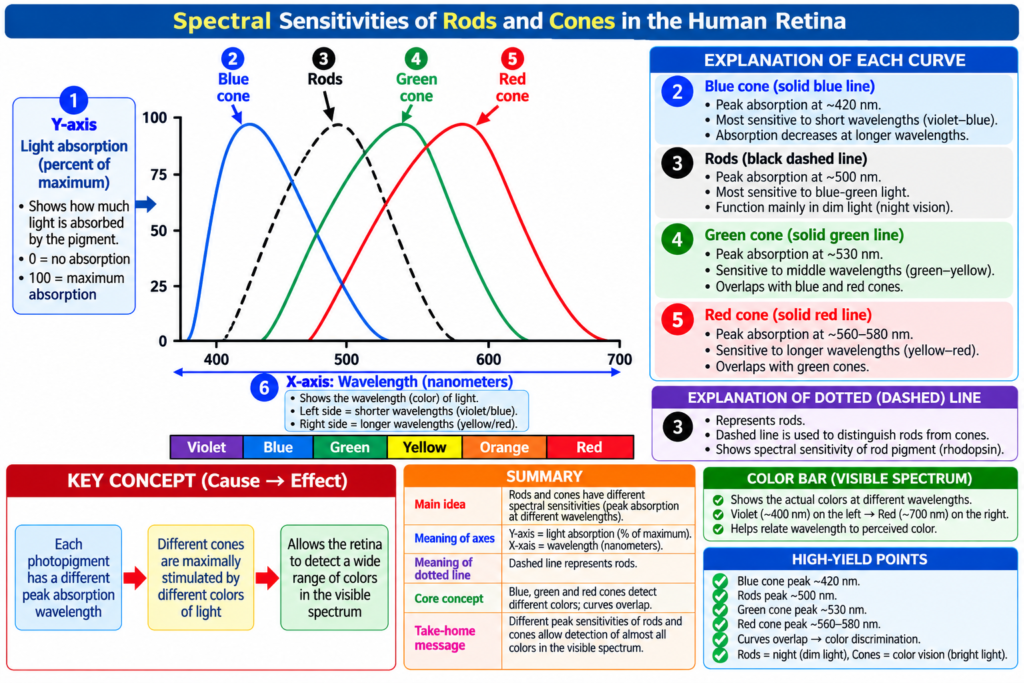
Spectral Sensitivities of the Three Types of Cones
Spectral sensitivity: The degree to which a cone responds to different wavelengths of light.
- Color vision tests show that the spectral sensitivities of the three types of cones are essentially similar to the light absorption curves of the three cone pigments.
- These sensitivity curves help explain most phenomena of color vision.
- These curves are shown in Fig. 51.8 and Fig. 51.10.
Interpretation of Color in the Nervous System
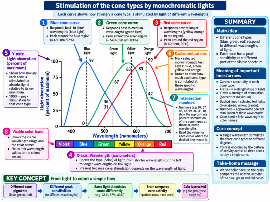
- The nervous system does not identify color simply from stimulation of one cone.
- It interprets the relative stimulation of the three types of cones.
Orange Light
- Wavelength = 580 nm
- Red cones → about 99
- Green cones → about 42
- Blue cones → 0
Therefore:
Red : Green : Blue = 99 : 42 : 0
→ Nervous system interprets this pattern as orange.
Blue Light
- Wavelength = 450 nm
- Red cones → 0
- Green cones → 0
- Blue cones → about 97
Therefore:
Red : Green : Blue = 0 : 0 : 97
→ Nervous system interprets this pattern as blue.
Yellow
Red : Green : Blue = 83 : 83 : 0
→ Interpreted as yellow.
Green
Red : Green : Blue = 31 : 67 : 36
→ Interpreted as green.
Key Concept
Different wavelength of light
→ produces a particular pattern of stimulation in red, green, and blue cones
→ nervous system compares the relative stimulation
→ a particular color is perceived.
Example:
580 nm light
→ Red = 99, Green = 42, Blue = 0
→ Ratio = 99 : 42 : 0
→ Orange sensation
PERCEPTION OF WHITE LIGHT
White light: The sensation produced when red, green, and blue cones are stimulated approximately equally.
- Approximately equal stimulation of all three cone types gives the sensation of white.
- There is no single wavelength corresponding to white light.
- White contains a combination of wavelengths from the spectrum.
- The perception of white can also be produced by using an appropriate combination of only three selected colors.
- These colors stimulate the three types of cones approximately equally.
Key Concept
Approximately equal stimulation of red + green + blue cones
→ white color sensation
White therefore does not depend on one special wavelength.
COLOR BLINDNESS
Color blindness: A condition in which a person cannot distinguish certain colors normally because one group of color-receptive cones is absent or deficient.
Red-Green Color Blindness
Definition: Difficulty distinguishing red from green because either the red-sensitive or green-sensitive cone system is missing.
- Normally, colors between approximately 525 and 675 nm include:
- Green
- Yellow
- Orange
- Red
- These colors are distinguished mainly by comparing the responses of red and green cones.
- If either the red or green cone type is missing:
- This comparison mechanism cannot work normally.
- Several colors become difficult to distinguish.
- Red and green are especially difficult to separate.
→ This condition is called red-green color blindness.
Protanopia
Protanopia: Red blindness caused by loss of red cones.
- Red cones are absent.
- The person has difficulty detecting red normally.
- The overall visual spectrum becomes noticeably shortened at its long-wavelength end.
- This occurs because red cones are unavailable to detect long-wavelength red light.
Key Concept
Loss of red cones
→ poor detection of long-wavelength red light
→ shortening of the visible spectrum at its long-wavelength end
→ protanopia
Deuteranopia
Deuteranopia: Green blindness caused by lack of green cones.
- Green cones are absent.
- Unlike protanopia, there is no abnormal shortening of the visual spectrum.
- Red cones remain available to detect long-wavelength red light.
- A person with deuteranopia may distinguish only two or three different hues.
- A person with normal vision can distinguish seven unique hues.
Key Concept
Loss of green cones
→ green blindness
→ red cones still detect long wavelengths
→ visual spectral width remains approximately normal
→ deuteranopia
Genetic Basis of Red-Green Color Blindness
- Red-green color blindness occurs almost exclusively in males.
- Genes associated with these cone systems are located on the X chromosome.
- Females have two X chromosomes.
- Color blindness is uncommon in females because at least one X chromosome usually contains a normal gene for each cone type.
- Males have only one X chromosome.
- Therefore, if the required gene is missing from that X chromosome, color blindness may occur.
- A male receives his X chromosome from his mother.
- Therefore, color blindness can pass from mother to son.
- Such a mother is described as a color blindness carrier.
- About 8% of women are color blindness carriers.
Carrier: A person who possesses the gene associated with the condition and can transmit it to offspring.
Key Concept
Mother carries the affected X chromosome
→ son receives his X chromosome from the mother
→ missing cone-related gene may be expressed
→ red-green color blindness can occur.
Blue Weakness
Blue weakness: A genetically inherited condition in which blue cones are underrepresented.
COLOR TEST CHARTS
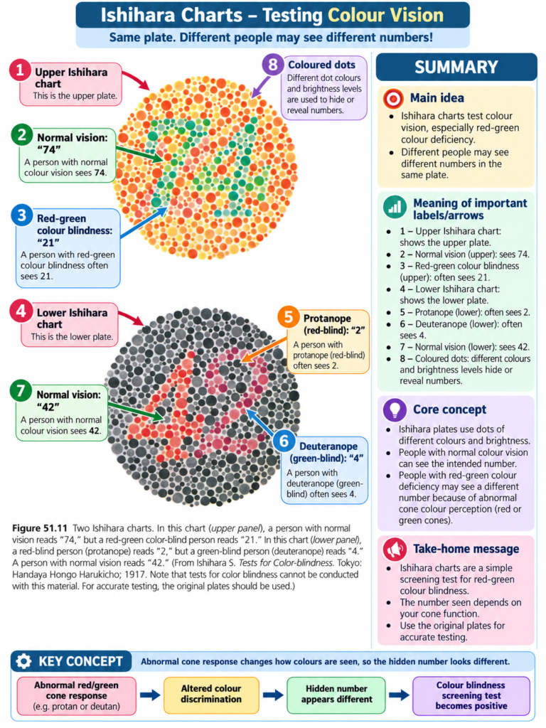
Color test charts: Spot charts containing mixtures of differently colored spots used as a rapid method for detecting color blindness.
- The charts contain spots of several different colors.
- People with normal color vision and people with specific color defects may see different numbers.
Fig. 51.11
Top Chart
- Normal color vision → reads 74
- Red-green color blindness → reads 21
Bottom Chart
- Normal color vision → reads 42
- Red blindness → reads 2
- Green blindness → reads 4
Key Concept
Different cone defects
→ different perception of colored spots
→ different numbers are recognized on the chart
→ helps identify color blindness.
CLINICAL IMPORTANCE
- Loss of a particular cone system can interfere with the ability to distinguish certain colors.
- Loss of red cones causes protanopia.
- Loss of green cones causes deuteranopia.
- Red-green color blindness occurs predominantly in males because the relevant genes are associated with the X chromosome.
- Color test charts provide a rapid method for detecting color vision abnormalities.
HIGH-YIELD POINTS
- Human color vision mainly depends on red, green, and blue cone systems.
- The nervous system interprets color according to the relative stimulation ratio of the three cone types.
- 99 : 42 : 0 → orange
- 0 : 0 : 97 → blue
- 83 : 83 : 0 → yellow
- 31 : 67 : 36 → green
- Approximately equal stimulation of all three cone types produces white.
- White does not correspond to a single wavelength.
- Loss of red cones → protanopia.
- Loss of green cones → deuteranopia.
- Protanopia shortens the long-wavelength end of the visible spectrum.
- Deuteranopia does not produce abnormal shortening of the visual spectrum.
- Red-green color blindness occurs almost exclusively in males.
- It can be transmitted from a carrier mother to her son.
- About 8% of women are carriers.
- Blue-cone abnormalities are rare.
- Spot charts can rapidly test color vision.
COMMON STUDENT MISTAKES
- Do not think that each perceived color is detected by only one cone type.
- Color depends on the relative stimulation of the three cone systems.
- Do not think that white has its own single wavelength.
- White results from approximately equal stimulation of the three cone types.
- Do not confuse:
- Protanopia = red cone loss
- Deuteranopia = green cone loss
- Do not assume both protanopia and deuteranopia shorten the visible spectrum.
- The shortening described in the text occurs with protanopia.
- Do not confuse blue weakness with red-green color blindness.
- Blue-cone deficiency is much rarer.
QUICK REVISION
Color detection
Red + Green + Blue cone stimulation pattern
→ nervous system compares the ratio
→ color sensation
White
Red ≈ Green ≈ Blue stimulation
→ white
Protanopia
Red cones absent
→ red blindness
→ long-wavelength spectrum shortened
Deuteranopia
Green cones absent
→ green blindness
→ spectral width not abnormally shortened
Inheritance
X chromosome gene abnormality
→ mainly affects males
→ may pass from carrier mother to son
Blue weakness
Reduced representation of blue cones
Color charts
Different color perception
→ different numbers recognized
→ rapid detection of color blindness
CONCEPTUAL SUMMARY
Color vision depends on the different responses of three types of cones to light.
Different wavelengths stimulate red, green, and blue cones in different proportions.
Light wavelength
→ different stimulation of three cone types
→ characteristic stimulation ratio
→ nervous system interprets the ratio
→ particular color is perceived.
For example:
580 nm
→ 99 : 42 : 0
→ orange
450 nm
→ 0 : 0 : 97
→ blue
When all three cone systems are stimulated approximately equally:
→ white is perceived
If one cone system is missing:
→ normal color comparison becomes difficult
→ color blindness can occur.
Red cone loss
→ protanopia
Green cone loss
→ deuteranopia
Underrepresentation of blue cones
→ blue weakness
Red-green color blindness is mainly associated with genes on the X chromosome, explaining why it occurs predominantly in males and may pass from a carrier mother to her son.
Color spot charts provide a rapid practical method for identifying these abnormalities.
Final Take-Home Concept
Different cone stimulation ratios = different colors
Equal stimulation of all three cone types = white
Missing cone system = impaired color discrimination
Reference Guyton and Hall Textbook of Medical Physiology 15th Edition, page. 654 Chapter51
