


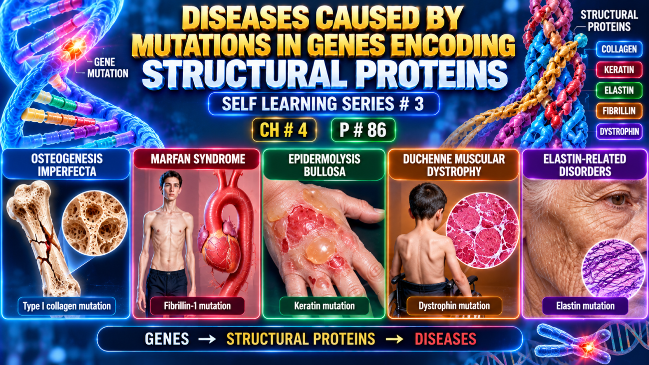
Marfan Syndrome
- Marfan syndrome is an autosomal dominant connective-tissue disorder.
- It mainly affects:
- skeleton
- eyes
- cardiovascular system
- It is caused by a defect in fibrillin, an extracellular glycoprotein.
Pathogenesis
- Fibrillin is produced by fibroblasts and is a major component of microfibrils in the extracellular matrix.
- Microfibrils act as a scaffold for tropoelastin → help form elastic fibers.
- Microfibrils are especially abundant in:
- aorta
- ligaments
- ciliary zonules supporting the lens
- Therefore, these tissues are particularly affected in Marfan syndrome.
- Fibrillin is encoded by the FBN1 gene on chromosome 15q21.
- Marfan syndrome is associated with mutations in FBN1.
- More than 1000 different FBN1 mutations have been described, so diagnosis is mainly based on clinical findings.
- The mutant fibrillin usually acts as a dominant-negative protein:
- abnormal fibrillin interferes with normal fibrillin
- → defective microfibril assembly.
- Prevalence is about 1 in 5000.
- About 70%–85% of cases are familial.
- Remaining cases result from new (de novo) FBN1 mutations.
- Some features result directly from weak connective tissue, but others are related to excessive TGF-β activity.
- Normally:
microfibrils bind/sequester TGF-β → limit its activity. - In Marfan syndrome:
↓ normal microfibrils → ↑ free TGF-β → excessive TGF-β signaling. - Excess TGF-β causes:
- abnormal vascular smooth muscle development
- weakening of the extracellular matrix
- Mutations in the TGF-β type II receptor can cause a related disorder called Marfan syndrome type 2 (MFS2).
- Cardiovascular complications may be reduced by:
- angiotensin receptor blockers
- β-blockers
- These help reduce the risk of serious cardiovascular complications.
- Clinical severity varies widely.
- Some patients mainly have cardiovascular disease, with only mild skeletal or eye changes.
- This variable expressivity is related to different FBN1 mutations.
KEY CONCEPT
- Marfan syndrome = autosomal dominant FBN1 mutation → abnormal fibrillin → defective microfibrils + excessive TGF-β activity.
- Main organs affected:
skeleton + eyes + aorta/cardiovascular system. - Dominant-negative effect → mutant fibrillin interferes with normal fibrillin.
CONCEPTUAL EXAMPLES
- FBN1 mutation → weak microfibrils in aorta → cardiovascular complications.
- Weak ciliary zonules → poor lens support → ocular abnormalities.
- ↓ Microfibril binding of TGF-β → ↑ TGF-β signaling → further connective-tissue damage.
MORPHOLOGY
- The most obvious changes in Marfan syndrome involve the skeleton.
- Typical skeletal features include:
- tall, slender body
- very long arms, legs, and fingers → arachnodactyly
- high-arched palate
- hyperextensible joints
- spinal deformities such as severe kyphoscoliosis
- chest deformity:
- pectus excavatum → depressed sternum
- pigeon-breast deformity
- The most characteristic eye abnormality is ectopia lentis:
- bilateral dislocation or subluxation of the lens
- caused by weak suspensory ligaments
- bilateral ectopia lentis strongly suggests Marfan syndrome
- The most serious abnormalities involve the cardiovascular system.
- In the aorta:
- elastic fibers in the tunica media fragment
- this predisposes to aortic dilation and aortic dissection
- these medial changes are called cystic medionecrosis
- similar changes can also occur with hypertension and aging
- Loss of support in the aortic media → dilation of the aortic valve ring → aortic incompetence.
- Cardiac valves, especially the mitral valve, may become excessively distensible and regurgitant.
- This can cause mitral valve prolapse
- and may lead to congestive cardiac failure
- Aortic rupture is the most common cause of death and can occur at any age.
- Less commonly, cardiac failure is the terminal event.
KEY CONCEPT
- Marfan morphology mainly affects:
skeleton + eyes + cardiovascular system. - Arachnodactyly + ectopia lentis + aortic disease are major characteristic findings.
- The most dangerous complication is:
aortic medial weakness → dilation/dissection → rupture.
CONCEPTUAL EXAMPLES
- Weak connective tissue in fingers → long, slender digits = arachnodactyly.
- Weak lens-supporting ligaments → lens shifts from normal position → ectopia lentis.
- Weak aortic media → wall dilates and may split → aortic dissection or rupture.
- Floppy mitral valve → backward blood flow → mitral regurgitation and possible heart failure.
Ehlers-Danlos Syndromes
- Ehlers-Danlos syndromes (EDS) are a group of single-gene connective-tissue disorders caused by defects in collagen synthesis or structure.
- Different genes may be affected, but all ultimately produce abnormal collagen.
- Inheritance may be:
- autosomal dominant
- autosomal recessive
- About 30 different collagen types exist, each with its own tissue distribution and gene.
- Therefore, mutations in different collagen genes help explain the clinical variation among EDS types.
- At least 13 clinical/genetic variants are recognized.
- Combined frequency is about 1 in 5000 births.
- Common features result from weak collagen:
- Joint hypermobility
- Skin, ligaments, and joints are commonly affected.
- Weak collagen → ↓ tensile strength → joints become excessively flexible.
- This also increases the risk of joint dislocation.
- Skin fragility
- Skin becomes highly stretchable and fragile.
- Minor injury may produce large, gaping wounds.
- Surgery is difficult because tissues lack normal tensile strength.
- Structural failure of tissues/organs
- Vascular EDS → rupture of large arteries or colon.
- Kyphoscoliotic EDS → ocular fragility, corneal rupture, retinal detachment.
- Classical EDS → may cause diaphragmatic hernia.
- Joint hypermobility
- Important molecular forms:
- Vascular EDS
- Mutation in COL3A1
- ↓ type III collagen
- Autosomal dominant
- Weak blood vessels and bowel wall → risk of rupture.
- Kyphoscoliotic EDS
- Deficiency of lysyl hydroxylase
- ↓ hydroxylation of lysine in type I and III collagen
- ↓ collagen cross-linking
- Autosomal recessive
- Common features → congenital scoliosis + ocular fragility.
- Classical EDS
- Mutations in COL5A1 or COL5A2
- ↓ type V collagen
- Autosomal dominant.
- Vascular EDS
KEY CONCEPT
- EDS = defective collagen → weak connective tissue.
- Main effects:
hypermobile joints + stretchy fragile skin + tissue/organ rupture. - COL3A1 → vascular EDS
- Lysyl hydroxylase deficiency → kyphoscoliotic EDS
- COL5A1/COL5A2 → classical EDS
CONCEPTUAL EXAMPLES
- Weak ligament collagen → joint bends excessively → hypermobility.
- Weak skin collagen → minor trauma causes a wide wound → skin fragility.
- Weak type III collagen in artery wall → vessel may rupture → vascular EDS.
Diseases Caused by Mutations in Genes Encoding Receptor Proteins or Channels
Familial Hypercholesterolemia
- Familial hypercholesterolemia (FH) is a receptor disease.
- In about 80%–85% of cases, it is caused by loss-of-function mutations in the LDL receptor gene.
- LDL receptors normally help transport and metabolize cholesterol.
- Defective LDL receptors → impaired cholesterol handling + loss of normal feedback control → markedly elevated cholesterol.
- High cholesterol → premature atherosclerosis → greatly increased risk of myocardial infarction.
- FH is one of the more common Mendelian disorders.
- Heterozygous FH occurs in about 1 in 500 people worldwide.
Normal Cholesterol Metabolism
- About 7% of body cholesterol circulates in plasma, mainly as LDL.
- Plasma cholesterol depends mainly on:
- cholesterol synthesis
- cholesterol breakdown/removal
- The liver plays a major role in both.
- Cholesterol comes from:
- diet
- endogenous synthesis
- Dietary triglycerides + cholesterol → packaged into chylomicrons in intestinal mucosa.
- Chylomicrons travel through:
intestinal lymphatics → blood. - In capillaries of muscle and fat:
- lipoprotein lipase hydrolyzes chylomicrons.
- Cholesterol-rich chylomicron remnants then go to the liver.
- In the liver:
- some cholesterol enters the metabolic pool
- some is excreted as free cholesterol or bile acids.
- Endogenous pathway begins in the liver (Fig. 4.5): Liver → VLDL → IDL → LDL
- Liver secretes triglyceride-rich VLDL into blood.
- In muscle and adipose capillaries:
- VLDL loses triglycerides → becomes IDL.
- IDL contains:
- less triglyceride
- more cholesterol ester
- apolipoproteins B-100 and E
- IDL then follows two paths:
- most is taken up by the liver through the LDL receptor
- some loses more triglyceride and apoE → becomes cholesterol-rich LDL
- The LDL receptor pathway removes about two-thirds of LDL.
- The remainder is handled mainly through scavenger receptors.
- LDL receptors bind apoB-100 and apoE, so they help transport both LDL and IDL.
- About 75% of LDL receptors are on hepatocytes → liver is central to LDL clearance.
- LDL uptake occurs as follows (Fig. 4.6):
- LDL binds LDL receptor on cell surface
- complex enters clathrin-coated pits
- moves into endosomes
- endosomes fuse with lysosomes
- LDL separates from receptor
- receptor is recycled back to the cell surface
- LDL is degraded → releases free cholesterol
- PCSK9 reduces LDL-receptor recycling by promoting degradation of internalized LDL receptors.
- Movement of cholesterol out of lysosomes requires NPC1 and NPC2.
- Free intracellular cholesterol has several feedback effects:
- inhibits HMG-CoA reductase → ↓ cholesterol synthesis
- stimulates formation of cholesterol esters → storage
- decreases synthesis of LDL receptors → limits further cholesterol uptake
- increases PCSK9 → decreases LDL-receptor recycling
- These mechanisms protect cells from excessive cholesterol accumulation.
- Scavenger receptors on monocytes and macrophages take up modified LDL, especially oxidized or acetylated LDL.
- Uptake through the scavenger pathway increases as plasma cholesterol rises.
KEY CONCEPT
- FH = defective LDL receptor → ↓ LDL clearance → ↑ plasma cholesterol → premature atherosclerosis.
- Normal pathway:
VLDL → IDL → LDL → LDL receptor → endocytosis → lysosomal breakdown → free cholesterol
- Intracellular cholesterol then:
↓ HMG-CoA reductase + ↓ LDL receptors + ↑ cholesterol storage + ↑ PCSK9
CONCEPTUAL EXAMPLES
- LDL receptor defective → LDL remains in blood → hypercholesterolemia.
- Persistently high LDL → cholesterol accumulates in arteries → premature atherosclerosis.
- ↑ PCSK9 → fewer recycled LDL receptors → less LDL removal from blood.
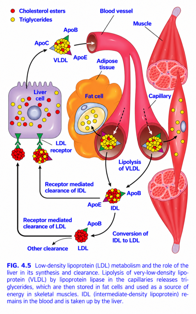
FIG. 4.5 — VLDL → IDL → LDL Metabolism
🧠 Simplest idea
Liver sends triglycerides out as VLDL → tissues remove triglycerides → VLDL becomes IDL → IDL either returns to liver or becomes LDL → LDL mainly carries cholesterol back to cells/liver.
Whole figure in one flow
Liver → VLDL → triglyceride removal → IDL → either liver uptake OR → LDL → LDL-receptor clearance🎨 COLOR KEY
- 🔴 Red circles = cholesterol esters
- 🟡 Yellow circles = triglycerides
- 🟣/green shapes on particles = apolipoproteins
- 🔴 Tubes = blood vessels/capillaries
- 🟠 Large cell = fat cell
- 🔴 elongated tissue = skeletal muscle
1️⃣ LIVER makes VLDL
🟣 Purple cell = liver cell
The liver packages mainly:
- lots of 🟡 triglycerides
- some 🔴 cholesterol esters
into:
VLDL = Very-Low-Density Lipoprotein
VLDL carries:
- ApoB
- ApoC
- ApoE
⬆️ Black arrow = VLDL is released from liver into blood.
Easy idea:
VLDL = liver’s triglyceride delivery truck.2️⃣ VLDL travels through blood
VLDL moves to capillaries supplying:
🟠 Adipose tissue
and
🔴 Skeletal muscle
⬇️ White dotted arrows show VLDL entering these capillary regions.
3️⃣ Lipolysis of VLDL
At capillary surfaces, lipoprotein lipase (LPL) breaks down VLDL triglycerides.
Important:
ApoC-II activates LPL.
So:
VLDL triglyceride
→ LPL
→ free fatty acids4️⃣ Fatty acids go to TWO places
🟠 Adipose tissue
Fatty acids enter fat cells.
➡️ re-formed into triglycerides
➡️ stored as fat
Think:
Adipose = STORAGE
🔴 Skeletal muscle
Fatty acids enter muscle.
➡️ oxidized
➡️ used to make energy
Think:
Muscle = BURNS fat
5️⃣ VLDL loses triglyceride → becomes IDL
After much triglyceride is removed:
VLDL → IDL
IDL = Intermediate-Density Lipoprotein
IDL has relatively:
- less 🟡 triglyceride
- more 🔴 cholesterol
and carries ApoB + ApoE.
6️⃣ IDL now has TWO possible fates
PATH A — IDL returns to liver
Black arrow:
IDL → liver
ApoE helps IDL bind hepatic receptors.
➡️ Receptor-mediated clearance of IDL
Memory:
ApoE = helps remnants Enter liver
7️⃣ PATH B — IDL → LDL
If more triglyceride is removed:
IDL → LDL
This is labeled:
Conversion of IDL to LDL
LDL becomes very rich in:
🔴 cholesterol esters
and mainly retains:
ApoB-100
8️⃣ LDL = cholesterol delivery particle
🔴 Red particle at bottom = LDL
Its main job:
Carry cholesterol to peripheral tissues and back to liver.
LDL binds the:
LDL receptor
through ApoB-100.
9️⃣ Receptor-mediated clearance of LDL
Green receptor on liver = LDL receptor
LDL ApoB-100
→ binds LDL receptor
→ LDL enters liver cell
→ cholesterol is cleared from blood.
Easy memory:
ApoB-100 binds LDL receptor.
🔟 Other clearance
Some LDL can also leave circulation through other uptake pathways.
But the major regulated route is:
LDL → LDL receptor → liver/cells
⭐ Arrow-by-arrow summary
Liver
⬇️
VLDL (TG-rich)
⬇️ LPL removes TG
↙️ ↘️
Fat → storage Muscle → energy
⬇️
IDL
↙️ ↘️
Liver uptake via ApoE → LDL
⬇️
ApoB-100 → LDL receptor
⬇️
LDL clearance Fastest exam recall
VLDL
TG-rich → made by liver → delivers triglycerides
IDL
VLDL remnant → ApoE helps liver uptake
LDL
Cholesterol-rich → ApoB-100 → LDL receptor
⭐ One-line memory
VLDL delivers TG → IDL is the remnant → LDL delivers cholesterol.✕Compare with Claude Opus 4.8
Pathogenesis
- In familial hypercholesterolemia (FH), defective LDL receptors impair:
- receptor expression on the cell surface
- LDL binding and endocytosis
- Result:
↓ LDL clearance from blood → ↑ plasma LDL cholesterol. - Loss of LDL receptors on hepatocytes also reduces IDL uptake by the liver.
- Therefore, more IDL is converted into LDL.
- So FH causes high cholesterol by two combined mechanisms:
↓ LDL catabolism + ↑ formation of LDL from IDL → severe hypercholesterolemia (Fig. 4.5). - Excess LDL is taken up increasingly by monocytes, macrophages, and vessel walls through scavenger receptors.
- This produces:
- xanthomas
- premature atherosclerosis
- Causes of FH include:
- LDL receptor mutations → about 80%–85%
- ApoB-100 loss-of-function mutations → about 5%–10%
- ApoB-100 is the ligand on LDL that binds the LDL receptor.
- PCSK9 gain-of-function mutations → about 1%–2%
- ↑ PCSK9 → ↑ LDL receptor degradation → ↓ receptor recycling
- All these abnormalities ultimately cause:
↓ hepatic LDL clearance → ↑ plasma LDL. - More than 2000 LDL receptor mutations are known.
- A common type causes abnormal receptor folding → receptor cannot reach the cell surface.
Clinical Features
- FH is an autosomal dominant disorder.
- Heterozygotes:
- plasma cholesterol rises about 2–3 times normal
- cholesterol is elevated from birth
- symptoms often appear in adulthood
- may develop:
- tendon xanthomas
- premature atherosclerosis
- coronary artery disease
- Homozygotes:
- plasma cholesterol may exceed 5 times normal
- disease is much more severe
- develop cutaneous xanthomas in childhood
- may die from myocardial infarction before age 20
- Statins lower cholesterol by inhibiting HMG-CoA reductase.
- This promotes increased production of LDL receptors (Fig. 4.6) → more LDL removed from blood.
- However, increased LDL receptors are accompanied by ↑ PCSK9, which promotes receptor degradation and reduces part of the statin effect.
- Therefore, resistant hypercholesterolemia may be treated with agents that inhibit PCSK9, including:
- PCSK9 antibodies
- siRNAs that reduce PCSK9 production
KEY CONCEPT
- FH = defective LDL clearance → very high LDL → xanthomas + premature atherosclerosis.
- Main mechanisms:
LDL receptor defect / ApoB defect / ↑ PCSK9 → ↓ hepatic LDL uptake. - Heterozygote → 2–3× cholesterol; homozygote → >5× and much more severe.
- Statins → ↓ HMG-CoA reductase → ↑ LDL receptors.
- PCSK9 inhibition → preserves LDL receptors → improves LDL clearance.
CONCEPTUAL EXAMPLES
- LDL receptor absent → LDL cannot enter liver efficiently → LDL accumulates in blood.
- Excess LDL enters macrophages → cholesterol deposits form → xanthomas and atherosclerosis.
- Gain-of-function PCSK9 → more LDL receptors destroyed → plasma LDL rises further.
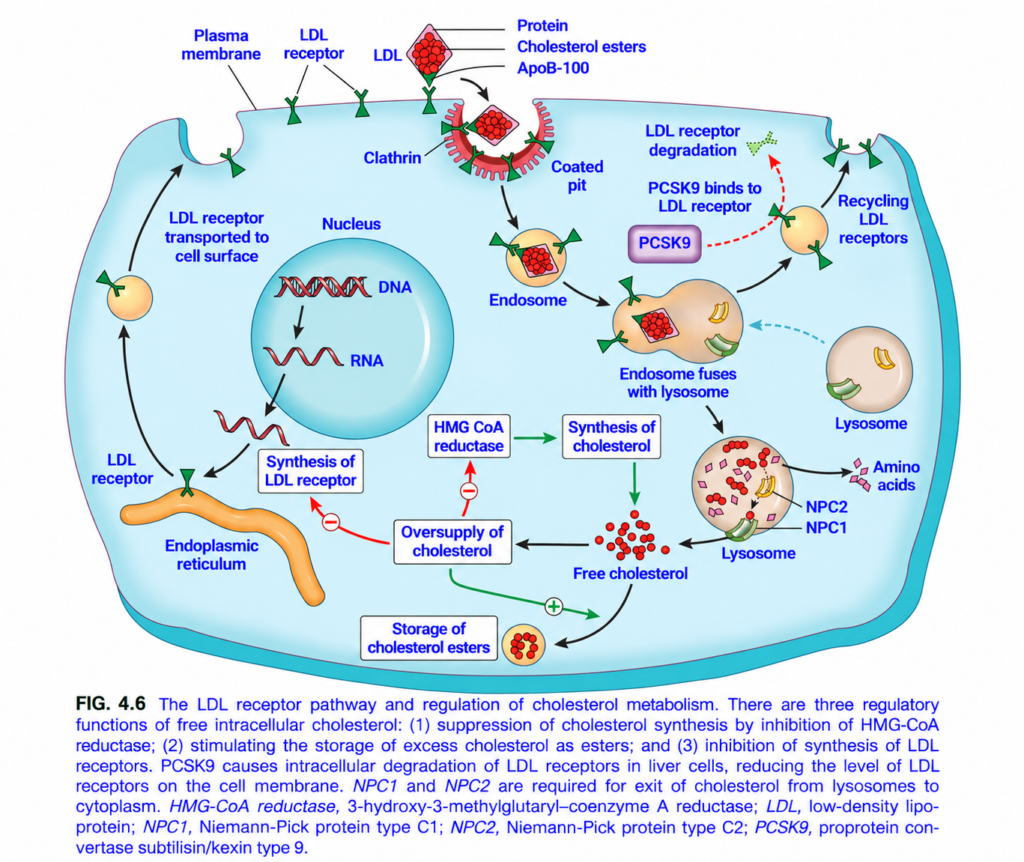
FIG. 4.6 — LDL Receptor Pathway & Cholesterol Regulation
🧠 Simplest idea
LDL brings cholesterol into the cell → LDL receptor is usually recycled → released cholesterol then tells the cell: “Stop making and importing more cholesterol; store the excess.”
Whole figure in one flow
LDL–ApoB100 → LDL receptor → clathrin-coated pit → endosome → lysosome → free cholesterol → ↓ cholesterol synthesis + ↓ LDL receptors + ↑ cholesterol storage
1️⃣ TOP — LDL binds LDL receptor
🔴 Cluster = cholesterol esters inside LDL
🟢 surface protein = ApoB-100
🟢 Y-shaped structure = LDL receptor
First step:
ApoB-100 on LDL → binds LDL receptor
🧠 Memory:
ApoB-100 = LDL’s “address label” for the LDL receptor.
2️⃣ Clathrin-coated pit
After LDL binds its receptor:
⬇️ Black arrow
The membrane folds inward into a:
Clathrin-coated pit
🩷 Red curved coat = clathrin
The pit pinches off and carries:
LDL + LDL receptor → inside the cell
3️⃣ Endosome
The vesicle becomes an:
Endosome
Inside the acidic endosome:
LDL separates from its LDL receptor.
Now the two take different paths.
4️⃣ 🟢 LDL receptor recycling
Most LDL receptors leave the endosome.
⬆️ Black arrow
Endosome → receptor-containing vesicle → plasma membrane
The receptor can be used again to capture another LDL particle.
Easy concept:
LDL receptor = reusable catcher
5️⃣ PCSK9 destroys LDL receptors
🟣 PCSK9 can bind the LDL receptor.
🔴 Dotted arrow shows:
PCSK9 + LDL receptor
→ prevents normal recycling
→ receptor goes to lysosome
→ LDL receptor degradation
Result:
↓ LDL receptors on liver surface
→ ↓ LDL removed from blood
→ ↑ blood LDL cholesterol
⭐ Important exam point:
PCSK9 raises plasma LDL by decreasing LDL-receptor recycling.
6️⃣ LDL goes to the lysosome
The endosome containing LDL fuses with:
🟤 Lysosome
Lysosomal enzymes break LDL apart.
LDL protein
→ amino acids
Cholesterol esters
→ free cholesterol
So:
LDL → lysosome → free intracellular cholesterol
7️⃣ NPC1 and NPC2
Inside the lysosome are:
- NPC1
- NPC2
These proteins help move cholesterol:
Lysosome → cytoplasm/cellular membranes
Easy idea:
NPC1/NPC2 = cholesterol exit system from lysosome
Defects cause cholesterol to become trapped in lysosomes.
8️⃣ Free cholesterol now controls the cell
🔴 Small red circles = free cholesterol
If cholesterol becomes abundant:
Three important feedback effects occur.
① 🔴 ↓ HMG-CoA reductase
HMG-CoA reductase is the key enzyme for making new cholesterol.
Excess cholesterol gives a negative feedback signal:
↑ intracellular cholesterol
→ inhibits HMG-CoA reductase
→ ↓ new cholesterol synthesis
🧠 Think:
“Already enough cholesterol → stop making more.”
② 🔴 ↓ LDL receptor synthesis
The nucleus normally makes RNA and protein for new LDL receptors.
But excess cholesterol inhibits this.
↑ intracellular cholesterol
→ ↓ LDL-receptor gene expression
→ fewer receptors produced
→ ↓ additional LDL entry
Think:
“Already full → stop importing more.”
③ 🟢 ↑ Cholesterol ester storage
Excess free cholesterol is converted into:
Cholesterol esters
and stored safely inside the cell.
↑ free cholesterol → ↑ esterification/storage
Think:
“Extra cholesterol → pack it away.”
9️⃣ LEFT SIDE — Making new LDL receptors
Inside nucleus:
DNA → RNA
⬇️
LDL receptor synthesized in ER
⬇️
receptor transported to cell surface
This supplies new receptors when the cell needs cholesterol.
🎨 Arrow / color guide
⭐ Three effects of HIGH intracellular cholesterol
1. ↓ Synthesis
↓ HMG-CoA reductase
2. ↓ Uptake
↓ LDL receptor synthesis
3. ↑ Storage
↑ Cholesterol ester formation
🧠 Fastest exam recall
LDL ApoB-100 → LDL receptor → endocytosis → lysosome → free cholesterol.
Then free cholesterol says:
STOP making it → ↓ HMG-CoA reductase
STOP importing it → ↓ LDL receptors
STORE extra → ↑ cholesterol esters
🔥 PCSK9 memory
PCSK9 destroys LDL receptors → fewer receptors → blood LDL rises.✕Compare with Claude Opus 4.8
