Granulomatous inflammation is a form of chronic inflammation characterized by collections of activated macrophages, often with T lymphocytes and sometimes central necrosis.
These inflammatory nodules are called granulomas because they have a granular appearance macroscopically.
Granulomas usually develop when the body tries to contain an offending agent that is difficult to eliminate.
Granulomatous inflammation may also develop around indigestible foreign bodies without a T-cell–mediated immune response.
These foreign bodies are not immunogenic, but they are too large to be completely phagocytosed by macrophages.
Therefore → foreign material persists → macrophages remain activated → foreign body granuloma forms.
Important causes of foreign body granulomas include:
Talc
Sutures
Other fibers
The foreign material is usually present in the center of the granuloma and may be identified microscopically, especially when it is refractile in polarized light.
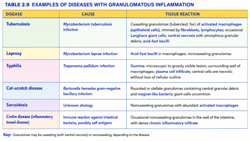
Recognition of a granuloma is important because only a limited number of conditions produce granulomatous inflammation (Table 2.9).
Tuberculosis is the classic infectious cause of granulomatous inflammation and should always be excluded when a granuloma is identified.
Other infections that may produce granulomas include syphilis and some fungal infections.
Their microscopic appearances may suggest the diagnosis, but the specific causative agent must be identified.
The cause may be confirmed by:
Special stains, such as acid-fast stains for M. tuberculosis
Microbial culture
Molecular techniques
Serologic studies, such as those used in syphilis
Granulomas may also occur in immune-mediated inflammatory diseases.
Important examples include:
Crohn disease
Sarcoidosis
KEY CONCEPT
Granuloma = activated macrophages + often T lymphocytes ± central necrosis.
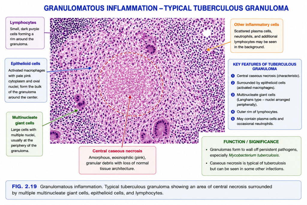
In routine hematoxylin and eosin (H&E) sections (Fig. 2.19), activated macrophages in granulomas have pink, granular cytoplasm with indistinct cell borders.
These activated macrophages are called epithelioid cells because they resemble epithelial cells.
Groups of epithelioid macrophages are often surrounded by a collar of lymphocytes.
In older granulomas → fibroblasts and connective tissue may form a rim around the granuloma.
Granulomas may also contain multinucleated giant cells, about 40–50 μm in diameter.
These giant cells are called Langhans giant cells and form by fusion of many activated macrophages.
In some infectious granulomas, especially Mycobacterium tuberculosis → hypoxia + free-radical injury → central necrosis.
Grossly, this necrotic center looks granular and cheese-like → called caseous necrosis.
Microscopically, caseous necrosis appears as amorphous, structureless, eosinophilic granular debris.
Granulomas in Crohn disease, sarcoidosis, and foreign-body reactions usually do not have central necrosis → called noncaseating granulomas.
During healing → granulomas may undergo fibrosis, which can sometimes become extensive.
Tuberculosis → central caseous necrosis → caseating granuloma.
Crohn disease / sarcoidosis / foreign body reaction → usually no central necrosis → noncaseating granuloma.
Older/healing granuloma → fibroblasts + fibrosis.
Conceptual Examples
Tuberculosis: Activated macrophages collect → epithelioid cells + giant cells form → central tissue dies → caseating granuloma.
Sarcoidosis: Epithelioid macrophages form granulomas but usually without central necrosis → noncaseating granuloma.
Old granuloma: Persistent inflammation → fibroblasts grow around granuloma → fibrosis and scar formation.
SYSTEMIC EFFECTS OF INFLAMMATION
Even when inflammation is localized, it can produce systemic effects because inflammatory cytokines enter the circulation.
These systemic reactions are commonly seen during severe infections such as influenza.
Bacterial products such as LPS and other inflammatory stimuli → stimulate production of cytokines.
The main cytokines responsible are TNF, IL-1, and IL-6; type I interferons also contribute.
Systemic effects are usually stronger in acute inflammation than in chronic inflammation because cytokine production is generally greater.
Fever: Body temperature usually rises by about 1–4°C, especially during infections.
Substances that cause fever are called pyrogens.
Infection → bacterial products such as LPS → leukocytes release IL-1 and TNF → increased PGE₂ production in the hypothalamus.
PGE₂ acts on the preoptic nucleus of the hypothalamus → raises the body’s temperature set point → fever.
NSAIDs, including aspirin → inhibit prostaglandin synthesis → reduce fever.
Fever may have a protective role, but the exact mechanism is uncertain.
Leukocytosis: Inflammation, especially bacterial infection, commonly causes an increase in circulating white blood cells.
Leukocyte counts commonly rise to about 15,000–20,000 cells/mL and may sometimes reach 40,000–100,000 cells/mL.
Very high leukocyte counts are called leukemoid reactions because they resemble the counts seen in leukemia.
Early leukocytosis occurs because TNF and IL-1 stimulate rapid release of leukocytes from the bone marrow reserve pool.
This increases immature neutrophils called band cells in blood → called a shift to the left.
Prolonged infection → macrophages, stromal cells, endothelial cells, and T lymphocytes release colony-stimulating factors (CSFs) → increase production of leukocytes in bone marrow.
Bacterial infections → neutrophilia.
Viral infections such as infectious mononucleosis, mumps, and German measles → lymphocytosis.
Allergies and parasitic infestations → eosinophilia.
Some infections, including typhoid fever, rickettsial infections, and certain viral and protozoal infections → decreased circulating leukocytes called leukopenia.
Acute-phase response: Inflammation stimulates production of acute-phase proteins, mainly by the liver.
Their blood levels may rise several hundred-fold during inflammation.
Important acute-phase proteins include:
C-reactive protein (CRP)
Fibrinogen
Serum amyloid A (SAA)
Cytokines stimulate hepatocytes to produce these proteins.
CRP and SAA bind microbial cell walls → may act as opsonins and activate complement → help host defense.
Fibrinogen reduces the negative surface charge of red blood cells → RBCs form stacks called rouleaux.
Rouleaux settle faster → increases the erythrocyte sedimentation rate (ESR).
Therefore, ESR is a simple marker of inflammation.
Acute-phase proteins are useful during acute inflammation, but prolonged production, especially of SAA, may lead to amyloidosis.
Increased CRP levels have been proposed as a marker of increased myocardial infarction risk in patients with coronary artery disease.
Inflammation also increases production of hepcidin → reduces iron availability → contributes to anemia of chronic inflammation.
Other systemic manifestations include:
Increased heart rate
Increased blood pressure
Decreased sweating
Rigors and chills
Anorexia
Somnolence
Malaise
Reduced sweating occurs because blood flow is redirected from the skin to deeper vascular beds → decreases heat loss.
Many symptoms such as anorexia, somnolence, and malaise are probably caused by cytokine effects on brain cells.
In severe bacterial infection (sepsis) → large amounts of bacteria and their products stimulate massive production of TNF, IL-1, and IL-6.
Very high cytokine levels → disseminated intravascular coagulation + hypotension + metabolic abnormalities, including insulin resistance and hyperglycemia.
This severe systemic response is called septic shock.
A similar systemic inflammatory response can occur without infection in conditions such as severe burns, trauma, and pancreatitis.
This is called systemic inflammatory response syndrome (SIRS).