


Hypoadrenalism (Adrenal Insufficiency)—Addison
Disease, Hyperadrenalism—Cushing Syndrome ,Primary Aldosteronism (Conn Syndrome),Congenital Adrenal Hyperplasia, Adrenogenital Syndrome
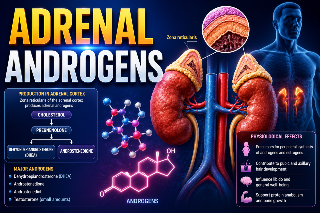
- The adrenal cortex continuously secretes small amounts of male sex hormones called adrenal androgens.
- The most important adrenal androgen is dehydroepiandrosterone (DHEA).
- Their secretion is especially important during fetal life.
- The adrenal cortex also secretes very small amounts of the female sex hormones progesterone and estrogens.
- Normally, adrenal androgens have only weak effects in humans.
- In childhood, they contribute partly to the early development of male sex organs.
- In females, they have mild effects before puberty and throughout life.
- They are responsible for much of the growth of pubic and axillary hair in females.
- In tissues outside the adrenal gland, some adrenal androgens are converted into testosterone.
- This conversion to testosterone explains much of their androgenic activity.
Abnormalities of Adrenocortical Secretion
Hypoadrenalism (Adrenal Insufficiency)—Addison Disease
- Addison disease occurs when the adrenal cortices cannot produce enough adrenocortical hormones.
- It usually results from atrophy or damage of the adrenal cortex.
- In about 80% of cases, the adrenal cortex is damaged by autoimmunity.
- Adrenal hypofunction can also result from tuberculosis or cancer invading the adrenal cortex.
- Adrenal insufficiency can also occur when the pituitary gland produces too little ACTH.
- Low ACTH causes decreased production of cortisol and aldosterone.
- With prolonged lack of ACTH stimulation, the adrenal glands may eventually atrophy.
- This is called secondary adrenal insufficiency and is more common than Addison disease.
- Addison disease is therefore also called primary adrenal insufficiency.
Mineralocorticoid Deficiency
- Lack of aldosterone greatly decreases sodium reabsorption by the renal tubules.
- Therefore, large amounts of sodium, chloride, and water are lost in urine.
- This causes a major decrease in extracellular fluid (ECF) volume and produces hyponatremia.
- At the same time, reduced secretion of potassium and hydrogen ions causes:
- Hyperkalemia
- Mild acidosis
- As ECF volume continues to fall:
- Plasma volume decreases
- Red blood cell concentration increases
- Cardiac output decreases
- Blood pressure decreases
- Severe volume loss can finally cause shock.
- Without treatment, complete loss of mineralocorticoid secretion may cause death within about 4 days to 2 weeks.
KEY CONCEPT
- Adrenal androgens = weak sex hormones from the adrenal cortex, especially DHEA.
- They contribute to pubic and axillary hair, particularly in females, and some are converted to testosterone.
- Addison disease = primary failure of the adrenal cortex → inadequate adrenal hormones.
- Low aldosterone → Na⁺ + water loss → ↓ ECF and blood volume → ↓ blood pressure → possible shock.
- At the same time, K⁺ and H⁺ are retained → hyperkalemia + mild acidosis.
Conceptual Examples
- Adrenal androgen example:
Adrenal androgens → act weakly themselves → some convert to testosterone → androgenic effects. - Aldosterone deficiency example:
↓ Aldosterone → ↓ renal Na⁺ reabsorption → Na⁺ + water lost in urine → ↓ ECF volume → ↓ blood pressure. - Potassium example:
↓ Aldosterone → ↓ K⁺ secretion → K⁺ stays in the body → hyperkalemia. - Severe deficiency example:
Severe fluid loss → ↓ plasma volume → ↓ cardiac output → ↓ blood pressure → shock.
Glucocorticoid Deficiency
- Loss of cortisol makes it difficult for a person with Addison disease to maintain normal blood glucose between meals.
- This happens because the body cannot produce enough glucose by gluconeogenesis.
- Lack of cortisol also decreases the mobilization of proteins and fats from tissues.
- Therefore, many metabolic functions become slower because the body cannot properly mobilize energy.
- Even when plenty of glucose and other nutrients are available, the muscles remain weak.
- This shows that glucocorticoids are needed not only for energy metabolism but also for maintaining other normal tissue metabolic functions.
- Inadequate glucocorticoids also make a person with Addison disease much less able to tolerate stress.
- Therefore, even a mild respiratory infection can become dangerous and may cause death.
Melanin Pigmentation
- Most people with Addison disease develop increased melanin pigmentation of the skin and mucous membranes.
- The pigmentation may be uneven and can appear as blotches.
- It is especially noticeable in thin areas such as:
- Mucous membranes of the lips
- Skin of the nipples
- ↓ Cortisol reduces the normal negative feedback on the hypothalamus and anterior pituitary.
- Therefore, secretion of ACTH becomes very high, along with increased MSH secretion.
- Large amounts of ACTH can stimulate melanocytes like MSH does.
- Thus, increased ACTH probably causes most of the increased pigmentation.
Treatment of People With Addison Disease
- Complete destruction of the adrenal glands without treatment can cause death within days to weeks.
- Death usually occurs because of severe weakness and circulatory shock.
- However, a person can survive for years if small amounts of mineralocorticoids and glucocorticoids are given every day.
Adrenal Crisis
- Normally, physical or mental stress causes secretion of large amounts of glucocorticoids.
- In Addison disease, glucocorticoid secretion cannot increase during stress.
- During severe stress such as:
- Trauma
- Disease
- Surgery
- The person may need 10 or more times the normal glucocorticoid amount to prevent death.
- This severe need for extra glucocorticoids, together with marked weakness during acute stress, is called an adrenal crisis (Addisonian crisis).
KEY CONCEPT
- ↓ Cortisol → ↓ gluconeogenesis → difficulty maintaining blood glucose between meals.
- ↓ Cortisol → ↓ protein and fat mobilization → poor energy mobilization + muscle weakness.
- ↓ Cortisol → poor ability to tolerate stress.
- ↓ Cortisol → ↓ negative feedback → ↑ ACTH + ↑ MSH → increased skin and mucosal pigmentation.
- Addison disease requires daily glucocorticoid + mineralocorticoid replacement.
- During severe stress, glucocorticoid requirements rise greatly; failure to meet this need can cause an adrenal crisis.
Conceptual Examples
- Blood glucose:
↓ Cortisol → ↓ gluconeogenesis → blood glucose becomes difficult to maintain between meals. - Pigmentation:
↓ Cortisol → ↓ negative feedback → ↑ ACTH → stimulation of melanocytes → increased pigmentation. - Stress:
Addison disease + surgery → glucocorticoids cannot rise normally → very high glucocorticoid requirement → risk of adrenal crisis. - Treatment:
Adrenal destruction → lack of adrenal hormones → daily mineralocorticoid + glucocorticoid replacement → long-term survival.
Hyperadrenalism—Cushing Syndrome
- Cushing syndrome results from excessive secretion of hormones by the adrenal cortex.
- Most abnormalities are caused by excess cortisol, although excess androgens may also contribute.
- Hypercortisolism can occur from several causes:
- Pituitary adenoma → ↑ ACTH → adrenal hyperplasia → ↑ cortisol.
- Abnormal hypothalamic function → ↑ CRH → ↑ ACTH → ↑ cortisol.
- Ectopic ACTH secretion from a tumor elsewhere in the body.
- Adrenal cortical adenoma → direct excess cortisol production.
- When excess cortisol results from excessive ACTH secretion by the anterior pituitary, it is called Cushing disease.
- Excess ACTH is the most common cause of Cushing syndrome.
- In ACTH-dependent disease:
- ↑ ACTH
- ↑ cortisol
- Primary adrenal overproduction of cortisol causes about 20%–25% of clinical cases.
- In primary adrenal disease:
- ↑ cortisol
- Cortisol produces negative feedback on the pituitary.
- Therefore, ACTH becomes low.
Dexamethasone Test
- Dexamethasone is a synthetic glucocorticoid used to help distinguish ACTH-dependent from ACTH-independent Cushing syndrome.
- In a pituitary ACTH-secreting adenoma or hypothalamic-pituitary dysfunction:
- Low-dose dexamethasone usually does not suppress ACTH normally.
- Very high-dose dexamethasone can suppress ACTH in most patients with Cushing disease.
- In primary adrenal cortisol overproduction:
- Cortisol is produced independently of ACTH.
- Therefore, ACTH is usually low or undetectable.
- The dexamethasone test is not always completely accurate.
- Some pituitary ACTH-secreting tumors may suppress ACTH after dexamethasone.
- Some tumors outside the pituitary, such as certain lung carcinomas, produce ACTH and do not respond normally to glucocorticoid negative feedback.
- Therefore, the dexamethasone test is mainly used as a first step in differentiating causes of Cushing syndrome.
Glucocorticoid Treatment as a Cause
- Cushing syndrome can also develop when large doses of glucocorticoids are taken for a long time.
- For example, patients receiving glucocorticoids for chronic inflammatory diseases such as rheumatoid arthritis may develop features of Cushing syndrome.
Characteristic Features
- Fat is mobilized from the lower part of the body.
- At the same time, more fat is deposited in the thorax and upper abdomen.
- This produces a characteristic buffalo-like torso.
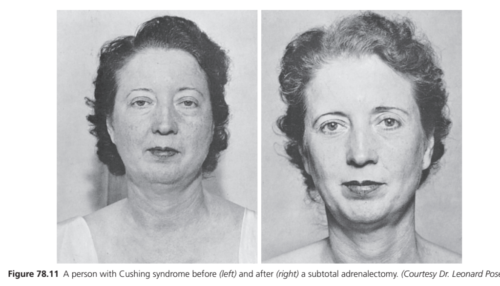
- Excess steroids also produce an edematous, rounded face.
- This characteristic appearance is called a “moon face” (Fig. 78.11).
- Androgenic effects may produce:
- Acne
- Hirsutism = excess facial hair growth
- About 80% of patients develop hypertension.
- This hypertension is thought to result mainly from the mineralocorticoid effects of excess cortisol.
KEY CONCEPT
- Cushing syndrome = excess cortisol effects in the body.
- Pituitary ↑ ACTH → ↑ cortisol = Cushing disease.
- Primary adrenal cortisol excess → ↑ cortisol + ↓ ACTH.
- Dexamethasone helps distinguish ACTH-dependent from ACTH-independent causes.
- Typical features include upper-body fat deposition, moon face, acne/hirsutism, and hypertension.
Conceptual Examples
- Pituitary cause:
Pituitary adenoma → ↑ ACTH → adrenal stimulation → ↑ cortisol → Cushing disease. - Adrenal cause:
Adrenal adenoma → ↑ cortisol → negative feedback → ↓ ACTH. - Dexamethasone:
Pituitary Cushing disease + very high-dose dexamethasone → ACTH can usually be suppressed. - Body appearance:
Fat moves away from the lower body and accumulates in the thorax and upper abdomen → buffalo-like torso. - Blood pressure:
Excess cortisol → mineralocorticoid-like effects → hypertension.
Effects of Cushing Syndrome on Carbohydrate and Protein Metabolism
- Excess cortisol in Cushing syndrome can markedly increase blood glucose.
- After meals, blood glucose may reach about 200 mg/dL, which can be nearly twice normal.
- This mainly occurs because cortisol causes:
- ↑ Gluconeogenesis → more glucose is formed.
- ↓ Glucose utilization by tissues → less glucose is used.
- Excess glucocorticoids also cause marked protein breakdown.
- Tissue proteins decrease in almost all parts of the body, except the liver.
- Plasma proteins also remain relatively unaffected.
- Loss of muscle protein causes severe muscle weakness.
- Reduced protein synthesis in lymphoid tissues suppresses the immune system.
- Therefore, patients become highly susceptible to infections.
- Loss of collagen fibers in subcutaneous tissues makes the skin and underlying tissues weak and easy to tear.
- This produces large purplish striae.
- Reduced protein deposition in bones causes severe osteoporosis.
- As a result, the bones become weak.
Treatment of People With Cushing Syndrome
- Treatment depends on the cause of excess cortisol.
- If an adrenal tumor is responsible, the tumor can be removed.
- If excess ACTH is the cause, treatment aims to reduce ACTH secretion.
- An enlarged pituitary gland or a small ACTH-secreting pituitary tumor may be:
- Surgically removed, or
- Destroyed by radiation.
- When surgery is not possible, drugs may be used to reduce hormone production.
- Drugs that block steroid synthesis include:
- Metyrapone
- Ketoconazole
- Aminoglutethimide
- Drugs that inhibit ACTH secretion may also be used.
- If ACTH cannot be adequately reduced, partial or total removal of both adrenal glands may be required.
- After adrenal removal, adrenal steroid replacement is given to correct the hormone deficiency that develops.
KEY CONCEPT
- Excess cortisol → ↑ gluconeogenesis + ↓ glucose use → hyperglycemia.
- Excess cortisol → ↑ protein breakdown → muscle weakness.
- ↓ Lymphoid proteins → suppressed immunity → increased infections.
- ↓ Collagen → thin, fragile tissues → purplish striae.
- ↓ Bone protein → osteoporosis → weak bones.
- Treatment aims to remove the hormone-producing tumor or reduce ACTH/cortisol production.
Conceptual Examples
- Blood glucose:
Excess cortisol → ↑ glucose production + ↓ tissue glucose use → high blood glucose. - Muscle:
Excess cortisol → protein breakdown → loss of muscle protein → severe weakness. - Skin:
↓ Collagen → weak subcutaneous tissue → easy tearing → purplish striae. - Bone:
↓ Protein deposition in bone → osteoporosis → weak bones. - Treatment:
ACTH-secreting pituitary tumor → remove or destroy tumor → ↓ ACTH → ↓ cortisol.
Primary Aldosteronism (Conn Syndrome)
- Primary aldosteronism (Conn syndrome) occurs when the adrenal cortex secretes excessive aldosterone.
- It may result from:
- A small tumor of zona glomerulosa cells.
- Adrenal cortical hyperplasia that mainly secretes aldosterone.
- Excess aldosterone produces:
- Hypokalemia
- Mild metabolic alkalosis
- Slight increase in extracellular fluid (ECF) volume
- Slight increase in blood volume
- Small rise in plasma Na⁺, usually <4–6 mEq/L
- Almost always hypertension
- Severe hypokalemia may occasionally cause muscle paralysis.
- Low extracellular K⁺ depresses action potential transmission in nerve fibers, leading to paralysis.
- An important diagnostic finding is low plasma renin concentration.
- The sequence is: ↑ Aldosterone → ↑ ECF volume + ↑ arterial pressure → suppression of renin → ↓ plasma renin
- Treatment may include:
- Surgical removal of an aldosterone-secreting tumor.
- Removal of most adrenal tissue when hyperplasia is responsible.
- Blocking mineralocorticoid receptors with spironolactone or eplerenone.
Congenital Adrenal Hyperplasia
- Congenital adrenal hyperplasia (CAH) is a group of autosomal-recessive genetic disorders.
- It causes enlargement of the adrenal cortex, especially:
- Zona fasciculata
- Zona reticularis
- CAH causes impaired production of cortisol and aldosterone.
- More than 95% of cases are caused by 21β-hydroxylase deficiency.
- Normally, 21β-hydroxylase helps convert: 17-hydroxyprogesterone → 11-deoxycortisol
- Therefore, deficiency of this enzyme causes:
- ↓ Cortisol
- ↓ Aldosterone
- ↓ Cortisol removes normal negative feedback on the hypothalamus and pituitary: ↓ Cortisol → ↑ CRH → ↑ ACTH
- ↑ ACTH then causes:
- Adrenal hyperplasia
- Accumulation of cortisol precursors such as 17-hydroxyprogesterone
- Increased production of androgenic steroids
- These androgenic hormones do not suppress CRH and ACTH by negative feedback.
- Severity depends on how much 21β-hydroxylase activity remains.
- Nonclassic CAH:
- Some enzyme activity remains.
- Usually diagnosed in later childhood or adulthood.
- Causes increased adrenal androgen production.
- Produces minimal salt wasting.
- Classic CAH:
- Little or no 21β-hydroxylase activity.
- Usually diagnosed in infancy or early childhood.
- It is the more severe form.
- Untreated patients may develop severe salt wasting with:
- ↓ Blood volume
- Hypotension
- Hyponatremia
- Hyperkalemia
- Metabolic acidosis
- Adrenal crisis
- These patients require glucocorticoid and mineralocorticoid therapy to survive.
Adrenogenital Syndrome
- Adrenogenital syndrome may occur when an adrenocortical tumor secretes excessive androgens.
- Excess androgens cause strong masculinizing effects.
- In females, these effects may include:
- Growth of a beard
- Deepening of the voice
- Possible baldness when genetically susceptible
- Masculine distribution of body and pubic hair
- Enlargement of the clitoris
- Increased protein deposition in skin and especially muscles, producing more masculine features
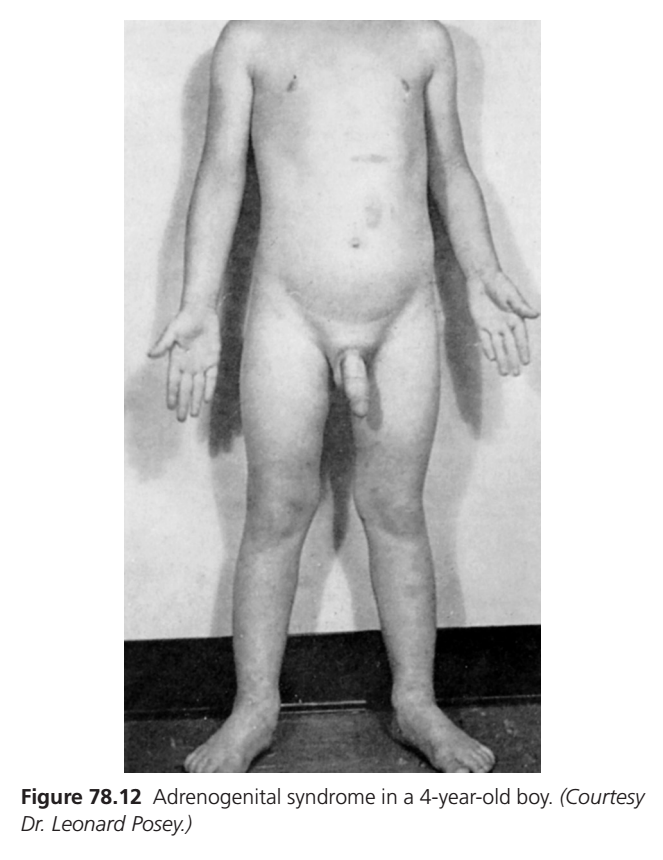
- In a prepubertal male, a virilizing adrenal tumor causes similar masculinizing effects plus rapid development of the male sexual organs (Fig. 78.12).
- In an adult male, the disorder may be difficult to recognize because normal testicular testosterone already produces strong masculine characteristics.
- In adrenogenital syndrome, urinary 17-ketosteroids may rise to 10–15 times normal.
- This marked increase can help in diagnosis.
KEY CONCEPT
- Conn syndrome:
↑ Aldosterone → Na⁺/water retention + K⁺ loss → hypertension + hypokalemia + ↓ renin. - CAH due to 21β-hydroxylase deficiency:
↓ Cortisol + ↓ aldosterone → ↓ negative feedback → ↑ ACTH → adrenal hyperplasia + ↑ androgen production. - Classic CAH:
Severe enzyme deficiency → salt wasting + hypotension + hyponatremia + hyperkalemia + metabolic acidosis + adrenal crisis. - Adrenogenital syndrome:
Excess adrenal androgens → masculinization, especially obvious in females and prepubertal males.
Conceptual Examples
- Conn syndrome:
↑ Aldosterone → ↑ Na⁺ and water retention → ↑ blood volume → hypertension → renin becomes low. - Hypokalemia:
↑ Aldosterone → ↑ K⁺ loss → ↓ extracellular K⁺ → impaired nerve action potential transmission → muscle paralysis. - CAH:
21β-hydroxylase deficiency → ↓ cortisol → ↑ ACTH → adrenal enlargement + ↑ androgen production. - Classic CAH:
↓ Aldosterone → salt and water loss → ↓ blood volume → hypotension and adrenal crisis. - Adrenogenital syndrome:
Adrenal tumor → ↑ androgens → masculinizing changes + markedly increased urinary 17-ketosteroids.
Bibliography
Auchus RJ. Approaching primary aldosteronism as a common disease.
Endocr Pract. 2023;29:994–998.
Auer MK, Nordenström A, Lajic S, Reisch N. Congenital adrenal
hyperplasia. Lancet. 2023;401:227–244.
Charoensri S, Auchus RJ. Therapeutic management of congenital forms
of endocrine hypertension. Eur J Endocrinol. 2023;189:R11–R22.
Claahsen-van der Grinten HL, Speiser PW, Ahmed SF, Arlt W, Auchus
RJ, et al. Congenital adrenal hyperplasia—current insights in
pathophysiology, diagnostics, and management. Endocr Rev.
2022;43:91–159
