


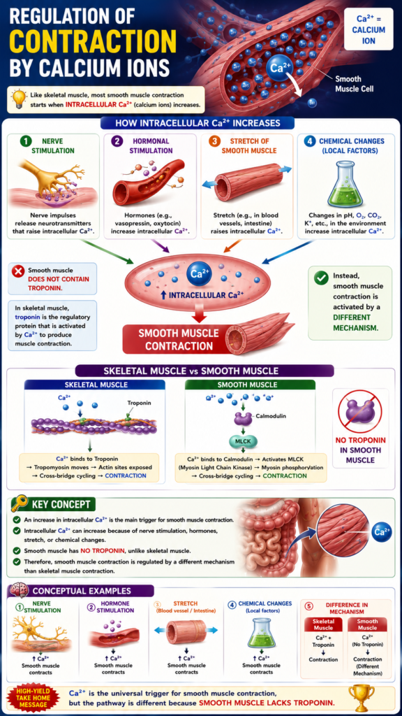
- Like skeletal muscle, most smooth muscle contraction starts when intracellular Ca²⁺ (calcium ions) increases.
- The increase in intracellular Ca²⁺ can occur by:
- Nerve stimulation of the smooth muscle fiber.
- Hormonal stimulation.
- Stretch of the smooth muscle fiber.
- Changes in the chemical environment around the fiber.
- Smooth muscle does not contain troponin.
- In skeletal muscle, troponin is the regulatory protein that is activated by Ca²⁺ to produce muscle contraction.
- Instead, smooth muscle contraction is activated by a different mechanism.
KEY CONCEPT
- An increase in intracellular Ca²⁺ is the main trigger for smooth muscle contraction.
- Intracellular Ca²⁺ can increase because of nerve stimulation, hormones, stretch, or chemical changes.
- Smooth muscle has no troponin, unlike skeletal muscle.
- Therefore, smooth muscle contraction is regulated by a different mechanism than skeletal muscle contraction.
Conceptual Examples
- Nerve stimulation → ↑ Intracellular Ca²⁺ → Smooth muscle contracts.
- Hormone stimulation → ↑ Intracellular Ca²⁺ → Smooth muscle contracts.
- Stretch of a blood vessel or intestine → ↑ Intracellular Ca²⁺ → Smooth muscle contracts.
- Skeletal muscle: Ca²⁺ + Troponin → Contraction.
- Smooth muscle: Ca²⁺ → No troponin → Contraction occurs by a different mechanism.
Ca²⁺ Combines With Calmodulin to Cause Activation of Myosin Kinase and Phosphorylation of the Myosin Head
- Instead of troponin, smooth muscle contains a large amount of another regulatory protein called calmodulin (Fig. 8.3).
- Calmodulin is similar to troponin in its amino acid sequence and 3-dimensional structure.
- However, calmodulin starts muscle contraction in a different way.
- Calmodulin starts contraction by activating the myosin cross-bridges.
- This activation and muscle contraction occur in the following sequence:
- 1. The amount of Ca²⁺ inside the cytosol of smooth muscle increases.
- This increase occurs because Ca²⁺ enters from the extracellular fluid through Ca²⁺ channels.
- It can also occur because Ca²⁺ is released from the sarcoplasmic reticulum.
- 2. The Ca²⁺ ions bind reversibly to calmodulin.
- 3. The Ca²⁺–calmodulin complex joins with and activates myosin light chain kinase.
- Myosin light chain kinase is a phosphorylating enzyme.
- 4. One light chain of each myosin head, called the regulatory chain, becomes phosphorylated by myosin light chain kinase.
- If the regulatory chain is not phosphorylated, the myosin head cannot attach to and detach from the actin filament.
- If the regulatory chain is phosphorylated, the myosin head can repeatedly bind to the actin filament.
- The myosin head then goes through repeated cycles of pulling on the actin filament.
- These repeated pulls produce smooth muscle contraction.
KEY CONCEPT
- Smooth muscle uses calmodulin instead of troponin to start contraction.
- Ca²⁺ binds to calmodulin.
- The Ca²⁺–calmodulin complex activates myosin light chain kinase (MLCK).
- MLCK phosphorylates the regulatory light chain of the myosin head.
- Phosphorylated myosin can bind to actin and produce repeated contractions.
- Without phosphorylation, the myosin head cannot cycle with actin, so contraction does not occur.
Conceptual Examples
- Ca²⁺ enters the smooth muscle cell → Binds to calmodulin → Activates MLCK → Myosin is phosphorylated → Myosin binds to actin → Smooth muscle contracts.
- No Ca²⁺ or no phosphorylation → Myosin cannot bind effectively to actin → No smooth muscle contraction.
Figure Number: Fig. 8.3
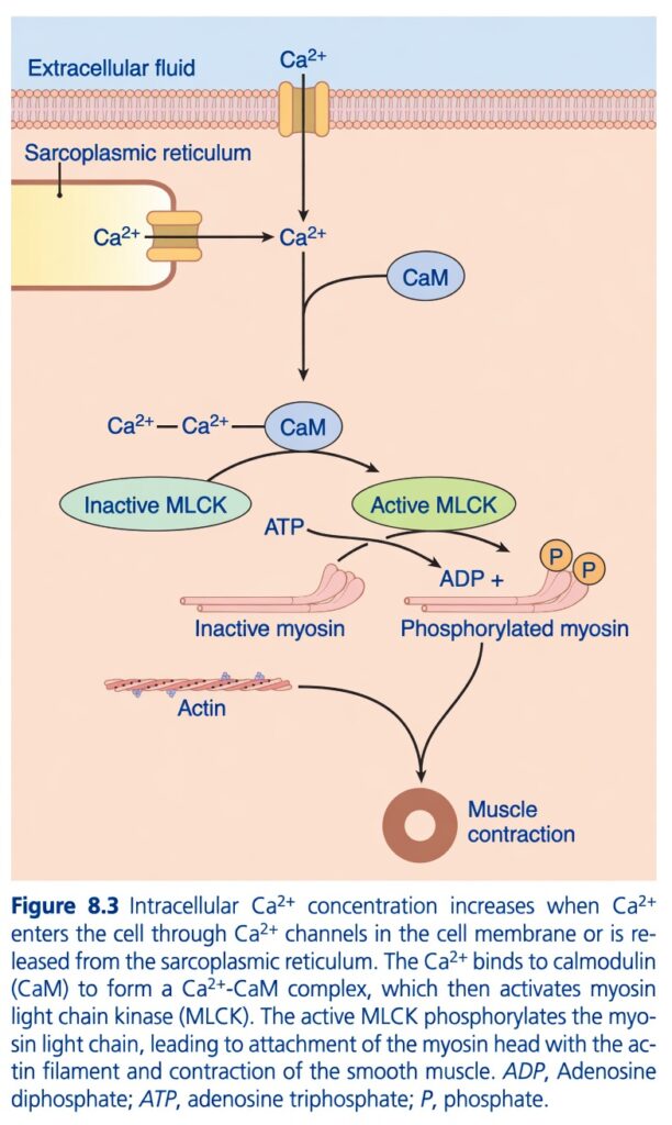
Figure 8.3: Activation of Myosin Light Chain Kinase (MLCK) by Ca²⁺–Calmodulin in Smooth Muscle
Easiest and Most Conceptual Explanation
⭐ One-Line Concept
Smooth muscle contracts only when Ca²⁺ binds to calmodulin, which activates MLCK. MLCK then phosphorylates myosin, allowing myosin to bind actin and produce contraction.
First Understand the Whole Story
Think of smooth muscle like a locked machine.
- Actin = Railway track
- Myosin = Engine
- MLCK = Key that starts the engine
- Calmodulin = Key holder
- Ca²⁺ = Person carrying the key
Without Ca²⁺, the engine (myosin) cannot move.
Step 1: Where Does Ca²⁺ Come From?
The figure shows two sources of Ca²⁺.
Source 1: Extracellular Fluid
Ca²⁺ enters the cell through Ca²⁺ channels in the cell membrane.
Extracellular fluid
↓
Ca²⁺ channel
↓
Ca²⁺ enters the cytoplasm
Source 2: Sarcoplasmic Reticulum (SR)
The SR stores Ca²⁺ inside the cell.
When stimulated,
the SR releases Ca²⁺ into the cytoplasm.
Sarcoplasmic reticulum
↓
Releases Ca²⁺
↓
Cytoplasm
Concept
Both sources increase intracellular Ca²⁺ concentration.
Step 2: Ca²⁺ Binds to Calmodulin (CaM)
The figure shows:
Ca²⁺ + CaM
↓
Ca²⁺–CaM Complex
What is Calmodulin (CaM)?
Calmodulin is a calcium-binding regulatory protein.
It acts as the calcium sensor in smooth muscle.
Concept
Unlike skeletal muscle, smooth muscle does not use troponin.
Instead,
Ca²⁺ binds to calmodulin.
Step 3: Ca²⁺–Calmodulin Activates MLCK
The figure shows:
Inactive MLCK
↓
Ca²⁺–CaM binds
↓
Active MLCK
What is MLCK?
MLCK = Myosin Light Chain Kinase
It is an enzyme.
Its job is to activate myosin.
Concept
Without Ca²⁺–CaM,
MLCK remains inactive.
Step 4: MLCK Uses ATP
The figure shows:
ATP
↓
ADP + Phosphate (P)
Why?
MLCK uses energy from ATP.
It transfers the phosphate to myosin.
This process is called phosphorylation.
Step 5: Myosin Becomes Phosphorylated
The figure shows:
Inactive myosin
↓
MLCK
↓
Phosphorylated myosin
(two phosphate groups are shown to indicate phosphorylation)
What is Phosphorylation?
It means adding a phosphate group to the myosin light chain.
Concept
Phosphorylation switches myosin ON.
Before phosphorylation:
❌ Myosin cannot pull actin.
After phosphorylation:
✅ Myosin becomes active.
Step 6: Active Myosin Binds to Actin
The figure shows:
Phosphorylated myosin
Actin
↓
Interaction between actin and myosin
Concept
Now myosin can attach to actin.
Cross-bridges form.
Step 7: Muscle Contracts
Once myosin binds actin,
cross-bridge cycling begins.
↓
Sliding occurs.
↓
Smooth muscle contracts.
Understanding Every Arrow
Arrow 1
Extracellular fluid
↓
Ca²⁺ enters through membrane channels.
Arrow 2
Sarcoplasmic reticulum
↓
Ca²⁺ is released into the cytoplasm.
Arrow 3
Ca²⁺
↓
Binds calmodulin (CaM).
Arrow 4
Ca²⁺–CaM
↓
Activates inactive MLCK.
Arrow 5
ATP
↓
MLCK uses ATP.
↓
ATP becomes ADP.
Arrow 6
Inactive myosin
↓
Phosphorylation
↓
Active (phosphorylated) myosin.
Arrow 7
Phosphorylated myosin
↓
Binds actin.
Arrow 8
Actin + active myosin
↓
Smooth muscle contraction.
Understanding Every Label
Extracellular Fluid
Contains Ca²⁺ outside the cell.
Sarcoplasmic Reticulum
Stores Ca²⁺ inside the cell.
Ca²⁺
The signal that starts contraction.
CaM (Calmodulin)
Calcium-binding protein.
Acts as the calcium sensor.
Ca²⁺–CaM Complex
Active complex that switches on MLCK.
Inactive MLCK
Cannot phosphorylate myosin.Active MLCK
Phosphorylates myosin light chains.
ATP
Provides energy.
ADP
Produced after ATP is used.
Inactive Myosin
Cannot interact effectively with actin.
Phosphorylated Myosin
Activated myosin that can bind actin.
Actin
Thin filament.
Provides the track for myosin movement.
Muscle Contraction
Final result of actin–myosin interaction.
Easy Story
Imagine a factory.
Myosin
A machine that is switched OFF.
MLCK
The worker who can switch the machine ON.
Calmodulin
The worker’s assistant.
Calcium
The manager who gives permission.
The sequence is:
Manager (Ca²⁺)
↓
Assistant (Calmodulin)
↓
Worker (MLCK)
↓
Turns ON the machine (Myosin)
↓
Machine grabs the conveyor belt (Actin)
↓
The factory starts working (Muscle contracts).
Simple Flow Diagram
Ca²⁺ enters the cell
(from extracellular fluid or sarcoplasmic reticulum)
↓
Intracellular Ca²⁺ increases
↓
Ca²⁺ binds calmodulin (CaM)
↓
Ca²⁺–CaM complex forms
↓
Inactive MLCK becomes active
↓
MLCK uses ATP
↓
Myosin light chain is phosphorylated
↓
Myosin becomes active
↓
Myosin binds actin
↓
Cross-bridge cycling begins
↓
Smooth muscle contraction
High-Yield Summary Table
| Step | Event | Result |
|---|---|---|
| 1 | Ca²⁺ enters from extracellular fluid or is released from the sarcoplasmic reticulum | Intracellular Ca²⁺ increases |
| 2 | Ca²⁺ binds calmodulin (CaM) | Ca²⁺–CaM complex forms |
| 3 | Ca²⁺–CaM activates MLCK | MLCK becomes active |
| 4 | MLCK uses ATP | ATP is converted to ADP while transferring phosphate |
| 5 | MLCK phosphorylates the myosin light chain | Myosin becomes active |
| 6 | Active myosin binds actin | Cross-bridges form |
| 7 | Cross-bridge cycling occurs | Smooth muscle contracts |
Key Concept (Figure 8.3)
Figure 8.3 shows the mechanism of smooth muscle contraction. An increase in intracellular Ca²⁺, either by entry from the extracellular fluid or release from the sarcoplasmic reticulum, allows Ca²⁺ to bind calmodulin (CaM). The Ca²⁺–calmodulin complex activates myosin light chain kinase (MLCK). Active MLCK uses ATP to phosphorylate the myosin light chain, converting inactive myosin into an active form that can bind actin. The interaction between phosphorylated myosin and actin produces cross-bridge cycling, resulting in smooth muscle contraction. Unlike skeletal muscle, smooth muscle uses calmodulin instead of troponin to initiate contraction.
Most of the Ca²⁺ That Causes Smooth Muscle Contraction Comes From Extracellular Fluid
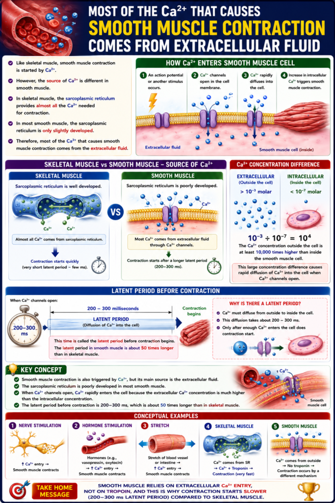
- Like skeletal muscle, smooth muscle contraction is started by Ca²⁺.
- However, the source of Ca²⁺ is different in smooth muscle.
- In skeletal muscle, the sarcoplasmic reticulum provides almost all the Ca²⁺ needed for contraction.
- In most smooth muscle, the sarcoplasmic reticulum is only slightly developed.
- Therefore, most of the Ca²⁺ that causes smooth muscle contraction comes from the extracellular fluid.
- The Ca²⁺ enters the smooth muscle cell when an action potential or another stimulus occurs.
- The Ca²⁺ concentration in the extracellular fluid is greater than 10⁻³ molar.
- The Ca²⁺ concentration inside the smooth muscle cell is less than 10⁻⁷ molar.
- Because the extracellular Ca²⁺ concentration is much higher, Ca²⁺ rapidly diffuses into the cell when Ca²⁺ channels open.
- This diffusion of Ca²⁺ takes about 200–300 milliseconds.
- This time is called the latent period before contraction begins.
- The latent period in smooth muscle is about 50 times longer than in skeletal muscle.
KEY CONCEPT
- Smooth muscle contraction is also triggered by Ca²⁺, but its main source is the extracellular fluid.
- The sarcoplasmic reticulum is poorly developed in most smooth muscle.
- When Ca²⁺ channels open, Ca²⁺ rapidly enters the cell because the extracellular Ca²⁺ concentration is much higher than the intracellular concentration.
- The latent period before contraction is 200–300 ms, which is about 50 times longer than in skeletal muscle.
Conceptual Examples
- Skeletal muscle: Most Ca²⁺ comes from the sarcoplasmic reticulum → Contraction starts quickly.
- Smooth muscle: Most Ca²⁺ comes from the extracellular fluid through Ca²⁺ channels → Contraction starts after a 200–300 ms latent period.
- Ca²⁺ concentration difference:
- Outside the cell: > 10⁻³ molar
- Inside the cell: < 10⁻⁷ molar
- Result: Ca²⁺ naturally moves from outside to inside when Ca²⁺ channels open.
Figure Number: None mentioned in the provided text.
Equation/Calculation
- Extracellular Ca²⁺ concentration: > 10⁻³ molar
- Intracellular Ca²⁺ concentration: < 10⁻⁷ molar
Conceptual comparison:
- 10−3÷10−7=104
- This means the Ca²⁺ concentration outside the cell is at least 10,000 times higher than inside the smooth muscle cell.
- This large concentration difference causes rapid diffusion of Ca²⁺ into the cell when Ca²⁺ channels open.
Role of Sarcoplasmic Tubules and Caveolae in Smooth Muscle Contraction
- Fig. 8.4 shows a few slightly developed sarcoplasmic tubules near the cell membrane in some large smooth muscle cells.
- Small inward folds of the cell membrane are called caveolae.
- The caveolae lie next to the sarcoplasmic tubules.
- The caveolae act as a simple (rudimentary) equivalent of the transverse (T) tubule system of skeletal muscle.
- When an action potential enters the caveolae, it is believed to trigger the release of Ca²⁺ from the nearby sarcoplasmic tubules.
- This occurs in the same basic way that action potentials in the T tubules of skeletal muscle cause the release of Ca²⁺ from the longitudinal sarcoplasmic tubules.
- In general, the more developed the sarcoplasmic reticulum in a smooth muscle fiber, the faster the muscle contracts.
KEY CONCEPT
- Smooth muscle has slightly developed sarcoplasmic tubules near the cell membrane (Fig. 8.4).
- Caveolae are small membrane invaginations that act like a simple T-tubule system.
- An action potential entering the caveolae helps release Ca²⁺ from nearby sarcoplasmic tubules.
- A more developed sarcoplasmic reticulum allows faster smooth muscle contraction.
Conceptual Examples
- Action potential → Caveolae → Ca²⁺ release from nearby sarcoplasmic tubules → Smooth muscle contraction.
- Smooth muscle with more sarcoplasmic reticulum → Faster contraction.
- Skeletal muscle: T tubules carry the action potential.
- Smooth muscle: Caveolae perform a similar role.
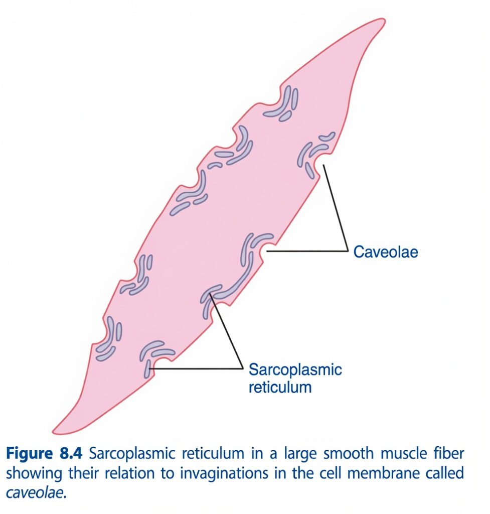
Figure 8.4: Sarcoplasmic Tubules and Caveolae in Smooth Muscle
Easiest and Most Conceptual Explanation
⭐ One-Line Concept
Smooth muscle has very little sarcoplasmic reticulum (SR) and no T-tubules. Instead, it uses caveolae (small membrane pockets) to bring extracellular Ca²⁺ close to the SR, allowing Ca²⁺ to enter the cell and start muscle contraction.
First Understand the Whole Story
Imagine a smooth muscle cell as a small house.
- The cell membrane is the wall of the house.
- The sarcoplasmic reticulum (SR) is a small water tank inside the house.
- The caveolae are small windows in the wall.
When the body wants the muscle to contract:
- Ca²⁺ comes through the windows (caveolae).
- Some Ca²⁺ also comes from the water tank (SR).
- Together they increase intracellular Ca²⁺ and start contraction.
Understanding Every Part of the Figure
1. Smooth Muscle Fiber (Pink Area)
The large pink structure represents one smooth muscle cell (fiber).
Inside this cell are:
- Sarcoplasmic reticulum
- Cytoplasm
- Caveolae along the cell membrane
Concept
This entire figure shows one smooth muscle cell.
2. Caveolae
The small inward folds (pockets) of the cell membrane are called caveolae.
They are small invaginations (indentations) of the plasma membrane.
What do caveolae do?
They:
Concept
Think of caveolae as small doors or windows through which Ca²⁺ enters the cell.
3. Sarcoplasmic Reticulum (SR)
The blue curved structures inside the cell are the sarcoplasmic reticulum (SR).What is the SR?
It is a network inside the cell that stores Ca²⁺.
When stimulated, it releases Ca²⁺ into the cytoplasm.
Concept
Think of the SR as a small internal calcium storage tank.
4. Relationship Between Caveolae and SR
Notice that each caveola lies very close to a sarcoplasmic reticulum tubule.
This close arrangement is the main message of the figure.
Why is this important?
When Ca²⁺ enters through caveolae:
↓
It immediately reaches the nearby SR.
↓
The SR can release even more Ca²⁺.
↓
Intracellular Ca²⁺ rises rapidly.
↓
Smooth muscle contracts.
Concept
The caveolae and SR work together like two neighboring calcium sources.
Why Doesn’t Smooth Muscle Have T-Tubules?
Skeletal Muscle
Has:
✅ T-tubules
Large SR
Very fast contractionmooth Muscle
Has:
❌ No T-tubules
Small SR
Uses caveolae instead
Concept
Caveolae are the functional replacement (rudimentary analog) of T-tubules in smooth muscle.
They perform a similar job of bringing the contraction signal from the cell surface toward the cell interior.
Why Is the SR Small?
Smooth muscle depends much more on extracellular Ca²⁺ than skeletal muscle.
Therefore,
it does not need a large calcium storage system.
Concept
Smooth muscle obtains Ca²⁺ from:
- Extracellular fluid (main source)
- Sarcoplasmic reticulum (additional source)
Sequence of Events
The figure represents this sequence:
Body stimulates smooth muscle
↓
Ca²⁺ enters through caveolae
↓
Ca²⁺ reaches the nearby sarcoplasmic reticulum
↓
SR releases additional Ca²⁺
↓
Intracellular Ca²⁺ increases
↓
Ca²⁺ binds calmodulin
↓
MLCK is activated
↓
Myosin is phosphorylated
↓
Actin and myosin interact
↓
Smooth muscle contracts
Understanding Every Label
Caveolae
- Small membrane invaginations.
- Functional replacement for T-tubules in smooth muscle.
- Allow rapid entry of extracellular Ca²⁺.
- Positioned very close to the SR.
Sarcoplasmic Reticulum
- Intracellular calcium store.
- Releases Ca²⁺ during stimulation.
- Less developed than in skeletal muscle.
Easy Story
Imagine a house.
The SR is a small water tank inside.
The caveolae are windows in the wall.
When water (Ca²⁺) is needed:
- Some enters through the windows (caveolae).
- Some comes from the tank (SR).
Both supplies combine to provide enough Ca²⁺ for contraction.
Simple Flow Diagram
Nerve or hormone stimulates smooth muscle
↓
Ca²⁺ enters through caveolae
↓
Ca²⁺ reaches the nearby sarcoplasmic reticulum
↓
SR releases additional Ca²⁺
↓
Intracellular Ca²⁺ increases
↓
Ca²⁺ binds calmodulin
↓
MLCK is activated
↓
Myosin is phosphorylated
↓
Actin–myosin interaction
↓
Smooth muscle contraction
High-Yield Comparison
| Feature | Smooth Muscle | Skeletal Muscle |
|---|---|---|
| T-tubules | ❌ Absent | ✅ Present |
| Caveolae | ✅ Present | ❌ Absent |
| Sarcoplasmic reticulum | Less developed | Highly developed |
| Main source of Ca²⁺ | Extracellular fluid + SR | Mainly SR |
| Calcium sensor | Calmodulin | Troponin |
High-Yield Summary Table
| Structure | Function | Easy Concept |
|---|---|---|
| Smooth muscle cell | Contracts when stimulated | The whole muscle fiber |
| Caveolae | Small membrane invaginations that help Ca²⁺ enter the cell | Small doors/windows for calcium |
| Sarcoplasmic reticulum | Stores and releases Ca²⁺ | Internal calcium tank |
| Caveolae + SR | Work together to increase intracellular Ca²⁺ | Two nearby calcium sources |
| Increased intracellular Ca²⁺ | Activates calmodulin → MLCK → contraction | Starts smooth muscle contraction |
Key Concept (Figure 8.4)
Figure 8.4 shows that smooth muscle lacks T-tubules and instead contains caveolae, which are small invaginations of the cell membrane. These caveolae lie very close to the slightly developed sarcoplasmic reticulum (SR). During stimulation, Ca²⁺ enters through the caveolae and can also be released from the nearby SR, increasing intracellular Ca²⁺ concentration. This rise in Ca²⁺ activates the calmodulin–MLCK pathway, leading to smooth muscle contraction. Thus, caveolae act as a rudimentary analog (functional replacement) of T-tubules in smooth muscle by helping transmit the contraction signal from the cell surface to the cell interior.
Smooth Muscle Contraction Depends on Extracellular Ca²⁺ Concentration
- In skeletal muscle, changing the extracellular Ca²⁺ concentration from normal has very little effect on the force of contraction.
- In most smooth muscle, this is not true.
- If the extracellular Ca²⁺ concentration decreases to about 1/3 to 1/10 of the normal level, smooth muscle contraction usually stops.
- Therefore, the force of smooth muscle contraction depends greatly on the extracellular Ca²⁺ concentration.
KEY CONCEPT
- Skeletal muscle: Force of contraction is not greatly affected by changes in extracellular Ca²⁺.
- Smooth muscle: Force of contraction strongly depends on extracellular Ca²⁺.
- If extracellular Ca²⁺ falls to about 1/3–1/10 of normal, smooth muscle usually cannot contract.
Conceptual Examples
- Normal extracellular Ca²⁺ → Smooth muscle contracts normally.
- Extracellular Ca²⁺ falls to 1/3–1/10 of normal → Smooth muscle contraction stops.
- Skeletal muscle: Even if extracellular Ca²⁺ changes, contraction is usually maintained because most Ca²⁺ comes from the sarcoplasmic reticulum.
- Smooth muscle: It depends mainly on extracellular Ca²⁺, so a fall in extracellular Ca²⁺ greatly reduces contraction.
Figure Number: None mentioned in the provided text.
Equation/Calculation
If the normal extracellular Ca²⁺ concentration = 1 (100%):
- 1/3 of normal = 33.3% of normal
- 1/10 of normal = 10% of normal
Conceptual meaning:
- Extracellular Ca²⁺ = 100% (normal) → Smooth muscle contracts normally.
- Extracellular Ca²⁺ = 33.3% to 10% of normal → Smooth muscle contraction usually stops.
A Ca²⁺ Pump Is Required to Cause Smooth Muscle Relaxation
- After smooth muscle contracts, Ca²⁺ must be removed from the intracellular fluid for the muscle to relax.
- This removal is carried out by a Ca²⁺ pump.
- The Ca²⁺ pump transports Ca²⁺ out of the smooth muscle cell into the extracellular fluid or into the sarcoplasmic reticulum, if it is present (Fig. 8.5).
- The Ca²⁺ pump requires ATP to work.
- This pump works slowly compared with the fast Ca²⁺ pump of the sarcoplasmic reticulum in skeletal muscle.
- Therefore, one smooth muscle contraction usually lasts for several seconds.
- In contrast, skeletal muscle contraction lasts only hundredths to tenths of a second.
KEY CONCEPT
- Smooth muscle relaxes only after Ca²⁺ is removed from the intracellular fluid.
- A Ca²⁺ pump moves Ca²⁺ out of the cell or into the sarcoplasmic reticulum (Fig. 8.5).
- The Ca²⁺ pump uses ATP.
- Because this pump is slow, smooth muscle contractions last much longer than skeletal muscle contractions.
Conceptual Examples
- Smooth muscle contraction ends → Ca²⁺ pump removes Ca²⁺ → Smooth muscle relaxes.
- Smooth muscle: Slow Ca²⁺ pump → Contraction lasts seconds.
- Skeletal muscle: Fast Ca²⁺ pump → Contraction lasts only hundredths to tenths of a second.
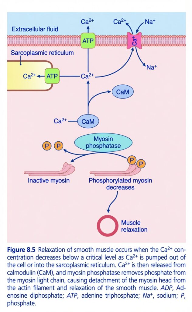
Figure 8.5: Relaxation of Smooth Muscle
Easiest and Most Conceptual Explanation
⭐ One-Line Concept
Smooth muscle relaxes when intracellular Ca²⁺ decreases. As Ca²⁺ is removed, calmodulin becomes inactive, myosin phosphatase removes phosphate from myosin, and the muscle relaxes.
First Understand the Whole Story
Imagine smooth muscle contraction is like switching on a machine.
- Ca²⁺ is the ON switch.
- Removing Ca²⁺ turns the switch OFF.
When Ca²⁺ leaves the cell:
- Calmodulin (CaM) stops working.
- Myosin phosphatase removes phosphate from myosin.
- Myosin can no longer bind strongly to actin.
- The muscle relaxes.
Understanding Every Part of the Figure
1. Intracellular Ca²⁺ Must Decrease
The central message of the figure is:
Relaxation begins when intracellular Ca²⁺ falls below a critical level.
How does Ca²⁺ decrease?
The figure shows three pathways.
Pathway 1: Ca²⁺ Pump Moves Ca²⁺ Outside the Cell
(Top center)
The green box labeled ATP represents the Ca²⁺-ATPase pump in the cell membrane.
What happens?
- Uses ATP (energy).
- Pumps Ca²⁺ from the cytoplasm → extracellular fluid.
Easy Concept
Think of this pump as a drain pump that removes excess calcium from the cell.
Pathway 2: Ca²⁺ Pump Moves Ca²⁺ into the Sarcoplasmic Reticulum
(Left side)
Another ATP-dependent Ca²⁺ pump is present in the sarcoplasmic reticulum (SR).
What happens?
- Uses ATP.
- Pumps Ca²⁺ back into the SR.
- Stores calcium for future contractions.
Easy Concept
The SR is like a calcium storage tank.
After contraction, the pump returns calcium to this tank.
Pathway 3: Na⁺–Ca²⁺ Exchanger
(Top right)
The pink protein is the Na⁺–Ca²⁺ exchanger.
What happens?
- Na⁺ enters the cell.
- Ca²⁺ leaves the cell.
This exchanger does not directly use ATP.
Instead, it uses the Na⁺ concentration gradient created by the Na⁺–K⁺ pump.
Easy Concept
Think of it as a trade system:
- 1 Ca²⁺ goes out.
- Na⁺ comes in.
Result of These Three Pathways
All three pathways reduce intracellular Ca²⁺.
↓
Less free Ca²⁺ remains inside the cell.
2. Ca²⁺ Leaves Calmodulin (CaM)
The blue oval labeled CaM is calmodulin.
During contraction:
Ca²⁺ + Calmodulin
↓
Ca²⁺–Calmodulin complex
↓
Activates MLCK
↓
Contraction
During relaxation:
Ca²⁺ decreases
↓
Ca²⁺ separates from calmodulin
↓
Calmodulin becomes inactive.
Easy Concept
Without Ca²⁺, calmodulin loses its “helper” and can no longer activate contraction.
3. Myosin Phosphatase
The large green oval is myosin phosphatase.
This enzyme is the main relaxation enzyme.
What does it do?
It removes phosphate (P) from phosphorylated myosin.
This process is called dephosphorylation.
Easy Concept
Think of myosin phosphatase as an eraser.
It erases the phosphate that MLCK had added during contraction.
4. Phosphorylated Myosin Decreases
During contraction:
MLCK adds phosphate to myosin.
↓
Myosin becomes active.
↓
Contraction occurs.
During relaxation:
Myosin phosphatase removes phosphate.
↓
Active myosin decreases.
↓
Myosin becomes inactive.
Easy Concept
No phosphate = inactive myosin.
Inactive myosin cannot produce contraction.5. Inactive Myosin
The left pink myosin molecule is inactive myosin.
It has:
❌ No phosphate attached.
Therefore,
it cannot attach effectively to actin.
6. Muscle Relaxation
The final result is shown at the bottom.
When:
- Ca²⁺ decreases,
- Calmodulin becomes inactive,
- Myosin is dephosphorylated,
↓
The myosin heads detach from actin.
↓
Cross-bridge cycling stops.
↓
The smooth muscle relaxes.
Complete Sequence
Smooth muscle finishes contracting
↓
Ca²⁺ is pumped out of the cell
↓
Ca²⁺ is pumped into the SR
↓
Na⁺–Ca²⁺ exchanger removes more Ca²⁺
↓
Intracellular Ca²⁺ decreases
↓
Ca²⁺ dissociates from calmodulin
↓
MLCK becomes inactive
↓
Myosin phosphatase removes phosphate from myosin
↓
Myosin becomes inactive
↓
Actin–myosin interaction stops
↓
Smooth muscle relaxes
Why Is ATP Needed During Relaxation?
Many students think ATP is needed only for contraction.
Actually:
ATP is also essential for relaxation because it powers the Ca²⁺-ATPase pumps, which remove Ca²⁺ from the cytoplasm.
Easy Concept
No ATP → Ca²⁺ cannot be removed efficiently → the muscle cannot relax properly.
Difference Between Contraction and Relaxation
| Contraction | Relaxation |
|---|---|
| Ca²⁺ increases | Ca²⁺ decreases |
| Calmodulin binds Ca²⁺ | Ca²⁺ leaves calmodulin |
| MLCK becomes active | MLCK becomes inactive |
| Myosin is phosphorylated | Myosin is dephosphorylated |
| Cross-bridges form | Cross-bridges detach |
| Muscle contracts | Muscle relaxes |
Easy Story
Imagine a factory.
During contraction:
- Calcium workers enter the factory.
- They activate the manager (calmodulin).
- The manager turns on MLCK.
- MLCK adds phosphate tags to myosin.
- The machines start working.
During relaxation:
- The calcium workers leave.
- The manager loses power.
- Myosin phosphatase removes the phosphate tags.
- The machines stop.
- The factory becomes quiet.
Simple Flow Diagram
Smooth muscle contraction ends
↓
Ca²⁺ is pumped out of the cell (Ca²⁺-ATPase)
↓
Ca²⁺ is pumped into the SR (SERCA pump)
↓
Na⁺–Ca²⁺ exchanger removes additional Ca²⁺
↓
Intracellular Ca²⁺ decreases
↓
Ca²⁺ dissociates from calmodulin
↓
MLCK becomes inactive
↓
Myosin phosphatase removes phosphate from myosin
↓
Myosin becomes inactive
↓
Cross-bridges detach
↓
Smooth muscle relaxation
High-Yield Summary Table
| Structure | Function | Easy Concept |
|---|---|---|
| Ca²⁺-ATPase (cell membrane) | Pumps Ca²⁺ out of the cell using ATP | Removes calcium from the cell |
| Ca²⁺-ATPase (SR) | Pumps Ca²⁺ into the SR using ATP | Stores calcium for future use |
| Na⁺–Ca²⁺ exchanger | Exchanges intracellular Ca²⁺ for extracellular Na⁺ | Trades Ca²⁺ out for Na⁺ in |
| Calmodulin (CaM) | Loses Ca²⁺ when intracellular Ca²⁺ falls | Turns off the contraction signal |
| Myosin phosphatase | Removes phosphate from myosin | The relaxation enzyme |
| Inactive myosin | Cannot bind effectively to actin | Stops contraction |
| Final result | Smooth muscle relaxes | Ca²⁺ removal switches the muscle OFF |
Key Concept (Figure 8.5)
Figure 8.5 shows that smooth muscle relaxation occurs when intracellular Ca²⁺ concentration decreases below a critical level. Ca²⁺ is removed from the cytoplasm by ATP-dependent Ca²⁺ pumps that transport it out of the cell or back into the sarcoplasmic reticulum, and by the Na⁺–Ca²⁺ exchanger, which extrudes Ca²⁺ in exchange for Na⁺ entry. As Ca²⁺ falls, it dissociates from calmodulin (CaM), causing MLCK to become inactive. Myosin phosphatase then dephosphorylates the myosin light chain, allowing myosin heads to detach from actin. Cross-bridge cycling stops, and the smooth muscle relaxes.
Myosin Phosphatase Is Important in Cessation of Contraction
- Smooth muscle relaxes when the Ca²⁺ channels close.
- The Ca²⁺ pump then removes Ca²⁺ from the cytosolic fluid of the smooth muscle cell.
- When the Ca²⁺ level falls below a critical level, the contraction process automatically reverses.
- However, phosphorylation of the myosin head does not reverse automatically.
- A separate enzyme called myosin phosphatase (Fig. 8.5) is needed for this step.
- Myosin phosphatase is present in the cytosol of the smooth muscle cell.
- It removes (splits off) the phosphate from the regulatory light chain of the myosin head.
- After the phosphate is removed, the attachment–detachment cycling of myosin stops.
- As a result, muscle contraction ends.
- Therefore, the time required for smooth muscle relaxation depends largely on the amount of active myosin phosphatase present in the cell.
KEY CONCEPT
- Smooth muscle relaxes when Ca²⁺ channels close and the Ca²⁺ pump removes intracellular Ca²⁺.
- When Ca²⁺ falls below a critical level, most contraction processes reverse.
- Myosin phosphatase is required to remove the phosphate from the regulatory light chain of myosin.
- Removal of the phosphate stops myosin cycling, so contraction ends.
- More active myosin phosphatase → Faster relaxation.
- Less active myosin phosphatase → Slower relaxation.
Conceptual Examples
- Ca²⁺ channels close → Ca²⁺ pump removes Ca²⁺ → Myosin phosphatase removes phosphate from myosin → Myosin stops pulling actin → Smooth muscle relaxes.
- High myosin phosphatase activity → Muscle relaxes quickly.
- Low myosin phosphatase activity → Muscle relaxation is delayed.
Possible Mechanism for Regulating the Latch Phenomenon
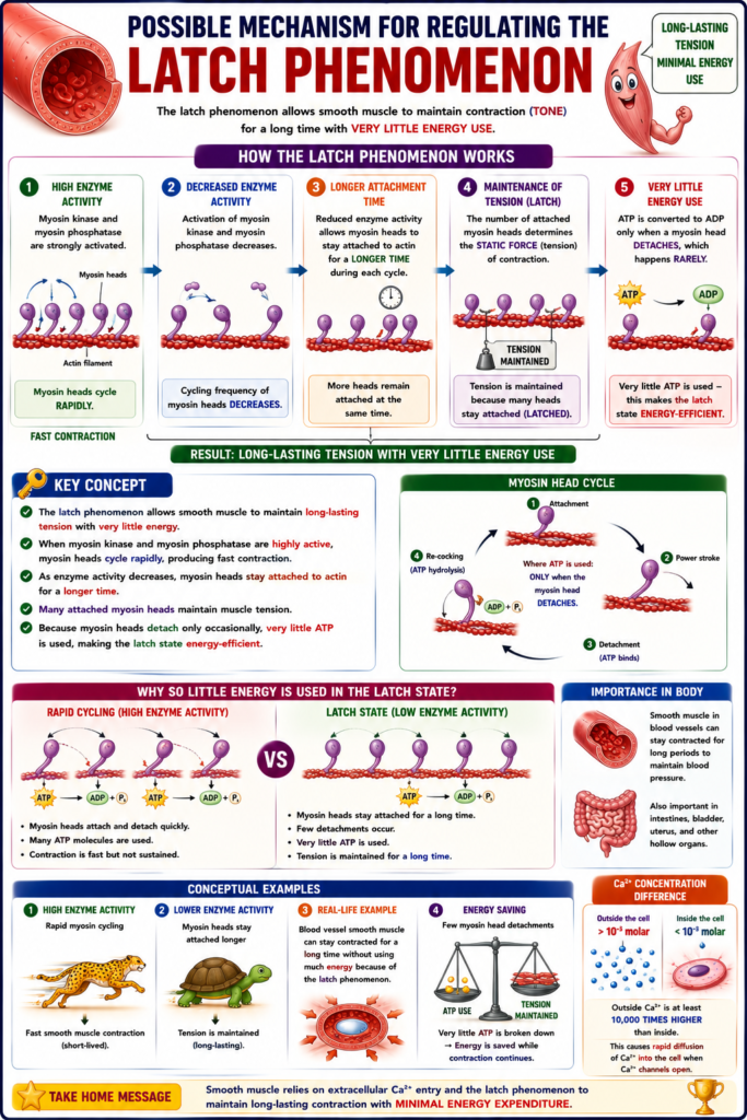
- The latch phenomenon is very important in smooth muscle.
- It allows smooth muscle to maintain contraction (tone) for a long time.
- This occurs with very little energy use.
- Many mechanisms have been suggested to explain the latch phenomenon.
- One of the simplest mechanisms is described below.
- When myosin kinase and myosin phosphatase are both strongly activated, the myosin heads cycle rapidly.
- As a result, the speed of muscle contraction is high.
- When the activation of these enzymes decreases, the cycling frequency of the myosin heads also decreases.
- At the same time, reduced enzyme activity allows the myosin heads to stay attached to the actin filament for a longer time during each cycle.
- Therefore, many myosin heads remain attached to actin at the same time.
- The number of attached myosin heads determines the static force (tension) of contraction.
- Because many heads remain attached, muscle tension is maintained (latched).
- Very little energy is used because ATP is converted to ADP only when a myosin head detaches, which happens rarely.
KEY CONCEPT
- The latch phenomenon allows smooth muscle to maintain long-lasting tension with very little energy.
- When myosin kinase and myosin phosphatase are highly active, myosin heads cycle rapidly, producing fast contraction.
- As enzyme activity decreases, myosin heads stay attached to actin for a longer time.
- Many attached myosin heads maintain muscle tension.
- Because myosin heads detach only occasionally, very little ATP is used, making the latch state energy-efficient.
Conceptual Examples
- High enzyme activity → Rapid myosin cycling → Fast smooth muscle contraction.
- Lower enzyme activity → Myosin heads stay attached longer → Tension is maintained.
- Blood vessel smooth muscle can stay contracted for a long time without using much energy because of the latch phenomenon.
- Few myosin head detachments → Very little ATP is broken down → Energy is saved while contraction continues.
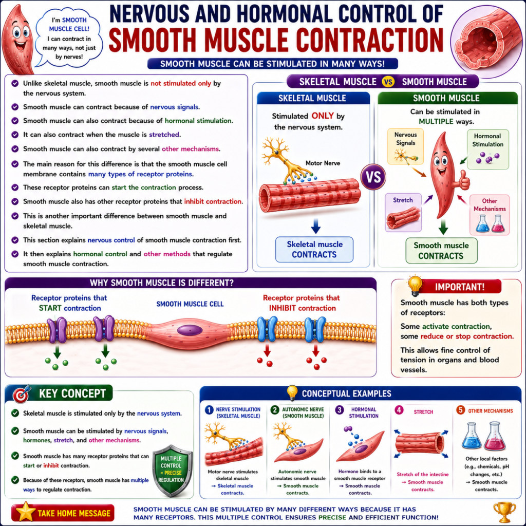
NERVOUS AND HORMONAL CONTROL OF SMOOTH MUSCLE CONTRACTION
- Unlike skeletal muscle, smooth muscle is not stimulated only by the nervous system.
- Smooth muscle can contract because of nervous signals.
- Smooth muscle can also contract because of hormonal stimulation.
- It can also contract when the muscle is stretched.
- Smooth muscle can also contract by several other mechanisms.
- The main reason for this difference is that the smooth muscle cell membrane contains many types of receptor proteins.
- These receptor proteins can start the contraction process.
- Smooth muscle also has other receptor proteins that inhibit contraction.
- This is another important difference between smooth muscle and skeletal muscle.
- This section explains nervous control of smooth muscle contraction first.
- It then explains hormonal control and other methods that regulate smooth muscle contraction.
KEY CONCEPT
- Skeletal muscle is stimulated only by the nervous system.
- Smooth muscle can be stimulated by:
- Nervous signals
- Hormones
- Muscle stretch
- Other mechanisms
- Smooth muscle has many receptor proteins that can start or inhibit contraction.
- Because of these receptors, smooth muscle has multiple ways to regulate contraction.
Conceptual Examples
- Motor nerve stimulates skeletal muscle → Skeletal muscle contracts.
- Autonomic nerve stimulates smooth muscle → Smooth muscle contracts.
- Hormone binds to a smooth muscle receptor → Smooth muscle contracts.
- Stretch of the intestine → Smooth muscle contracts.
- Some receptors activate contraction, while other receptors reduce or stop contraction.
MADE BY SELF LEARNING CEO AND FOUNDER DR SHEEN
