


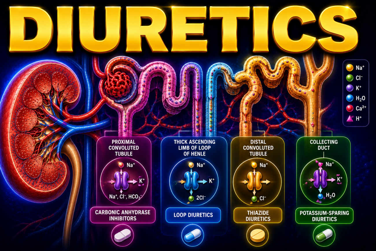
- Diuretics are drugs that increase urine production by reducing the reabsorption of water and electrolytes.
- Understanding how diuretics work helps explain the factors that control urine volume and electrolyte excretion.
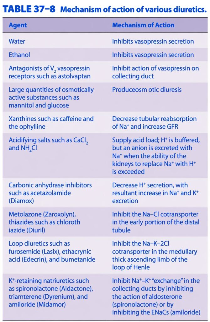
- The different mechanisms of diuretics are summarized in Table 37–8.
- Water, alcohol, osmotic diuretics, xanthines, and acidifying salts have limited clinical use.
- Vasopressin antagonists are currently being studied in clinical trials.
- Many other diuretics are widely used in medical practice.
- Carbonic anhydrase inhibitors are moderately effective diuretics.
- They reduce H⁺ secretion by decreasing the formation of carbonic acid.
- Reduced H⁺ secretion increases Na⁺ excretion.
- These drugs also reduce HCO₃⁻ (bicarbonate) reabsorption.
- Because H⁺, K⁺, and Na⁺ compete with each other, decreased H⁺ secretion increases K⁺ secretion and excretion.
- Furosemide and other loop diuretics block the Na⁺–K⁺–2Cl⁻ cotransporter in the thick ascending limb of the loop of Henle.
- Loop diuretics produce a marked increase in the excretion of Na⁺ (natriuresis) and K⁺ (kaliuresis).
- Thiazide diuretics block the Na⁺–Cl⁻ cotransporter in the distal tubule.
- Thiazides produce less diuresis than loop diuretics.
- Both loop diuretics and thiazides increase the amount of Na⁺ and water reaching the collecting ducts.
- This increases K⁺ secretion and excretion.
- With long-term use, loop diuretics and thiazides commonly cause K⁺ depletion and hypokalaemia unless K⁺ intake is supplemented.
- K⁺-sparing diuretics act in the collecting ducts.
- They reduce K⁺ loss by either:
- Blocking the action of aldosterone.
- Blocking epithelial sodium channels (ENaCs).
Table: Table 37–8
KEY CONCEPT
- Diuretics increase urine formation by decreasing Na⁺ and water reabsorption at different sites of the nephron. Carbonic anhydrase inhibitors increase Na⁺, HCO₃⁻, and K⁺ excretion. Loop diuretics block the Na⁺–K⁺–2Cl⁻ cotransporter, while thiazides block the Na⁺–Cl⁻ cotransporter. Both can cause hypokalaemia with prolonged use. K⁺-sparing diuretics act in the collecting ducts by blocking aldosterone or ENaCs, helping prevent excessive K⁺ loss.
KEY CONCEPT
- Diuretics increase urine formation by reducing Na⁺ and water reabsorption at different parts of the nephron. Carbonic anhydrase inhibitors increase Na⁺, HCO₃⁻, and K⁺ excretion. Loop diuretics block the Na⁺–K⁺–2Cl⁻ cotransporter, while thiazides block the Na⁺–Cl⁻ cotransporter. Both loop diuretics and thiazides increase K⁺ loss and may cause hypokalaemia with long-term use.
EFFECTS OF DISORDERED KIDNEY FUNCTION
- Many different kidney diseases produce similar abnormalities.
- The kidneys produce renin, which plays an important role in regulating blood pressure.
- The relationship between the kidneys, renin, and hypertension is discussed separately.
- Many kidney diseases cause abnormal substances to appear in the urine.
- Protein may be present in the urine (proteinuria).
- White blood cells (leukocytes) may be present in the urine.
- Red blood cells may be present in the urine (hematuria).
- Casts may also be present in the urine.
- Casts are protein-containing materials that form inside the renal tubules and are later washed into the bladder.
- Kidney disease can reduce the kidney’s ability to concentrate urine.
- Kidney disease can also reduce the kidney’s ability to dilute urine.
- Kidney disease may lead to uremia due to the accumulation of waste products in the blood.
- Kidney disease may cause acidosis because the kidneys cannot remove enough acid from the body.
- Kidney disease may also cause abnormal retention of Na⁺.
Clinical Box: Clinical Box 37–3
KEY CONCEPT
- Many kidney diseases share common features, including protein, white blood cells, red blood cells, and casts in the urine. They can also impair the kidney’s ability to concentrate or dilute urine and may lead to uremia, acidosis, and abnormal Na⁺ retention.
CLINICAL BOX 37–3
Proteinuria
- In many kidney diseases, the permeability of the glomerular capillaries increases.
- As a result, more protein than normal passes into the urine.
- This condition is called proteinuria.
- Most of the protein lost in the urine is albumin.
- Therefore, this condition is commonly called albuminuria.
- Changes in the electrical charges of the glomerular membrane contribute to albuminuria.
- In some kidney diseases, especially nephrosis, the amount of protein lost in the urine can be very large.
- The liver may not be able to replace the lost plasma proteins fast enough.
- This leads to hypoproteinaemia (low plasma protein levels).
- Low plasma protein reduces the plasma oncotic pressure.
- As oncotic pressure falls, plasma volume decreases.
- The plasma volume may become dangerously low.
- At the same time, fluid moves into the tissues, causing oedema.
- A harmless (benign) condition called orthostatic albuminuria can also cause proteinuria.
- Its exact cause is not fully understood but is thought to involve changes in renal blood flow (renal haemodynamics).
- In some otherwise healthy people, protein appears in the urine only while they are standing.
- When these individuals lie down, their urine becomes free of protein.
KEY CONCEPT
- Proteinuria is the presence of excess protein in the urine due to increased permeability of the glomerular capillaries. Most urinary protein is albumin (albuminuria). Severe protein loss, especially in nephrosis, causes hypoproteinaemia, reduced plasma oncotic pressure, decreased plasma volume, and oedema. In orthostatic albuminuria, protein appears in the urine only when standing and disappears when lying down.
LOSS OF CONCENTRATING & DILUTING ABILITY
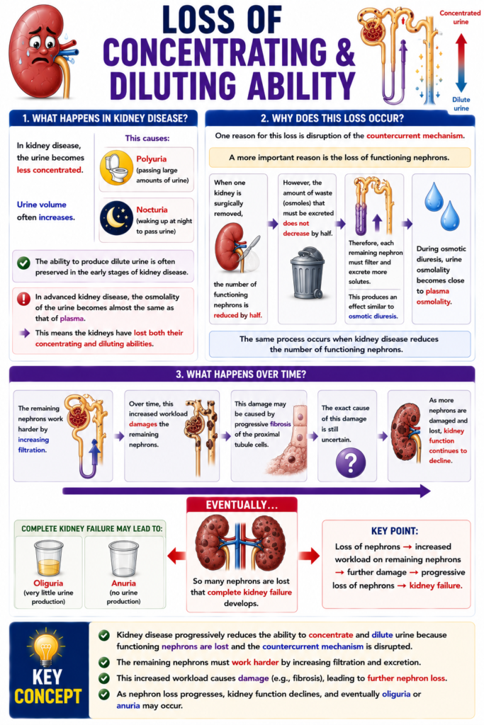
- In kidney disease, the urine becomes less concentrated.
- Urine volume often increases.
- This causes:
- Polyuria (passing large amounts of urine).
- Nocturia (waking up at night to pass urine).
- The ability to produce dilute urine is often preserved in the early stages of kidney disease.
- In advanced kidney disease, the osmolality of the urine becomes almost the same as that of plasma.
- This means the kidneys have lost both their concentrating and diluting abilities.
- One reason for this loss is disruption of the countercurrent mechanism.
- A more important reason is the loss of functioning nephrons.
- When one kidney is surgically removed, the number of functioning nephrons is reduced by half.
- However, the amount of waste (osmoles) that must be excreted does not decrease by half.
- Therefore, each remaining nephron must filter and excrete more solutes.
- This produces an effect similar to osmotic diuresis.
- During osmotic diuresis, urine osmolality becomes close to plasma osmolality.
- The same process occurs when kidney disease reduces the number of functioning nephrons.
- The remaining nephrons work harder by increasing filtration.
- Over time, this increased workload damages the remaining nephrons.
- This damage may be caused by progressive fibrosis of the proximal tubule cells.
- The exact cause of this damage is still uncertain.
- As more nephrons are damaged and lost, kidney function continues to decline.
- Eventually, so many nephrons are lost that complete kidney failure develops.
- Complete kidney failure may lead to:
- Oliguria (very little urine production).
- Anuria (no urine production).
KEY CONCEPT
- Kidney disease progressively reduces the ability to concentrate and dilute urine because functioning nephrons are lost and the countercurrent mechanism is disrupted. The remaining nephrons must work harder, leading to further nephron damage, progressive kidney failure, and eventually oliguria or anuria.
UREMIA
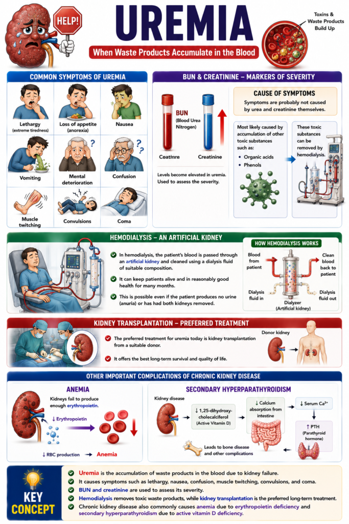
- Uremia develops when waste products from protein metabolism accumulate in the blood.
- Common symptoms of uremia include:
- Lethargy (extreme tiredness)
- Loss of appetite (anorexia)
- Nausea
- Vomiting
- Mental deterioration
- Confusion
- Muscle twitching
- Convulsions
- Coma
- Blood urea nitrogen (BUN) and creatinine levels become elevated in uremia.
- The levels of BUN and creatinine are used to assess the severity of uremia.
- The symptoms of uremia are probably not caused by urea and creatinine themselves.
- Instead, they are most likely caused by the accumulation of other toxic substances, such as organic acids or phenols.
- The toxic substances causing uremia can be removed by hemodialysis.
- In hemodialysis, the patient’s blood is passed through an artificial kidney and cleaned using a dialysis fluid of suitable composition.
- Hemodialysis can keep patients alive and in reasonably good health for many months.
- This is possible even if the patient produces no urine (anuria) or has had both kidneys removed.
- The preferred treatment for uremia today is kidney transplantation from a suitable donor.
- Chronic kidney disease also commonly causes anemia.
- This anemia occurs mainly because the kidneys fail to produce enough erythropoietin.
- Chronic kidney disease can also cause secondary hyperparathyroidism.
- This occurs because of a deficiency of 1,25-dihydroxycholecalciferol (active vitamin D).
KEY CONCEPT
- Uremia is the accumulation of waste products in the blood due to kidney failure. It causes symptoms such as lethargy, nausea, confusion, muscle twitching, convulsions, and coma. BUN and creatinine are used to assess its severity. Hemodialysis removes toxic waste products, while kidney transplantation is the preferred long-term treatment. Chronic kidney disease also commonly causes anemia due to erythropoietin deficiency and secondary hyperparathyroidism due to active vitamin D deficiency.
ACIDOSIS
- Acidosis is common in chronic kidney disease.
- It occurs because the kidneys cannot remove enough acid produced from digestion and normal body metabolism.
- In renal tubular acidosis, there is a specific defect in the kidney’s ability to produce acidic urine.
- Most other kidney functions remain normal in renal tubular acidosis.
- In most cases of chronic kidney disease, the kidneys are still able to produce maximally acidic urine.
- However, acidosis still develops because the total amount of H⁺ that can be secreted is reduced.
- The reduced H⁺ secretion occurs because the diseased renal tubules produce less NH₄⁺ (ammonium).
- Since NH₄⁺ helps remove H⁺ from the body, decreased NH₄⁺ production reduces acid excretion.
- As a result, H⁺ accumulates in the body, causing acidosis.
KEY CONCEPT
- Acidosis is a common complication of chronic kidney disease because the kidneys cannot excrete enough acid. In renal tubular acidosis, the main defect is the inability to produce acidic urine, while in chronic kidney disease, urine is maximally acidified but total H⁺ excretion is reduced due to decreased NH₄⁺ production by the renal tubules.
ABNORMAL Na⁺ HANDLING
- Many patients with kidney disease retain too much Na⁺ in the body.
- Excess Na⁺ retention causes oedema (fluid accumulation in the tissues).
- There are at least three main causes of Na⁺ retention in kidney disease.
- First cause: Acute glomerulonephritis
- This disease mainly affects the glomeruli.
- The amount of Na⁺ filtered by the kidneys decreases markedly.
- Less filtered Na⁺ leads to Na⁺ retention.
- Second cause: Nephrotic syndrome
- Aldosterone secretion increases.
- Higher aldosterone increases Na⁺ reabsorption.
- The plasma protein level is low.
- Fluid moves from the plasma into the interstitial tissues.
- Plasma volume decreases.
- The fall in plasma volume activates the renin–angiotensin system.
- This increases aldosterone secretion.
- More aldosterone causes further Na⁺ retention.
- Third cause: Heart failure
- Heart failure also causes Na⁺ retention and oedema.
- Kidney disease increases the risk of heart failure.
- One reason is that kidney disease often causes hypertension (high blood pressure).
KEY CONCEPT
- Abnormal Na⁺ handling in kidney disease leads to excessive Na⁺ retention and oedema. The three main causes are reduced Na⁺ filtration in acute glomerulonephritis, increased aldosterone secretion in nephrotic syndrome through activation of the renin–angiotensin system, and heart failure, which is often associated with kidney disease and hypertension.
MADE BY SELF LEARNING CEO AND FOUNDER DR SHEEN
