


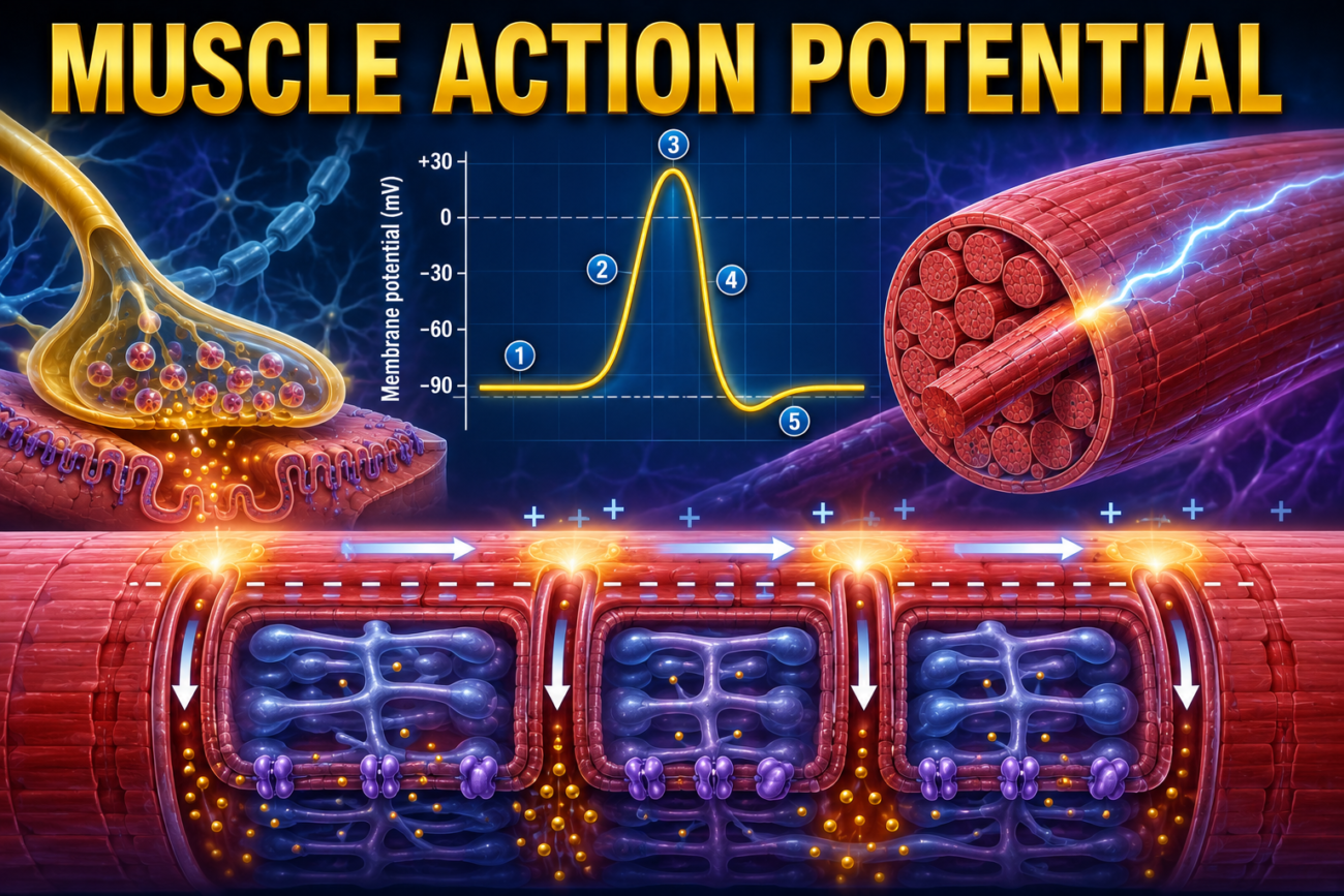
- The basic process of initiation and conduction of action potentials in skeletal muscle fibers is almost the same as in nerve fibers.
- The main differences are quantitative (numerical) differences.
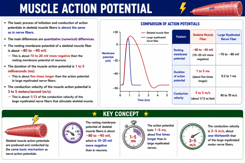
- The resting membrane potential of a skeletal muscle fiber is about −80 to −90 mV.
- This is about 10 to 20 mV more negative than the resting membrane potential of neurons.
- The duration of the muscle action potential is 1 to 5 milliseconds (ms).
- This is about five times longer than the action potential in large myelinated nerve fibers.
- The conduction velocity of the muscle action potential is 3 to 5 meters/second (m/s).
- This is about 1/13 of the conduction velocity of the large myelinated nerve fibers that stimulate skeletal muscle.
KEY CONCEPT
- Skeletal muscle action potentials are produced and conducted by the same basic mechanism as nerve action potentials.
- The resting membrane potential of skeletal muscle fibers is about −80 to −90 mV, which is 10–20 mV more negative than in neurons.
- The action potential lasts 1–5 ms, about five times longer than in large myelinated nerves.
- The conduction velocity is 3–5 m/s, about one-thirteenth that of the large myelinated motor nerve fibers.
Action Potentials Spread to the Interior of the Muscle Fiber Via Transverse Tubules
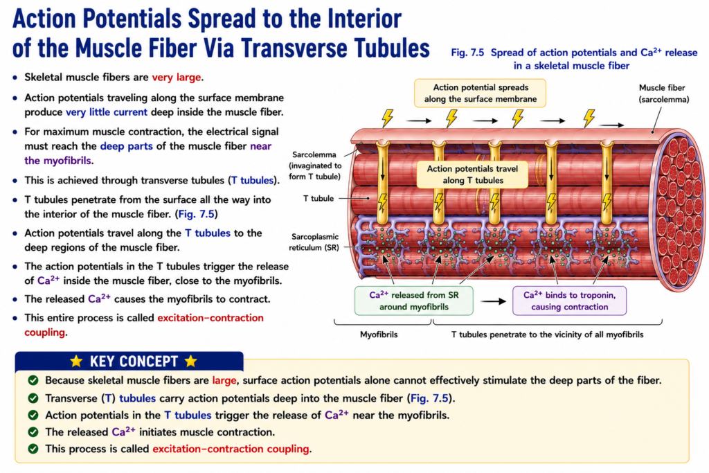
- Skeletal muscle fibers are very large.
- Action potentials traveling along the surface membrane produce very little current deep inside the muscle fiber.
- For maximum muscle contraction, the electrical signal must reach the deep parts of the muscle fiber near the myofibrils.
- This is achieved through transverse tubules (T tubules).
- T tubules penetrate from the surface all the way into the interior of the muscle fiber. (Fig. 7.5)
- Action potentials travel along the T tubules to the deep regions of the muscle fiber.
- The action potentials in the T tubules trigger the release of Ca²⁺ inside the muscle fiber, close to the myofibrils.
- The released Ca²⁺ causes the myofibrils to contract.
- This entire process is called excitation-contraction coupling.
KEY CONCEPT
- Because skeletal muscle fibers are large, surface action potentials alone cannot effectively stimulate the deep parts of the fiber.
- Transverse (T) tubules carry action potentials deep into the muscle fiber (Fig. 7.5).
- Action potentials in the T tubules trigger the release of Ca²⁺ near the myofibrils.
- The released Ca²⁺ initiates muscle contraction.
- This process is called excitation-contraction coupling.
EXCITATION-CONTRACTION COUPLING
Transverse Tubule–Sarcoplasmic Reticulum System
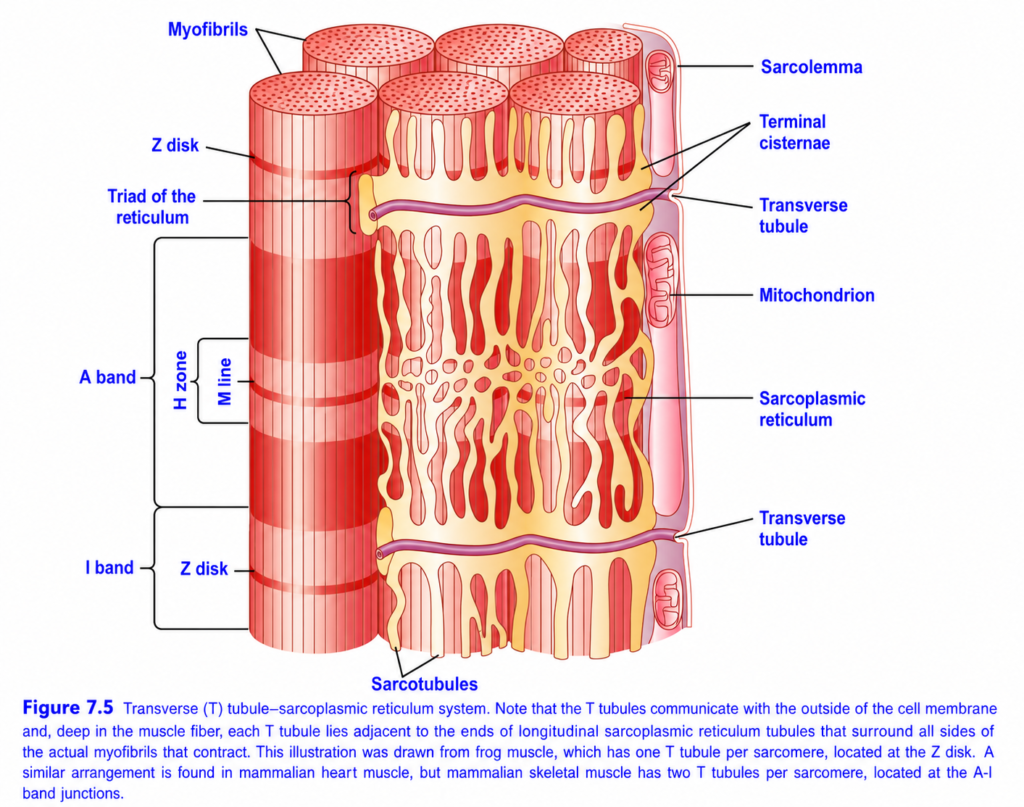
- Fig. 7.5 shows the myofibrils surrounded by the transverse (T) tubule–sarcoplasmic reticulum system.
- The T tubules are small tubes that run across (transverse to) the myofibrils.
- The T tubules begin at the cell membrane.
- They extend completely through the muscle fiber from one side to the other.
- The T tubules branch repeatedly and form complete planes of tubules.
- These branches interlace among all the individual myofibrils.
- At their origin from the cell membrane, the T tubules are open to the outside of the muscle fiber.
- Therefore, the T tubules communicate directly with the extracellular fluid surrounding the muscle fiber.
- The lumens of the T tubules contain extracellular fluid.
- Thus, the T tubules are internal extensions of the cell membrane.
- When an action potential spreads along the muscle fiber membrane, the same electrical change also spreads through the T tubules into the deep interior of the muscle fiber.
- The electrical currents around the T tubules trigger muscle contraction.
- Fig. 7.5 also shows the sarcoplasmic reticulum.
- The sarcoplasmic reticulum has two major parts:
- Large chambers called terminal cisternae, which lie next to the T tubules.
- Long longitudinal tubules, which surround all surfaces of the contracting myofibrils.
KEY CONCEPT
- The T tubules and sarcoplasmic reticulum together form the excitation-contraction coupling system (Fig. 7.5).
- T tubules are small transverse tubes that begin at the cell membrane and extend through the entire muscle fiber.
- They branch extensively, communicate with the extracellular fluid, and are internal extensions of the cell membrane.
- Action potentials travel along the T tubules into the deep interior of the muscle fiber, where they initiate muscle contraction.
- The sarcoplasmic reticulum consists of terminal cisternae beside the T tubules and longitudinal tubules surrounding the myofibrils.
Transverse (T) Tubule–Sarcoplasmic Reticulum System (Figure 7.5)
Easiest & Most Conceptual Summary
This figure explains how an electrical signal (action potential) quickly reaches the inside of a muscle fiber and causes calcium release, leading to muscle contraction.
It shows the T-tubule–Sarcoplasmic Reticulum (SR) system, also called the Excitation–Contraction (E–C) Coupling System.
Main Concept
Muscle action potential → T-tubules carry the signal deep into the muscle → Sarcoplasmic reticulum releases Ca²⁺ → Actin and myosin interact → Muscle contracts
Overall View of the Figure
The figure contains three major structures working together:
- Myofibrils → Contractile units (produce force)
- T-tubules → Carry the electrical signal inside the muscle
- Sarcoplasmic Reticulum (SR) → Stores and releases calcium
Together, they ensure that every myofibril contracts at the same time.
Understanding Each Label
1. Myofibrils
The large red cylinders are myofibrils.
Function
They are the contractile machinery of skeletal muscle.
Each myofibril contains repeating sarcomeres, which shorten during contraction.
Components inside each sarcomere
- Actin (thin filaments)
- Myosin (thick filaments)
Concept
Think of myofibrils as the engines that actually produce force.
2. Sarcolemma
The sarcolemma is the cell membrane of the muscle fiber.
Function
- Receives the muscle action potential.
- Conducts the electrical impulse over the surface of the muscle.
Concept
Think of the sarcolemma as the outer electrical wire of the muscle.
3. Transverse (T) Tubules
The T-tubules are deep invaginations (folds) of the sarcolemma.
They extend from the surface deep into the muscle fiber.
Function
Carry the electrical signal from the surface to the center of the muscle.
Why are they needed?
Skeletal muscle fibers are very thick.
If the electrical signal traveled only along the surface:
- The outer part would contract first.
- The inner part would contract much later.
The T-tubules deliver the signal deep inside, so all myofibrils receive it almost simultaneously.
Easy Concept
Think of T-tubules as electrical tunnels carrying the impulse to the center of the muscle.
4. Sarcoplasmic Reticulum (SR)
The yellow network surrounding each myofibril is the sarcoplasmic reticulum.
It is a specialized smooth endoplasmic reticulum.
Function
Stores calcium ions (Ca²⁺).
Important Point
At rest, most calcium is stored inside the SR.
5. Sarcotubules
The sarcotubules are the longitudinal portions of the sarcoplasmic reticulum.
Function
- Surround the myofibrils.
- Transport calcium within the SR.
- Connect the terminal cisternae.
Think of them as the pipelines of the SR.
6. Terminal Cisternae
These are the enlarged sacs of the sarcoplasmic reticulum located on either side of a T-tubule.
Function
Store a large amount of calcium.
When stimulated,
they rapidly release calcium into the muscle cell.
Concept
Think of terminal cisternae as calcium storage tanks.
7. Triad
The figure labels the Triad of the Reticulum.
A Triad consists of:
Terminal Cisternae
        │
     T-Tubule
        │
Terminal Cisternae
Formula
Triad = 1 T-tubule + 2 Terminal Cisternae
Importance
This is the site where excitation is converted into contraction (Excitation–Contraction Coupling).
8. Mitochondria
The oval structures are mitochondria.
Function
Produce ATP.
ATP is required for:
- Muscle contraction
- Muscle relaxation
- Calcium pumps
- Active transport
Without ATP,
muscles cannot contract or relax properly.
9. Sarcomere Landmarks
The left side of the figure shows one sarcomere.
Z Disc
Marks the boundary of one sarcomere.
A sarcomere extends:
Z Disc -------- Z DiscI Band
Contains:
- Thin (actin) filaments only.
Appearance
Light band.
A Band
Contains:
- Entire length of myosin.
- Area where actin overlaps myosin.
Appearance
Dark band.
H Zone
Located in the center of the A band.
Contains:
- Myosin only.
No actin overlap.
M Line
Located in the center of the H zone.
Function
Holds the thick (myosin) filaments together.
How Excitation–Contraction Coupling Occurs
Step 1
A muscle action potential spreads over the sarcolemma.
↓
Step 2
The action potential enters the T-tubules.
↓
Step 3
The T-tubules rapidly carry the electrical signal deep into the muscle fiber.
↓
Step 4
The signal reaches the triads.
↓
Step 5
The terminal cisternae of the SR release Ca²⁺.
↓
Step 6
Calcium binds to troponin on the thin filament.
↓
Step 7
Tropomyosin moves away from the myosin-binding sites on actin.
↓
Step 8
Myosin binds to actin.
↓
Step 9
Cross-bridge cycling occurs.
↓
Step 10
Sarcomeres shorten.
↓
Step 11
The whole muscle contracts.
How Relaxation Occurs
After the action potential ends:
Easy Flow Diagram
Muscle action potential
          ↓
Sarcolemma
          ↓
T-Tubules
          ↓
Triad
          ↓
Terminal cisternae release Ca²⁺
          ↓
Calcium binds troponin
          ↓
Actin–myosin interaction
          ↓
Sarcomere shortens
          ↓
Muscle contraction
Why Are T-Tubules Important?
Without T-tubules:
- Only the outer part of the muscle fiber would receive the electrical signal first.
- The inner myofibrils would contract later.
- Contraction would be weak and uncoordinated.
With T-tubules:
- Every myofibril receives the signal almost simultaneously.
- The entire muscle fiber contracts together.
- Contraction becomes rapid, strong, and synchronized.
Clinical Correlation
Diseases affecting the T-tubule–SR system or calcium handling can cause:
- Muscle weakness
- Poor coordination of contraction
- Delayed relaxation
- Reduced muscle force
Important Note from the Figure
The caption explains a species difference:
- Frog skeletal muscle has one T-tubule per sarcomere, located at the Z disc.
- Mammalian skeletal muscle has two T-tubules per sarcomere, located at the junction of the A band and I band (A–I junctions).
This arrangement in mammals allows even more efficient excitation–contraction coupling.
Important Points from Figure 7.5
- Myofibrils contain the contractile proteins actin and myosin.
- The sarcolemma is the muscle fiber membrane that conducts the action potential.
- T-tubules are invaginations of the sarcolemma that carry the electrical signal deep into the muscle fiber.
- The sarcoplasmic reticulum (SR) surrounds each myofibril and serves as the major intracellular calcium store.
- Terminal cisternae are enlarged portions of the SR that release Ca²⁺ when stimulated.
- A triad consists of one T-tubule flanked by two terminal cisternae and is the key site of excitation–contraction coupling.
- Mitochondria provide ATP required for contraction, relaxation, and calcium pumping.
- The Z disc, I band, A band, H zone, and M line are structural landmarks of the sarcomere.
- In mammalian skeletal muscle, there are two T-tubules per sarcomere, located at the A–I junctions, ensuring rapid and synchronized contraction.
KEY CONCEPT (Figure 7.5)
The T-tubule–sarcoplasmic reticulum system links the electrical signal in the muscle membrane to mechanical contraction. The sarcolemma carries the muscle action potential, and the T-tubules rapidly transmit it deep into the muscle fiber. At the triad (one T-tubule plus two terminal cisternae), the action potential triggers the sarcoplasmic reticulum to release Ca²⁺. Calcium binds to troponin, permitting actin–myosin interaction and sarcomere shortening, which produces muscle contraction. After contraction, calcium is pumped back into the SR, allowing the muscle to relax. This specialized arrangement ensures rapid, powerful, and synchronized contraction of the entire muscle fiber.
Release of Ca²⁺ By the Sarcoplasmic Reticulum
- The sarcoplasmic reticulum stores a high concentration of Ca²⁺ inside its vesicular tubules.
- When an action potential travels through the adjacent T tubule, Ca²⁺ is released from the sarcoplasmic reticulum.
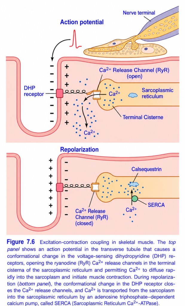
- Figs. 7.6 and 7.7 show that the action potential in the T tubule causes electrical current to flow into the terminal cisternae of the sarcoplasmic reticulum, where they are closely attached to the T tubule.
- As the action potential reaches the T tubule, the voltage change is detected by dihydropyridine receptors (DHPRs).
- The dihydropyridine receptors are linked to calcium release channels, called ryanodine receptor (RyR) channels, in the adjacent terminal cisternae of the sarcoplasmic reticulum. (Fig. 7.6)
- Activation of the dihydropyridine receptors opens the ryanodine receptor calcium release channels.
- These calcium release channels open in both the terminal cisternae and the attached longitudinal tubules.
- The channels remain open for a few milliseconds.
- During this time, large amounts of Ca²⁺ are released into the sarcoplasm surrounding the myofibrils.
- The released Ca²⁺ initiates muscle contraction.
KEY CONCEPT
- The sarcoplasmic reticulum stores a high concentration of Ca²⁺ in its vesicular tubules.
- An action potential in the T tubule triggers Ca²⁺ release from the sarcoplasmic reticulum (Figs. 7.6 and 7.7).
- The voltage change is detected by dihydropyridine receptors (DHPRs), which are linked to ryanodine receptor (RyR) calcium release channels (Fig. 7.6).
- Activation of DHPRs opens RyR channels in the terminal cisternae and longitudinal tubules.
- The channels remain open for a few milliseconds, releasing Ca²⁺ into the sarcoplasm around the myofibrils.
- The released Ca²⁺ initiates muscle contraction.
Transverse (T) Tubule–Sarcoplasmic Reticulum System (Figure 7.5)
Easiest & Most Conceptual Summary
This figure explains how an electrical signal (action potential) quickly reaches the inside of a muscle fiber and causes calcium release, leading to muscle contraction.
It shows the T-tubule–Sarcoplasmic Reticulum (SR) system, also called the Excitation–Contraction (E–C) Coupling System.
Main Concept
Muscle action potential → T-tubules carry the signal deep into the muscle → Sarcoplasmic reticulum releases Ca²⁺ → Actin and myosin interact → Muscle contracts
Overall View of the Figure
The figure contains three major structures working together:
- Myofibrils → Contractile units (produce force)
- T-tubules → Carry the electrical signal inside the muscle
- Sarcoplasmic Reticulum (SR) → Stores and releases calcium
Together, they ensure that every myofibril contracts at the same time.
Understanding Each Label
1. Myofibrils
The large red cylinders are myofibrils.
Function
They are the contractile machinery of skeletal muscle.
Each myofibril contains repeating sarcomeres, which shorten during contraction.
Components inside each sarcomere
- Actin (thin filaments)
- Myosin (thick filaments)
Concept
Think of myofibrils as the engines that actually produce force.
2. Sarcolemma
The sarcolemma is the cell membrane of the muscle fiber.
Function
- Receives the muscle action potential.
- Conducts the electrical impulse over the surface of the muscle.
Concept
Think of the sarcolemma as the outer electrical wire of the muscle.
3. Transverse (T) Tubules
The T-tubules are deep invaginations (folds) of the sarcolemma.
They extend from the surface deep into the muscle fiber.
Function
Carry the electrical signal from the surface to the center of the muscle.
Why are they needed?
Skeletal muscle fibers are very thick.
If the electrical signal traveled only along the surface:
- The outer part would contract first.
- The inner part would contract much later.
The T-tubules deliver the signal deep inside, so all myofibrils receive it almost simultaneously.
Easy Concept
Think of T-tubules as electrical tunnels carrying the impulse to the center of the muscle.
4. Sarcoplasmic Reticulum (SR)
The yellow network surrounding each myofibril is the sarcoplasmic reticulum.
It is a specialized smooth endoplasmic reticulum.
Function
Stores calcium ions (Ca²⁺).
Important Point
At rest, most calcium is stored inside the SR.
5. Sarcotubules
The sarcotubules are the longitudinal portions of the sarcoplasmic reticulum.
Function
- Surround the myofibrils.
- Transport calcium within the SR.
- Connect the terminal cisternae.
Think of them as the pipelines of the SR.
6. Terminal Cisternae
These are the enlarged sacs of the sarcoplasmic reticulum located on either side of a T-tubule.
Function
Store a large amount of calcium.
When stimulated,
they rapidly release calcium into the muscle cell.
Concept
Think of terminal cisternae as calcium storage tanks.
7. Triad
The figure labels the Triad of the Reticulum.
A Triad consists of:
Terminal Cisternae
        │
     T-Tubule
        │
Terminal Cisternae
Formula
Triad = 1 T-tubule + 2 Terminal Cisternae
Importance
This is the site where excitation is converted into contraction (Excitation–Contraction Coupling).
8. Mitochondria
The oval structures are mitochondria.
Function
Produce ATP.
ATP is required for:
- Muscle contraction
- Muscle relaxation
- Calcium pumps
- Active transport
Without ATP,
muscles cannot contract or relax properly.
9. Sarcomere Landmarks
The left side of the figure shows one sarcomere.
Z Disc
Marks the boundary of one sarcomere.
A sarcomere extends:
Z Disc -------- Z DiscI Band
Contains:
- Thin (actin) filaments only.
Appearance
Light band.
A Band
Contains:
- Entire length of myosin.
- Area where actin overlaps myosin.
Appearance
Dark band.
H Zone
Located in the center of the A band.
Contains:
- Myosin only.
No actin overlap.
M Line
Located in the center of the H zone.
Function
Holds the thick (myosin) filaments together.
How Excitation–Contraction Coupling Occurs
Step 1
A muscle action potential spreads over the sarcolemma.
↓
Step 2
The action potential enters the T-tubules.
↓
Step 3
The T-tubules rapidly carry the electrical signal deep into the muscle fiber.
↓
Step 4
The signal reaches the triads.
↓
Step 5
The terminal cisternae of the SR release Ca²⁺.
↓
Step 6
Calcium binds to troponin on the thin filament.
↓
Step 7
Tropomyosin moves away from the myosin-binding sites on actin.
↓
Step 8
Myosin binds to actin.
↓
Step 9
Cross-bridge cycling occurs.
↓
Step 10
Sarcomeres shorten.
↓
Step 11
The whole muscle contracts.
How Relaxation Occurs
After the action potential ends:
- Calcium pumps move Ca²⁺ back into the sarcoplasmic reticulum.
- Calcium dissociates from troponin.
- Tropomyosin again blocks the binding sites on actin.
- Cross-bridge cycling stops.
- The muscle relaxes.
Easy Flow Diagram
Muscle action potential
          ↓
Sarcolemma
          ↓
T-Tubules
          ↓
Triad
          ↓
Terminal cisternae release Ca²⁺
          ↓
Calcium binds troponin
          ↓
Actin–myosin interaction
          ↓
Sarcomere shortens
          ↓
Muscle contraction
Why Are T-Tubules Important?
Without T-tubules:
- Only the outer part of the muscle fiber would receive the electrical signal first.
- The inner myofibrils would contract later.
- Contraction would be weak and uncoordinated.
With T-tubules:
- Every myofibril receives the signal almost simultaneously.
- The entire muscle fiber contracts together.
- Contraction becomes rapid, strong, and synchronized.
Clinical Correlation
Diseases affecting the T-tubule–SR system or calcium handling can cause:
- Muscle weakness
- Poor coordination of contraction
- Delayed relaxation
- Reduced muscle force
Important Note from the Figure
The caption explains a species difference:
- Frog skeletal muscle has one T-tubule per sarcomere, located at the Z disc.
- Mammalian skeletal muscle has two T-tubules per sarcomere, located at the junction of the A band and I band (A–I junctions).
This arrangement in mammals allows even more efficient excitation–contraction coupling.Important Points from Figure 7.5
- Myofibrils contain the contractile proteins actin and myosin.
- The sarcolemma is the muscle fiber membrane that conducts the action potential.
- T-tubules are invaginations of the sarcolemma that carry the electrical signal deep into the muscle fiber.
- The sarcoplasmic reticulum (SR) surrounds each myofibril and serves as the major intracellular calcium store.
- Terminal cisternae are enlarged portions of the SR that release Ca²⁺ when stimulated.
- A triad consists of one T-tubule flanked by two terminal cisternae and is the key site of excitation–contraction coupling.
- Mitochondria provide ATP required for contraction, relaxation, and calcium pumping.
- The Z disc, I band, A band, H zone, and M line are structural landmarks of the sarcomere.
- In mammalian skeletal muscle, there are two T-tubules per sarcomere, located at the A–I junctions, ensuring rapid and synchronized contraction.
KEY CONCEPT (Figure 7.5)
The T-tubule–sarcoplasmic reticulum system links the electrical signal in the muscle membrane to mechanical contraction. The sarcolemma carries the muscle action potential, and the T-tubules rapidly transmit it deep into the muscle fiber. At the triad (one T-tubule plus two terminal cisternae), the action potential triggers the sarcoplasmic reticulum to release Ca²⁺. Calcium binds to troponin, permitting actin–myosin interaction and sarcomere shortening, which produces muscle contraction. After contraction, calcium is pumped back into the SR, allowing the muscle to relax. This specialized arrangement ensures rapid, powerful, and synchronized contraction of the entire muscle fiber.
Calcium Pump Removes Ca²⁺ From the Myofibrillar Fluid After Contraction Occurs
- Once Ca²⁺ is released from the sarcoplasmic reticulum, it diffuses around the myofibrils.
- Muscle contraction continues as long as the Ca²⁺ concentration around the myofibrils remains high.
- A continuously active calcium pump is present in the wall of the sarcoplasmic reticulum. (Fig. 7.6)
- This pump moves Ca²⁺ from the myofibrillar fluid back into the sarcoplasmic reticulum.
- The calcium pump is called SERCA (Sarcoplasmic Reticulum Ca²⁺-ATPase).
- SERCA can concentrate Ca²⁺ about 10,000 times inside the sarcoplasmic reticulum.
- The sarcoplasmic reticulum also contains a calcium-binding protein called calsequestrin.
- Each calsequestrin molecule can bind up to 40 Ca²⁺ ions.
Excitatory Pulse of Ca²⁺
- In the resting muscle, the cytosolic Ca²⁺ concentration is less than 10⁻⁷ molar.
- This Ca²⁺ level is too low to produce muscle contraction.
- Therefore, the troponin-tropomyosin complex continues to block the actin filaments, keeping the muscle relaxed.
- When the T tubule–sarcoplasmic reticulum system is fully excited, a large amount of Ca²⁺ is released.
- This raises the Ca²⁺ concentration in the myofibrillar fluid to about 2 × 10⁻⁴ molar.
- This is about a 500-fold increase from the resting level.
- This concentration is about 10 times higher than the amount needed for maximum muscle contraction.
- Immediately after the Ca²⁺ is released, SERCA pumps the Ca²⁺ back into the sarcoplasmic reticulum.
- In a typical skeletal muscle fiber, the entire Ca²⁺ pulse lasts about 1/20 of a second.
- In some skeletal muscle fibers, the Ca²⁺ pulse may last several times longer or several times shorter.
- In cardiac muscle, the Ca²⁺ pulse lasts about one-third of a second.
- The longer Ca²⁺ pulse in cardiac muscle is due to the longer duration of the cardiac action potential.
- Muscle contraction occurs during the Ca²⁺ pulse.
- If muscle contraction must continue for a long time, repeated action potentials are required to produce repeated Ca²⁺ pulses.
KEY CONCEPT
- After contraction begins, SERCA continuously pumps Ca²⁺ from the myofibrillar fluid back into the sarcoplasmic reticulum (Fig. 7.6).
- SERCA concentrates Ca²⁺ about 10,000-fold inside the sarcoplasmic reticulum.
- Calsequestrin stores Ca²⁺ inside the sarcoplasmic reticulum by binding up to 40 Ca²⁺ ions per molecule.
- The resting cytosolic Ca²⁺ concentration (<10⁻⁷ M) is too low for contraction, so the troponin-tropomyosin complex keeps the muscle relaxed.
- Excitation raises the Ca²⁺ concentration to about 2 × 10⁻⁴ M, a 500-fold increase and about 10 times the concentration needed for maximum contraction.
- The Ca²⁺ pulse lasts about 1/20 second in skeletal muscle and about one-third second in cardiac muscle.
- Sustained muscle contraction requires repeated action potentials that generate repeated Ca²⁺ pulses.
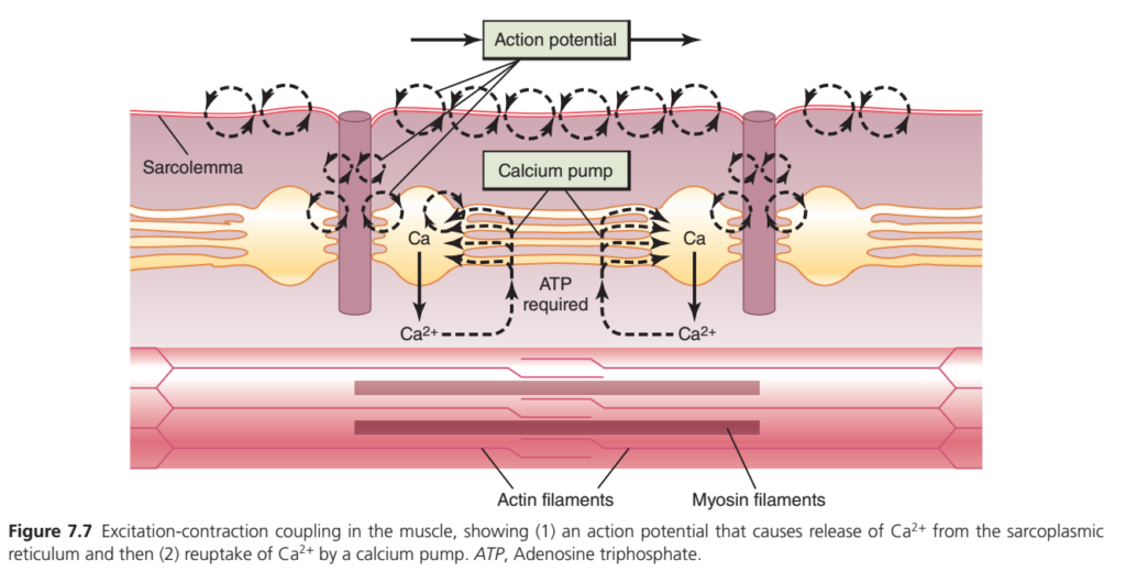
Excitation–Contraction Coupling (Figure 7.7)
Easiest & Most Conceptual Summary
This figure explains how an electrical signal (action potential) is converted into mechanical muscle contraction, and then how the muscle relaxes.
This process is called Excitation–Contraction Coupling (E–C Coupling).
Main Concept
Action Potential → Calcium Released → Actin & Myosin Interact → Muscle Contracts → Calcium Pump Returns Ca²⁺ → Muscle Relaxes
This is the complete cycle of one muscle contraction.
Overall Flow of the Figure
Action Potential
        ↓
Travels over sarcolemma
        ↓
Enters T-tubules
        ↓
Sarcoplasmic reticulum releases Ca²⁺
        ↓
Ca²⁺ binds troponin
        ↓
Actin and myosin interact
        ↓
Muscle contracts
        ↓
Ca²⁺ pump returns Ca²⁺ to SR (uses ATP)
        ↓
Muscle relaxes
Understanding Each Label
1. Action Potential (Starting Signal)
The box labeled Action Potential is the electrical signal that starts muscle contraction.
Where does it come from?
- Brain
- Spinal cord
- Motor neuron
- Neuromuscular junction
The action potential is generated on the muscle membrane after acetylcholine activates the muscle fiber.
Function
It tells the muscle,
“Start contracting!”
2. Sarcolemma
The sarcolemma is the membrane of the muscle fiber.
Function
It carries the action potential rapidly over the surface of the muscle.
The dotted arrows in the figure show the electrical impulse spreading across the sarcolemma.
Concept
Think of the sarcolemma as an electrical wire carrying the signal.
3. T-Tubules (Deep Electrical Pathways)
The vertical tubes extending downward from the sarcolemma are T-tubules.
Function
Carry the electrical signal deep inside the muscle fiber.
Why are they important?
Without T-tubules:
- Only the surface would receive the signal.
- Deep myofibrils would contract late.
With T-tubules:
- Every myofibril receives the signal almost simultaneously.
Easy Concept
They are electrical tunnels carrying the action potential into the center of the muscle.
4. Sarcoplasmic Reticulum (SR)
The yellow network around the T-tubules is the sarcoplasmic reticulum (SR).
Function
Stores large amounts of calcium ions (Ca²⁺).
Important Point
At rest,
almost all calcium is stored inside the SR.
5. Calcium Release
When the action potential reaches the T-tubules,
it signals the SR to open its calcium-release channels.
Result
Large amounts of Ca²⁺ are released into the muscle cytoplasm.
The downward arrows labeled Ca show this calcium release.
Concept
Think of the SR as a water tank, and calcium as the water.
When the valve opens, calcium quickly floods the muscle cell.
6. Calcium Activates Contraction
Released calcium binds to troponin on the thin filament.
This causes:
- Tropomyosin to move away.
- Myosin-binding sites on actin to become exposed.
Now,
myosin heads can attach to actin.
Cross-bridge cycling begins.
Result
The muscle contracts.
7. Actin Filaments
The thin pink filaments at the bottom are actin filaments.
Function
Provide binding sites for myosin during contraction.. Myosin Filaments
The thick dark filaments are myosin filaments.
Function
Pull actin toward the center of the sarcomere.
This shortens the sarcomere and produces contraction.
9. Calcium Pump (SERCA Pump)
After contraction,
the calcium pump transports Ca²⁺ back into the SR.
This pump is called the SERCA (Sarcoplasmic/Endoplasmic Reticulum Ca²⁺-ATPase) pump.
Function
Moves calcium from the cytoplasm back into the SR.
Important Point
This is an active transport process.
It requires energy.
10. ATP Required
The figure clearly states:
ATP required
Why?
The calcium pump cannot work without ATP.
ATP provides the energy needed to pump calcium back into the SR against its concentration gradient.
Result
Cytoplasmic calcium decreases.
11. Muscle Relaxation
When calcium returns to the SR:
- Troponin loses calcium.
- Tropomyosin moves back over the myosin-binding sites on actin.
- Cross-bridge formation stops.
- Muscle relaxes.
Complete Sequence of Excitation–Contraction Coupling
Step 1
Motor neuron stimulates the muscle.
↓
Step 2
A muscle action potential is generated.
↓
Step 3
The action potential spreads over the sarcolemma.
↓
Step 4
The action potential enters the T-tubules.
↓
Step 5
The T-tubules stimulate the sarcoplasmic reticulum.
↓
Step 6
The SR releases Ca²⁺.
↓
Step 7
Calcium binds to troponin.
↓
Step 8
Tropomyosin moves away from actin.
↓
Step 9
Myosin binds to actin.
↓
Step 10
Cross-bridge cycling shortens the sarcomere.
↓
Step 11
Muscle contracts.
↓
Step 12
SERCA pumps Ca²⁺ back into the SR (requires ATP).
↓
Step 13
Troponin releases calcium.
↓
Step 14
Tropomyosin again blocks the binding sites.
↓
Step 15
Muscle relaxes.
Easy Flow Diagram
Motor neuron
      ↓
Muscle action potential
      ↓
Sarcolemma
      ↓
T-tubules
      ↓
Sarcoplasmic reticulum
      ↓
Ca²⁺ released
      ↓
Ca²⁺ binds troponin
      ↓
Actin + Myosin interact
      ↓
Muscle contraction
      ↓
SERCA pump uses ATP
      ↓
Ca²⁺ returns to SR
      ↓
Muscle relaxation
Simple Real-Life Analogy
Imagine a factory with automatic doors.
- Action potential = Factory manager presses the start button.
- T-tubules = Electrical wiring carrying the signal inside the factory.
- Sarcoplasmic reticulum = Warehouse storing calcium.
- Calcium = Workers released from the warehouse.
- Actin & myosin = Machines that begin working.
- SERCA pump = Bus that takes the workers back to the warehouse after the job is finished.
- ATP = Fuel for the bus.
When the workers return to the warehouse, the machines stop, and the factory rests.
Important Points from Figure 7.7
- Excitation–contraction coupling links an electrical signal to mechanical contraction.
- A muscle action potential spreads over the sarcolemma and travels into the fiber through the T-tubules.
- The T-tubules stimulate the sarcoplasmic reticulum (SR) to release Ca²⁺ into the cytoplasm.
- Calcium binds to troponin, allowing actin and myosin to interact and produce muscle contraction.
- After contraction, the SERCA (Ca²⁺-ATPase) pump actively transports calcium back into the SR.
- ATP is required for calcium reuptake into the SR.
- Removal of calcium from the cytoplasm stops actin–myosin interaction, allowing the muscle to relax.
KEY CONCEPT (Figure 7.7)
Excitation–contraction coupling is the process by which a muscle action potential is converted into muscle contraction. The action potential spreads along the sarcolemma and through the T-tubules, triggering the sarcoplasmic reticulum to release Ca²⁺. Calcium binds to troponin, exposing binding sites on actin so that myosin can form cross-bridges and shorten the sarcomere, producing contraction. When the contraction is complete, the SERCA calcium pump uses ATP to transport calcium back into the sarcoplasmic reticulum. As cytoplasmic calcium falls, actin–myosin interaction stops, and the muscle relaxes. This coordinated sequence ensures rapid, efficient, and reversible skeletal muscle contraction.
Malignant Hyperthermia
- Malignant hyperthermia is a hypermetabolic crisis that can occur in susceptible individuals during anesthesia.
- It may be triggered by certain anesthetic drugs, including:
- Halothane
- Isoflurane
- Succinylcholine
- At least six genetic mutations have been identified that greatly increase the risk of developing malignant hyperthermia.
- These mutations mainly affect the genes for:
- Ryanodine receptors (RyR)
- Dihydropyridine receptors (DHPR)
- The exact mechanism by which anesthetic drugs interact with these abnormal receptors is not fully understood.
- However, these mutations cause uncontrolled release of Ca²⁺ from the sarcoplasmic reticulum into the muscle cell.
- The excess Ca²⁺ causes continuous and excessive contraction of skeletal muscle fibers.
- These sustained muscle contractions greatly increase the metabolic rate.
- The increased metabolism:
- Produces a large amount of heat.
- Causes cellular acidosis.
- Depletes the body’s energy stores.
- Symptoms of malignant hyperthermia include:
- Muscle rigidity
- High fever
- Rapid heart rate
- In severe cases, additional complications may occur, including:
- Rapid breakdown of skeletal muscle (rhabdomyolysis)
- High blood potassium (hyperkalemia) due to release of large amounts of potassium from damaged muscle cells
- Treatment of malignant hyperthermia includes:
- Rapid cooling of the patient
- Administration of dantrolene
- Dantrolene blocks the action of ryanodine receptors.
- As a result, Ca²⁺ release from the sarcoplasmic reticulum decreases.
- This reduces muscle contraction and helps control the condition.
KEY CONCEPT
- Malignant hyperthermia is a life-threatening hypermetabolic crisis triggered by certain anesthetics (halothane, isoflurane) or succinylcholine in genetically susceptible individuals.
- Mutations of ryanodine receptor (RyR) or dihydropyridine receptor (DHPR) genes cause uncontrolled Ca²⁺ release from the sarcoplasmic reticulum.
- Excess Ca²⁺ produces sustained muscle contraction, increased metabolic rate, heat production, cellular acidosis, and depletion of energy stores.
- Major symptoms are muscle rigidity, high fever, and rapid heart rate.
- Severe complications include rhabdomyolysis and hyperkalemia.
- Treatment consists of rapid cooling and dantrolene, which inhibits Ca²⁺ release by blocking ryanodine receptors.
MADE BY SELF LEARNING DR SHEEN CEO AND FOUNDER

So informative.. Thank you so much sir
It’s very easy to understand……Thank you so much sir