


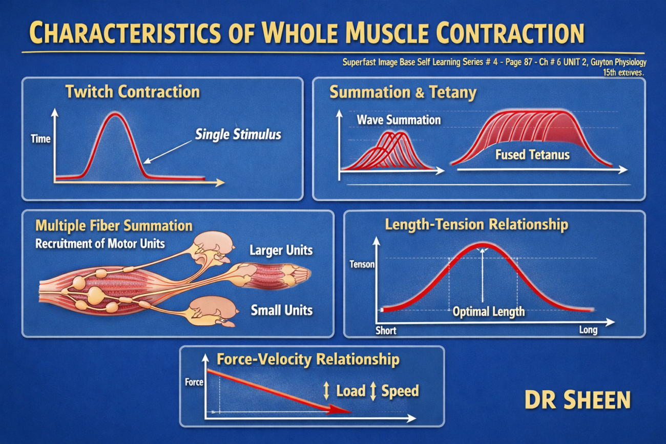
Isometric Contractions Do Not Shorten Muscle, Whereas Isotonic Contractions Shorten Muscle at a Constant Tension
- Many features of muscle contraction can be studied by producing a single muscle twitch.
- A single muscle twitch is a single, brief muscle contraction that lasts only a fraction of a second.
- A single muscle twitch can be produced by:
- Applying an electrical stimulus to the nerve supplying the muscle, or
- Applying a short electrical stimulus directly to the muscle.
- Muscle contraction is called isometric contraction when:
- The muscle develops tension.
- The muscle does not shorten during contraction.
- Muscle contraction is called isotonic contraction when:
- The muscle shortens during contraction.
- The tension remains constant throughout the contraction.
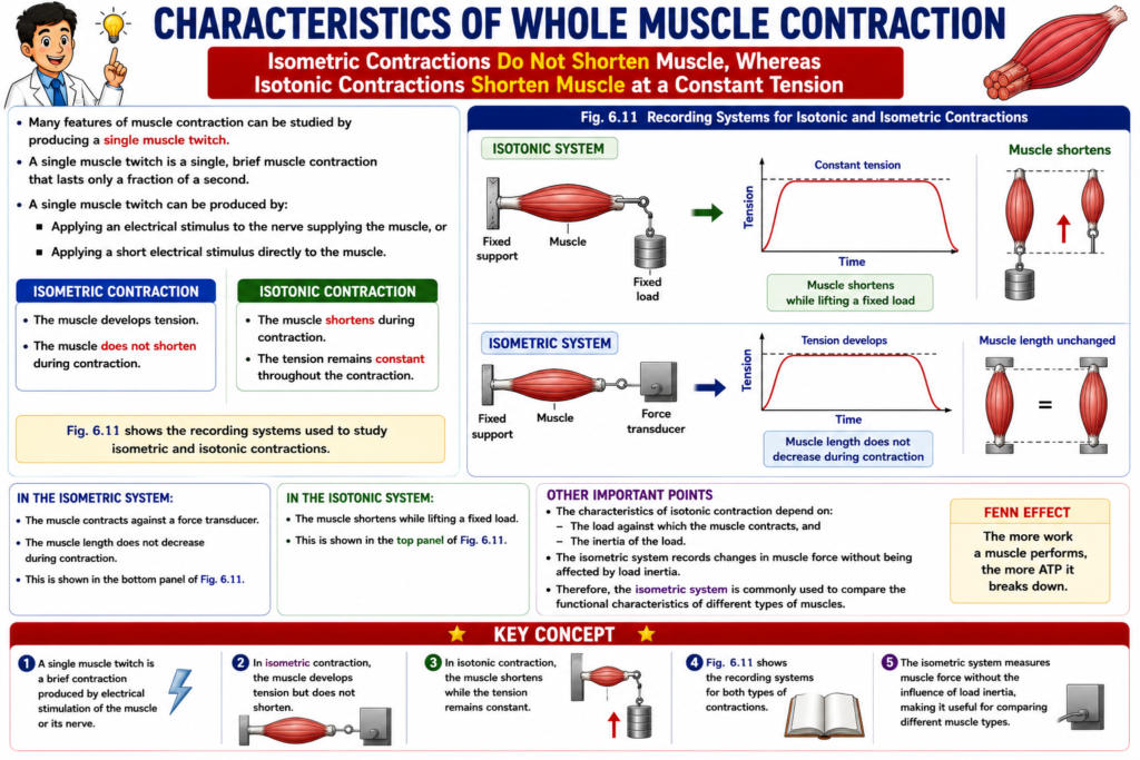
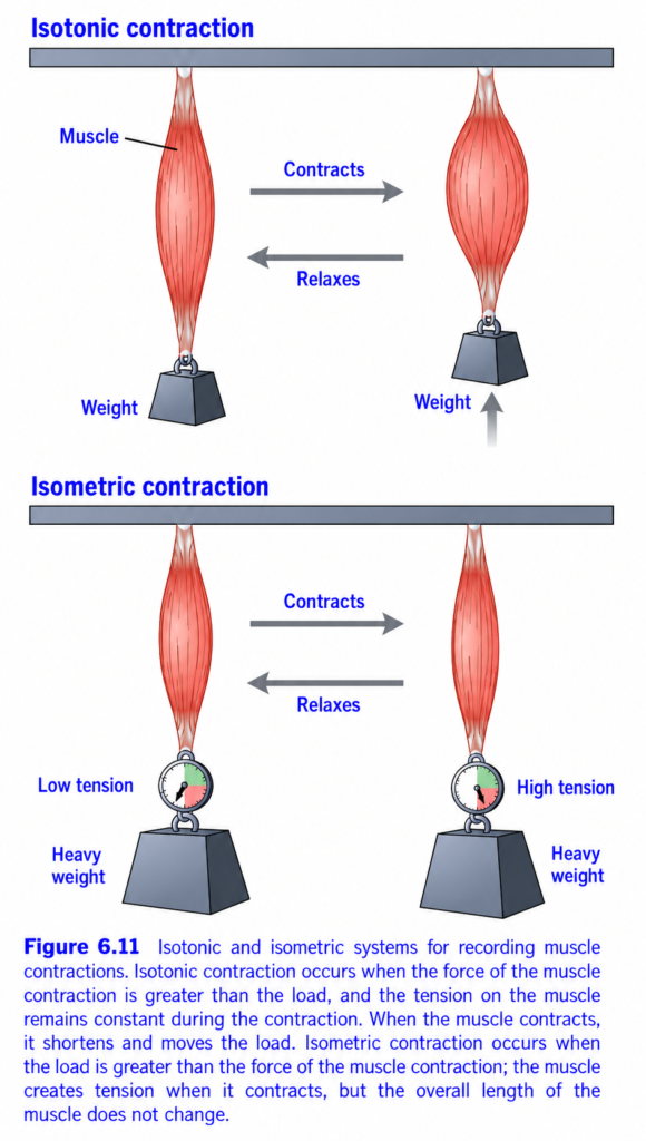
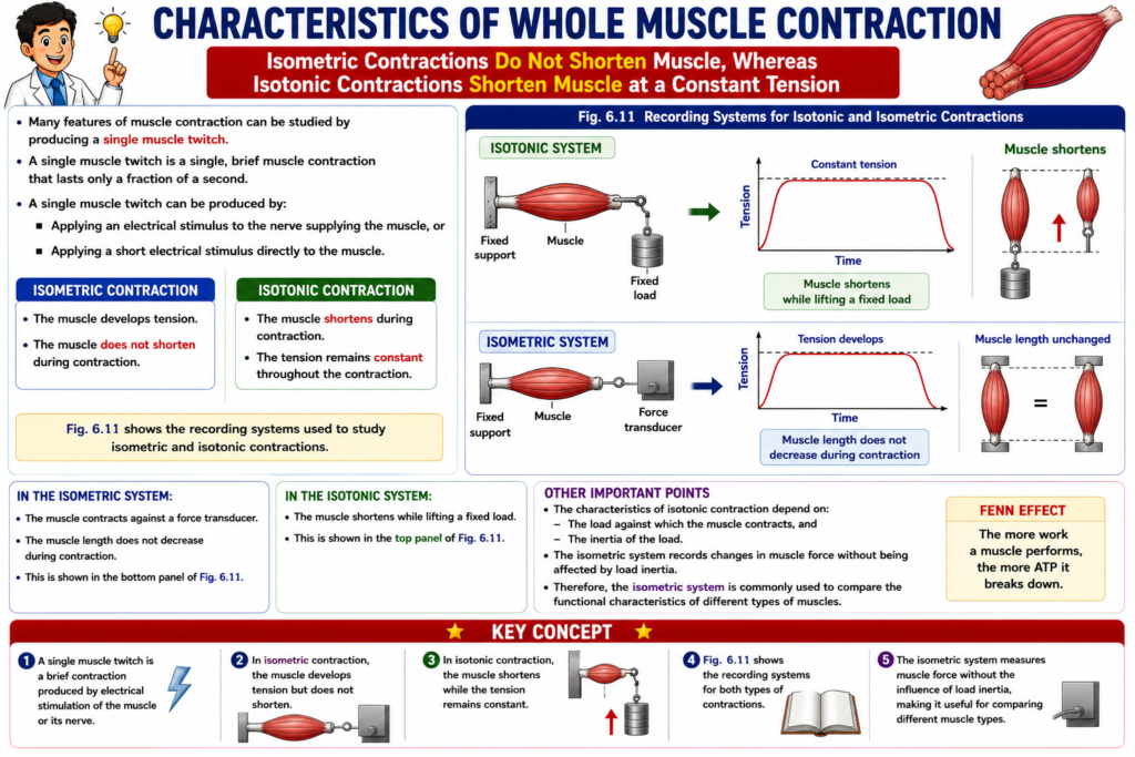
- Fig. 6.11 shows the recording systems used to study isometric and isotonic contractions.
- In the isometric system:
- The muscle contracts against a force transducer.
- The muscle length does not decrease during contraction.
- This is shown in the bottom panel of Fig. 6.11.
- In the isotonic system:
- The muscle shortens while lifting a fixed load.
- This is shown in the top panel of Fig. 6.11.
- The characteristics of isotonic contraction depend on:
- The load against which the muscle contracts, and
- The inertia of the load.
- The isometric system records changes in muscle force without being affected by load inertia.
- Therefore, the isometric system is commonly used to compare the functional characteristics of different types of muscles.
KEY CONCEPT
- A single muscle twitch is a brief contraction produced by electrical stimulation of the muscle or its nerve.
- In isometric contraction, the muscle develops tension but does not shorten.
- In isotonic contraction, the muscle shortens while the tension remains constant.
- Fig. 6.11 shows the recording systems for both types of contractions.
- The isometric system measures muscle force without the influence of load inertia, making it useful for comparing different muscle types.
Isotonic vs Isometric Muscle Contraction (Figure 6.11) – Easy Conceptual Summary
This figure compares the two basic types of skeletal muscle contraction:
- Isotonic Contraction (Muscle shortens and moves the load)
- Isometric Contraction (Muscle develops tension but does not shorten)
This is one of the most important concepts in muscle physiology and is frequently asked in MBBS examinations.
Basic Concept
When a muscle contracts, two things can change:
- Muscle length
- Muscle tension (force)
Depending on which one changes, muscle contraction is classified into:
PART A – Isotonic Contraction
The upper part of the figure shows isotonic contraction.
What is Isotonic Contraction?
Definition
Isotonic contraction is a contraction in which:
- Muscle shortens
- The load moves
- Muscle tension remains nearly constant
Understanding the Figure
Before Contraction
- The muscle is long.
- A weight is hanging from it.
- The weight is stationary.
During Contraction
The muscle contracts.
The muscle becomes shorter.
Because the muscle force becomes greater than the weight, the load is lifted upward.
The arrow beside the weight shows:
➡️ Weight moves upward.During Relaxation
When the muscle relaxes,
It lengthens again.
The weight returns downward.
Why does the muscle shorten?
The muscle produces enough force to overcome the external load.
Since:
Muscle force > Load
The muscle shortens and lifts the weight.
Easy Concept
Imagine lifting a water bottle.
Your biceps shortens.
The bottle moves upward.
This is an isotonic contraction.
Examples of Isotonic Contraction
- Lifting a book
- Walking
- Running
- Climbing stairs
- Picking up a glass
- Flexing the elbow
Key Point
Length changes.
Tension remains almost constant.
PART B – Isometric Contraction
The lower part of the figure shows isometric contraction.
What is Isometric Contraction?
Definition
Isometric contraction is a contraction in which:
- Muscle length does not change
- The load does not move
- Muscle tension increases
Understanding the Figure
Before Contraction
The muscle supports a heavy weight.
The tension gauge shows:
Low tension
During Contraction
The muscle contracts.
However,
The weight is too heavy to lift.
The muscle does not shorten.
Instead,
The tension gauge shows:
High tensionWhy doesn’t the weight move?
Because:
Load > Muscle force
The muscle develops force,
but the force is insufficient to lift the weight.
Therefore,
Only tension increases.
Muscle length remains the same.
Easy Concept
Imagine pushing against a brick wall.
Your muscles become tight.
You exert great force.
But the wall does not move.
Your muscles do not shorten.
This is isometric contraction.
Examples of Isometric Contraction
- Pushing against a wall
- Holding a heavy suitcase without moving it
- Standing upright (postural muscles)
- Carrying a heavy object without lifting it
- Plank exercise
Key Point
Length remains constant.
Tension increases.
Understanding the Tension Gauge
The tension gauge is shown only in the isometric contraction diagram.
Before Contraction
The gauge shows:
Low tension
After Contraction
The gauge shows:
High tension
This means:
The muscle is producing much more force,
even though its length has not changed.
Why Are the Two Contractions Different?
The difference depends on the relationship between:
- Muscle force
- External load
If Muscle Force > Load
The muscle shortens.
The load moves.
➡️ Isotonic contractionIf Muscle Force < Load
The muscle cannot shorten.
Only tension increases.
➡️ Isometric contraction
Comparison of Isotonic and Isometric Contraction
| Feature | Isotonic Contraction | Isometric Contraction |
|---|---|---|
| Muscle length | ↓ Shortens | No change |
| Muscle tension | Nearly constant | ↑ Increases |
| Load movement | Yes | No |
| Muscle force | Greater than the load | Less than or equal to the load |
| Mechanical work | Yes | No external mechanical work |
Daily Life Examples
Isotonic
Isometric
- Holding a heavy bag without moving
- Pushing a car that won’t move
- Holding a yoga plank
- Standing still
Clinical Importance
Isotonic Exercise
Improves:
- Muscle strength
- Joint movement
- Functional mobility
Examples:sometric Exercise
Improves:
- Muscle strength
- Postural stability
Used during rehabilitation when joint movement should be minimized.
Examples:
- Wall sit
- Static quadriceps exercises
- Plank
Quick Memory Table
| Feature | Isotonic | Isometric |
|---|---|---|
| Muscle shortens | ✔ Yes | ✘ No |
| Muscle tension increases | Slightly | ✔ Markedly |
| Weight moves | ✔ Yes | ✘ No |
| Muscle length changes | ✔ Yes | ✘ No |
| External work performed | ✔ Yes | ✘ No |
Easy Memory Trick
IsoTONIC = “Tone stays almost constant”
- Tension ≈ Constant
- Muscle shortens
- Weight moves
Think: “TONIC = Travels” → the load travels because the muscle shortens.
IsoMETRIC = “Measurements stay the same”
- Length stays the same
- Tension increases
- Weight does not move
Think: “METRIC = Measurement” → the muscle’s length measurement remains unchanged.
Relationship with the Previous Load–Velocity Graph
- Light or moderate load → Muscle can shorten → Isotonic contraction
- Load equals maximum muscle force → Shortening velocity becomes zero → Isometric contraction
Thus, Figure 6.11 illustrates the same principle shown in the previous load–velocity relationship graph.Key Concept
Muscle contraction can occur as either isotonic or isometric contraction depending on the relationship between muscle force and the external load. In isotonic contraction, the muscle generates a force greater than the load, allowing it to shorten and move the weight, while muscle tension remains relatively constant. This type of contraction performs external mechanical work and is seen during activities such as lifting objects and walking. In isometric contraction, the external load is greater than or equal to the force generated by the muscle, so the muscle develops tension without changing its length, and the load does not move. Although no external work is performed, muscle force increases substantially. Therefore, isotonic contraction changes muscle length, whereas isometric contraction changes muscle tension without changing muscle length.
Characteristics of Isometric Twitches Recorded From Different Muscles
- The human body contains skeletal muscles of many different sizes.
- The stapedius muscle in the middle ear is very small, measuring only a few millimeters in length and about 1 millimeter in diameter.
- The quadriceps muscle is very large, being about half a million times larger than the stapedius muscle.
- Muscle fibers also vary greatly in size.
- Some muscle fibers are about 10 micrometers in diameter, whereas others are about 80 micrometers in diameter.
- The energetics of muscle contraction differ considerably from one muscle to another.
- Therefore, the mechanical characteristics of muscle contraction also differ among different muscles.
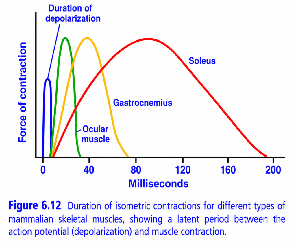
- Fig. 6.12 shows isometric contractions of three different skeletal muscles.
- The ocular muscle has an isometric contraction lasting less than 1/50 second.
- The gastrocnemius muscle has an isometric contraction lasting about 1/15 second.
- The soleus muscle has an isometric contraction lasting about 1/5 second.
- These different contraction durations are adapted to the specific functions of each muscle.
- The ocular muscles contract extremely rapidly.
- This rapid contraction helps keep the eyes fixed on specific objects, providing accurate vision.
- The gastrocnemius muscle contracts at a moderate speed.
- This provides sufficient speed for limb movements during running and jumping.
- The soleus muscle contracts slowly.
- It is mainly responsible for continuous, long-term support of the body against gravity.
KEY CONCEPT
- Different skeletal muscles vary greatly in size, muscle fiber diameter, and energy metabolism.
- Therefore, the mechanical characteristics of muscle contraction differ among muscles.
- Fig. 6.12 shows that the ocular muscle contracts in less than 1/50 second, the gastrocnemius muscle in about 1/15 second, and the soleus muscle in about 1/5 second.
- These contraction speeds are adapted to each muscle’s function: rapid eye movements, moderate-speed limb movements, and slow, sustained postural support.
Duration of Isometric Contractions in Different Skeletal Muscles (Figure 6.12) – Easy Conceptual Summary
This figure compares how long a single isometric muscle contraction (muscle twitch) lasts in different skeletal muscles.
It also shows an important concept:
A muscle action potential (depolarization) is very short, but the muscle contraction lasts much longer.
The graph compares three muscles:
- Ocular muscle (Eye muscle) – Fastest contraction
- Gastrocnemius muscle (Calf muscle) – Intermediate contraction
- Soleus muscle (Postural muscle) – Slowest contraction
Basic Concept
A muscle twitch is the response of a muscle to one action potential.
A muscle twitch has three phases:
- Latent period
- Contraction period
- Relaxation period
Understanding the Axes
X-axis (Milliseconds)
Shows time after stimulation.
Moving to the right means:
➡️ More time has passed.
Y-axis (Force of Contraction)
Shows the amount of force produced by the muscle.
Higher curve
➡️ Greater force
Lower curve
➡️ Less force
Understanding the Blue Curve (Depolarization)
The small blue spike at the beginning represents:
Action Potential (Depolarization)
What happens?
The muscle membrane is stimulated.
An action potential is generated.
The action potential lasts only 2–5 milliseconds.
Why is it so short?
Depolarization is simply an electrical signal.
Its purpose is to trigger muscle contraction.
It does not represent the contraction itself.
Easy Concept
Think of a switch turning on a fan.
Pressing the switch takes only a second.
The fan continues spinning much longer.
Similarly,
The action potential is very short,
but it starts a much longer muscle contraction.
Key Point
Electrical activity is much shorter than mechanical contraction.
What is the Latent Period?
Between the action potential and the beginning of muscle contraction,
there is a short delay.
This delay is called the:
Latent Period
Why does it occur?
Several events must happen before the muscle can contract:
- Action potential travels along the sarcolemma.
- Action potential enters the T-tubules.
- Sarcoplasmic reticulum releases Ca²⁺.
- Calcium binds to troponin.
- Tropomyosin moves away.
- Cross-bridges begin to form.
Only then does contraction start.
Easy Concept
Imagine pressing the start button on a car.
The engine does not move the car instantly.
A few events occur before the car starts moving.
That short delay is the latent period.
Understanding Each Muscle Curve
1. Green Curve — Ocular Muscle
(Eye muscles)
What happens?
This muscle contracts and relaxes very quickly.
The entire twitch lasts only about:
20–30 milliseconds
Why?
Eye muscles must move rapidly.
Examples:
- Reading
- Tracking moving objects
- Rapid eye movements
They contain many fast-twitch fibers (Type II fibers).
These fibers:
- Release calcium quickly.
- Reabsorb calcium quickly.
- Have rapid cross-bridge cycling.
Therefore,
The twitch is very short.
Easy Concept
Eye muscles are like a sports car:
- Start quickly
- Move quickly
- Stop quickly
Key Point
Ocular muscles have the shortest twitch duration.
2. Yellow Curve — Gastrocnemius Muscle
(Calf muscle)
What happens?
The contraction lasts about:
40–60 milliseconds
Why?
The gastrocnemius performs rapid but powerful movements such as:
- Running
- Jumping
- Climbing stairs
Its contraction is slower than eye muscles but faster than postural muscles.
It contains a mixture of:
- Fast-twitch fibers
- Slow-twitch fibers
Easy Concept
The gastrocnemius is like a family car:
Fast enough,
but not as fast as a sports car.
Key Point
Intermediate twitch duration.
3. Red Curve — Soleus Muscle
(Postural muscle)
What happens?
The contraction lasts about:
100–200 milliseconds
This is the longest twitch shown in the graph.
Why?
The soleus helps maintain:
- Standing posture
- Balance
- Long-term muscle tone
It contains mainly slow-twitch (Type I) fibers.
These fibers:
- Release calcium slowly.
- Reabsorb calcium slowly.
- Have slower cross-bridge cycling.
Therefore,
The contraction lasts much longer.
Easy Concept
The soleus is like a diesel engine.
It starts slowly,
but it works for a long time without tiring.
Key Point
Soleus has the longest twitch duration.
Why Are Different Muscles Different?
The duration of contraction depends mainly on:
1. Type of Muscle Fibers
Fast fibers:
- Contract rapidly.
- Relax rapidly.
Slow fibers:
- Contract slowly.
- Relax slowly.
2. Calcium Handling
Fast muscles:
- Release Ca²⁺ rapidly.
- Pump Ca²⁺ back rapidly.
Slow muscles:
- Release Ca²⁺ slowly.
- Remove Ca²⁺ slowly.
3. ATPase Activity
Fast fibers have:
High myosin ATPase activity.
Cross-bridge cycling is rapid.
Slow fibers have:
Low ATPase activity.
Cross-bridge cycling is slower.
Comparison of the Three Muscles
| Muscle | Twitch Duration | Fiber Type | Function |
|---|---|---|---|
| Ocular muscle | 20–30 ms | Mostly fast-twitch (Type II) | Rapid eye movements |
| Gastrocnemius | 40–60 ms | Mixed fast and slow fibers | Running, jumping |
| Soleus | 100–200 ms | Mostly slow-twitch (Type I) | Standing, posture |
Clinical Importance
Ocular Muscles
Need extremely rapid contractions to:
- Track moving objects.
- Maintain accurate vision.
Gastrocnemius
Provides:
- Speed
- Power
- Explosive movement
Useful for sprinting and jumping.
Soleus
Provides:
- Continuous muscle tone.
- Postural support.
- Resistance to fatigue.
Allows prolonged standing without tiring quickly.
Quick Memory Table
| Muscle | Speed | Fatigue Resistance | Main Function |
|---|---|---|---|
| Ocular | Fastest | Low | Eye movements |
| Gastrocnemius | Intermediate | Moderate | Running, jumping |
| Soleus | Slowest | Highest | Standing, posture |
Easy Memory Trick
Eye = Instant ⚡
- Fastest contraction
- Shortest twitch
Gastrocnemius = Athlete 🏃
- Medium speed
- Powerful movement
Soleus = Soldier 🪖
- Slow contraction
- Works all day
- Resists fatigue
Key Concept
This figure demonstrates that the electrical event (action potential or depolarization) in skeletal muscle lasts only 2–5 milliseconds, whereas the mechanical contraction (muscle twitch) lasts much longer. Between the action potential and the onset of contraction is the latent period, during which excitation–contraction coupling occurs: the action potential spreads along the sarcolemma and T-tubules, calcium is released from the sarcoplasmic reticulum, calcium binds to troponin, and cross-bridges begin to form. The duration of the twitch varies among muscles according to their function. Ocular muscles, composed mainly of fast-twitch (Type II) fibers, have the shortest twitch because they require rapid eye movements. The gastrocnemius has an intermediate-duration twitch, allowing both speed and power for activities such as running and jumping. The soleus, composed predominantly of slow-twitch (Type I) fibers, has the longest twitch, enabling sustained postural activity and excellent fatigue resistance. Thus, fast muscles contract briefly for speed, whereas slow muscles contract for longer to maintain posture and endurance.
Fast Versus Slow Muscle Fibers
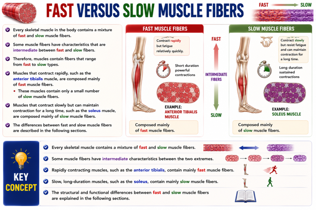
- Every skeletal muscle in the body contains a mixture of fast and slow muscle fibers.
- Some muscle fibers have characteristics that are intermediate between fast and slow fibers.
- Therefore, muscles contain fibers that range from fast to slow types.
- Muscles that contract rapidly, such as the anterior tibialis muscle, are composed mainly of fast muscle fibers.
- These muscles contain only a small number of slow muscle fibers.
- Muscles that contract slowly but can maintain contraction for a long time, such as the soleus muscle, are composed mainly of slow muscle fibers.
- The differences between fast and slow muscle fibers are described in the following sections.
KEY CONCEPT
- Every skeletal muscle contains a mixture of fast and slow muscle fibers.
- Some muscle fibers have intermediate characteristics between the two extremes.
- Rapidly contracting muscles, such as the anterior tibialis, contain mainly fast muscle fibers.
- Slow, long-duration muscles, such as the soleus, contain mainly slow muscle fibers.
- The structural and functional differences between fast and slow muscle fibers are explained in the following sections.
Slow Fibers (Type 1, Red Muscle)
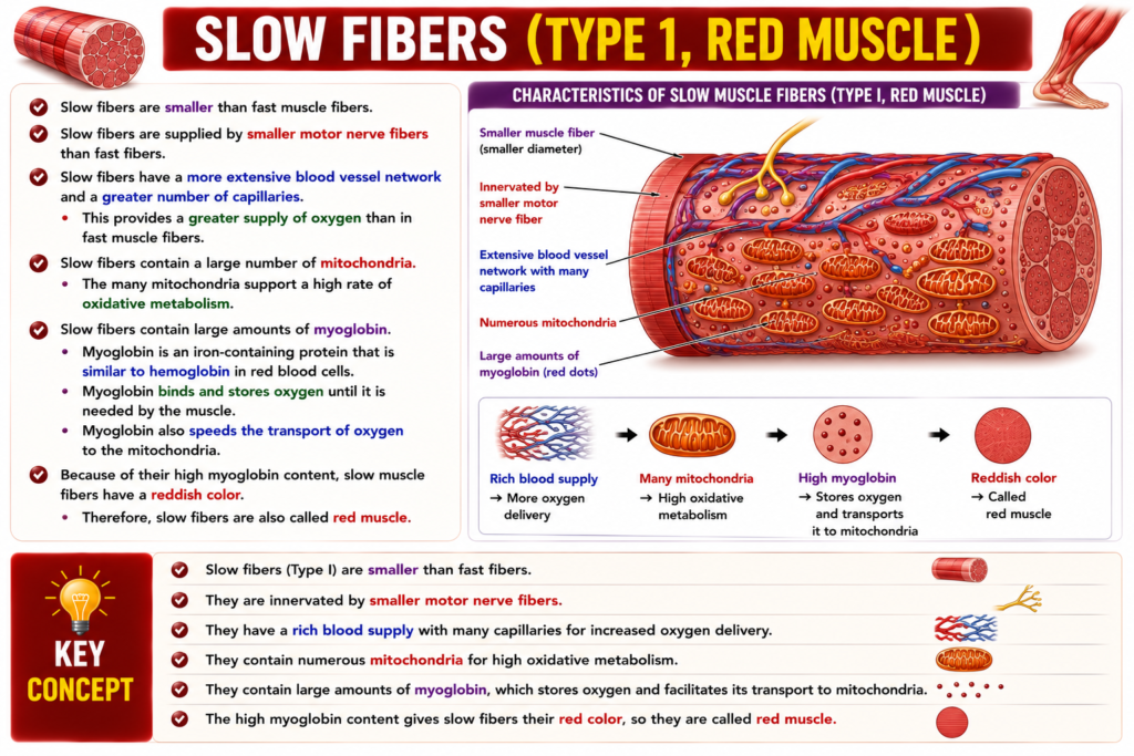
- Slow fibers are smaller than fast muscle fibers.
- Slow fibers are supplied by smaller motor nerve fibers than fast fibers.
- Slow fibers have a more extensive blood vessel network and a greater number of capillaries.
- This provides a greater supply of oxygen than in fast muscle fibers.
- Slow fibers contain a large number of mitochondria.
- The many mitochondria support a high rate of oxidative metabolism.
- Slow fibers contain large amounts of myoglobin.
- Myoglobin is an iron-containing protein that is similar to hemoglobin in red blood cells.
- Myoglobin binds and stores oxygen until it is needed by the muscle.
- Myoglobin also speeds the transport of oxygen to the mitochondria.
- Because of their high myoglobin content, slow muscle fibers have a reddish color.
- Therefore, slow fibers are also called red muscle.
KEY CONCEPT
- Slow fibers (Type I) are smaller than fast fibers.
- They are innervated by smaller motor nerve fibers.
- They have a rich blood supply with many capillaries for increased oxygen delivery.
- They contain numerous mitochondria for high oxidative metabolism.
- They contain large amounts of myoglobin, which stores oxygen and facilitates its transport to mitochondria.
- The high myoglobin content gives slow fibers their red color, so they are called red muscle.
Fast Fibers (Type II, White Muscle)
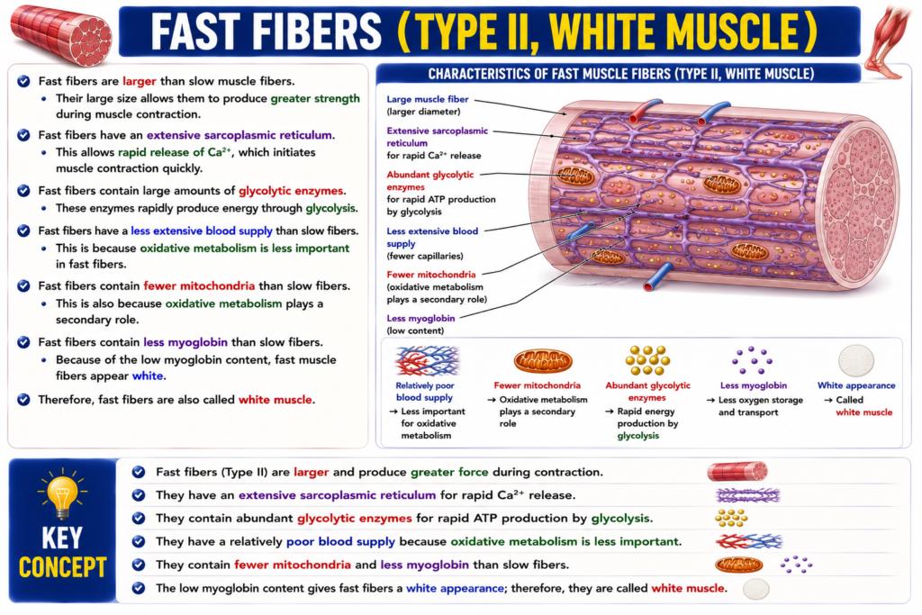
- Fast fibers are larger than slow muscle fibers.
- Their large size allows them to produce greater strength during muscle contraction.
- Fast fibers have an extensive sarcoplasmic reticulum.
- This allows rapid release of Ca²⁺, which initiates muscle contraction quickly.
- Fast fibers contain large amounts of glycolytic enzymes.
- These enzymes rapidly produce energy through glycolysis.
- Fast fibers have a less extensive blood supply than slow fibers.
- This is because oxidative metabolism is less important in fast fibers.
- Fast fibers contain fewer mitochondria than slow fibers.
- This is also because oxidative metabolism plays a secondary role.
- Fast fibers contain less myoglobin than slow fibers.
- Because of the low myoglobin content, fast muscle fibers appear white.
- Therefore, fast fibers are also called white muscle.
KEY CONCEPT
- Fast fibers (Type II) are larger and produce greater force during contraction.
- They have an extensive sarcoplasmic reticulum for rapid Ca²⁺ release.
- They contain abundant glycolytic enzymes for rapid ATP production by glycolysis.
- They have a relatively poor blood supply because oxidative metabolism is less important.
- They contain fewer mitochondria and less myoglobin than slow fibers.
- The low myoglobin content gives fast fibers a white appearance; therefore, they are called white muscle.
PREPARE AND MADE BY SELF LEARNING DR SHEEN
