


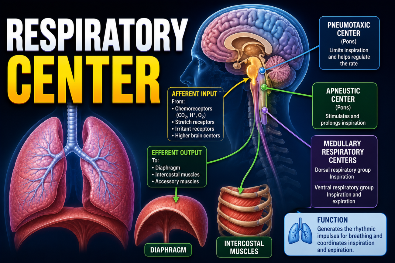
- The respiratory center consists of several groups of neurons.
- These neurons are located on both sides (bilaterally) in the medulla oblongata and pons of the brain stem.
- Fig. 42.1 shows the location of the respiratory center.
- The respiratory center is divided into three major groups of neurons:
- Dorsal respiratory group (DRG):
- Located in the dorsal part of the medulla.
- Mainly controls inspiration (breathing in).
- Ventral respiratory group (VRG):
- Located in the ventrolateral part of the medulla.
- Contains both inspiratory and expiratory neurons.
- Pneumotaxic center:
- Located dorsally in the upper part of the pons.
- Mainly controls the rate and depth of breathing.
- Dorsal respiratory group (DRG):
- The respiratory center generates and maintains the normal rhythm of breathing.
- It also adjusts breathing according to the body’s physiological needs.
- These adjustments are influenced by signals from:
- Peripheral chemoreceptors
- Mechanoreceptors
- Hormonal changes
- Other brain regions, including:
- Cerebral cortex
- Hypothalamus
- Together, these signals help regulate the rate and depth of respiration.
Fig. 42.1
- Respiratory center is located in the brain stem.
- It has three main parts:
- Dorsal Respiratory Group (DRG) → Inspiration
- Ventral Respiratory Group (VRG) → Inspiration + Expiration
- Pneumotaxic Center → Controls rate and depth of breathing
KEY CONCEPT
- The respiratory center is located in the medulla oblongata and pons of the brain stem. It consists of the dorsal respiratory group (inspiration), ventral respiratory group (inspiration and expiration), and pneumotaxic center (controls breathing rate and depth). Together, these centers generate the breathing rhythm and adjust respiration according to signals from receptors, hormones, and higher brain centers.
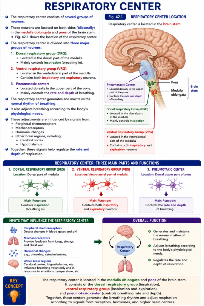
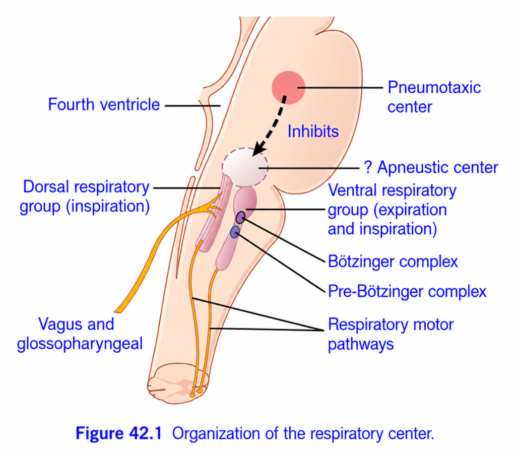
Figure 42.1: Organization of the Respiratory Center (Guyton) – Easiest Conceptual Summary for MBBS Students
🎯 One-Line Concept
Think of the respiratory center as the brain’s automatic breathing control room.
It continuously answers three questions:
Easy Memory Trick
Imagine a car:
- 🚗 DRG = Accelerator → Starts inspiration
- 🛑 Pneumotaxic Center = Brake → Stops inspiration
- ⚙ VRG = Extra Engine → Works during forceful breathing
- ❤️ Pre-Bötzinger Complex = Ignition Key → Creates breathing rhythm
1. Dorsal Respiratory Group (DRG)
Location: Dorsal medulla
Main Function
➡ Produces normal inspiration
It sends rhythmic nerve impulses to:
- Diaphragm
- External intercostal muscles
These muscles contract →
Chest expands →
Air enters lungs.
Easy Concept
Think:
DRG = Normal Breathing Generator
It works automatically even while sleeping.
Remember
DRG = Inspiration only. Ventral Respiratory Group (VRG)
Location: Ventrolateral medulla
Function
Normally almost silent during quiet breathing.
Becomes active during:
- Exercise
- Running
- Heavy breathing
- Coughing
It controls both:
- Inspiration
- Expiration
Especially forceful expiration.
Easy Concept
Imagine DRG is enough for walking.
But during sprinting…
VRG comes to help.
3. Pre-Bötzinger Complex
This tiny group of neurons is the natural pacemaker of breathing.
It automatically generates rhythmic electrical signals.
Without it,
No breathing rhythm.
Easy Concept
Like the SA node of the heart
↓
Pre-Bötzinger Complex
↓
SA Node
Both create automatic rhythms.
Memory
4. Bötzinger Complex
Mainly contains neurons that help produce expiration and inhibit inspiratory neurons when needed.
Easy Concept
Think of it as
“Turning inspiration OFF”
so expiration can occur smoothly.5. Pneumotaxic Center (Pontine Respiratory Group)
Location: Upper pons
Function
Stops inspiration.
It sends inhibitory signals to inspiratory neurons.
This prevents lungs from over-expanding.
Easy Concept
Imagine filling a balloon.
If nobody says “Stop”
the balloon bursts.
The pneumotaxic center says:
If Pneumotaxic Center is Strong
Stops inspiration early
↓
Short inspiration
↓
Rapid breathing
If Weak
Inspiration lasts longer
↓
Large tidal volume
↓
Slow breathing
Easy Memory
Pneumotaxic = “Stop Breathing In”
6. Apneustic Center (Lower Pons)
Guyton places a question mark because its exact role is less certain.
Proposed Function
Stimulates inspiration
Makes inspiration longer and deeper.
Normally,
its activity is inhibited by the pneumotaxic center.
Easy Concept
Think of it as saying:
“Keep breathing in…”
while the pneumotaxic center says:
“Stop now.”
Normal breathing is the balance between these two influences. Vagus and Glossopharyngeal Nerves
These nerves bring sensory information from the lungs and body to the respiratory center.
They report:
- Lung stretch
- Oxygen level
- Carbon dioxide level
- Blood pH
The respiratory center then adjusts breathing accordingly.
Easy Concept
Think of them as sensors that continuously update the brain.
8. Respiratory Motor Pathways
After the brain decides how to breathe,
signals travel through motor nerves to respiratory muscles.
Main muscles:
- Diaphragm
- External intercostals
- Accessory muscles (during forceful breathing)
Flow of Normal Breathing (Super Easy)
Pre-Bötzinger Complex
          ↓
Generates breathing rhythm
          ↓
DRG starts inspiration
          ↓
Diaphragm contracts
          ↓
Air enters lungs
          ↓
Pneumotaxic center stops inspiration
          ↓
Expiration occurs
          ↓
Cycle repeats automatically
During Exercise
Exercise
      ↓
VRG activated
      ↓
Accessory muscles contract
      ↓
Forceful inspiration
      ↓
Forceful expiration
      ↓
Breathing becomes deeper and faster
Clinical Correlation
Damage to DRG
- Abnormal or absent normal inspiratory rhythm.
Damage to Pneumotaxic Center
- Prolonged inspiration (apneustic breathing).
- Slow, deep breaths.
Damage to Pre-Bötzinger Complex
- Severe disturbance or loss of automatic breathing rhythm, which can be life-threatening.
High-Yield MBBS Summary Table
| Structure | Location | Main Function | Easy Memory |
|---|---|---|---|
| Dorsal Respiratory Group (DRG) | Dorsal medulla | Generates quiet inspiration | Accelerator |
| Ventral Respiratory Group (VRG) | Ventral medulla | Forceful inspiration & expiration | Extra engine during exercise |
| Pre-Bötzinger Complex | Medulla | Generates breathing rhythm | Pacemaker of breathing |
| Bötzinger Complex | Medulla | Helps expiration; inhibits inspiration | Turns inspiration OFF |
| Pneumotaxic Center | Upper pons | Stops inspiration | Brake |
| Apneustic Center | Lower pons | Promotes prolonged inspiration | Keeps inspiration ON |
| Vagus & Glossopharyngeal Nerves | Sensory input | Carry feedback from lungs and blood | Sensors |
| Respiratory Motor Pathways | Descending pathways | Activate respiratory muscles | Output wires |
🧠 Final Memory Story (30 Seconds)
Imagine breathing is driving a car:
- ❤️ Pre-Bötzinger Complex = turns the engine ON (creates the rhythm).
- 🚗 DRG = presses the accelerator, starting inspiration.
- 🛑 Pneumotaxic Center = applies the brake, stopping inspiration at the right time.
- 💪 VRG = adds turbo power during exercise or forceful breathing.
- 🔄 Bötzinger Complex = helps switch to expiration.
- 📡 Vagus & Glossopharyngeal Nerves = act as sensors, reporting lung stretch and blood gas levels.
- 🫁 Motor pathways = deliver the brain’s commands to the diaphragm and respiratory muscles.
Memory Formula:
Rhythm → Inspiration → Stop → Expiration → Repeat
Pre-Bötzinger → DRG → Pneumotaxic → VRG/Bötzinger → Continuous breathing
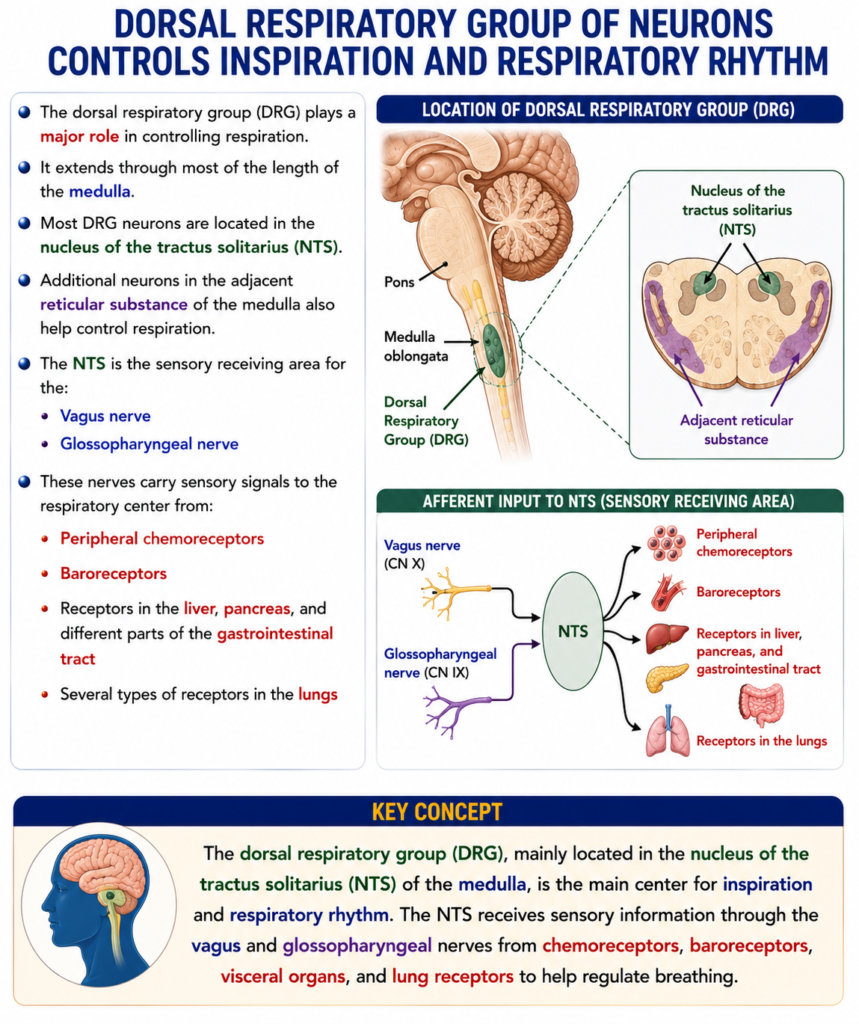
DORSAL RESPIRATORY GROUP OF NEURONS CONTROLS INSPIRATION AND RESPIRATORY RHYTHM
- The dorsal respiratory group (DRG) plays a major role in controlling respiration.
- It extends through most of the length of the medulla.
- Most DRG neurons are located in the nucleus of the tractus solitarius (NTS).
- Additional neurons in the adjacent reticular substance of the medulla also help control respiration.
- The NTS is the sensory receiving area for the:
- Vagus nerve
- Glossopharyngeal nerve
- These nerves carry sensory signals to the respiratory center from:
- Peripheral chemoreceptors
- Baroreceptors
- Receptors in the liver, pancreas, and different parts of the gastrointestinal tract
- Several types of receptors in the lungs
KEY CONCEPT
- The dorsal respiratory group (DRG), mainly located in the nucleus of the tractus solitarius (NTS) of the medulla, is the main center for inspiration and respiratory rhythm. The NTS receives sensory information through the vagus and glossopharyngeal nerves from chemoreceptors, baroreceptors, visceral organs, and lung receptors to help regulate breathing.
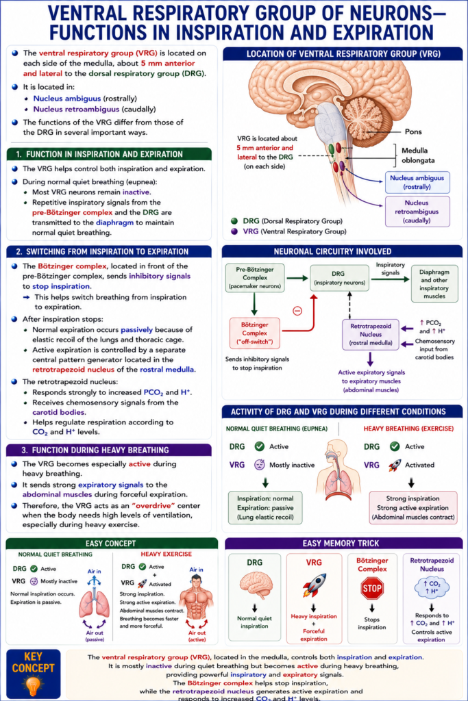
RHYTHMICAL INSPIRATORY DISCHARGES FROM THE DORSAL AND VENTRAL RESPIRATORY GROUPS
- The exact mechanism that generates the normal breathing rhythm is still not completely understood.
- The dorsal respiratory group (DRG) is believed to be partly responsible for generating the basic respiratory rhythm.
- Even if all peripheral nerves entering the medulla are cut, and the brain stem is cut above and below the medulla, the DRG still produces repetitive bursts of inspiratory nerve impulses.
- In primitive animals, one group of neurons:
- Excites another group of neurons.
- The second group then inhibits the first group.
- This cycle repeats continuously throughout life.
- A similar neuronal network is present in the human medulla.
- This network includes the:
- Dorsal respiratory group (DRG)
- Adjacent medullary neurons
- Together, these neurons help generate the basic rhythm of breathing.
- A small area in the rostral ventral respiratory group (VRG) is called the pre-Bötzinger complex.
- The pre-Bötzinger complex contains neurons that fire spontaneously.
- These neurons send signals to both the:
- Dorsal respiratory group (DRG)
- Ventral respiratory group (VRG)
- They help generate the basic respiratory rhythm.
- Some neurons in the pre-Bötzinger complex have pacemaker-like properties.
- If the pre-Bötzinger complex is removed, the normal respiratory rhythm stops.
- Therefore, the pre-Bötzinger complex is considered a key part of the respiratory central pattern generator.
- It may be responsible for activating the DRG inspiratory neurons to produce the inspiratory ramp signal.
KEY CONCEPT
- The dorsal respiratory group (DRG) helps generate the basic rhythm of breathing by producing repetitive inspiratory nerve impulses. The pre-Bötzinger complex in the rostral ventral respiratory group contains pacemaker-like neurons that are essential for generating this respiratory rhythm and are considered a major part of the respiratory central pattern generator.
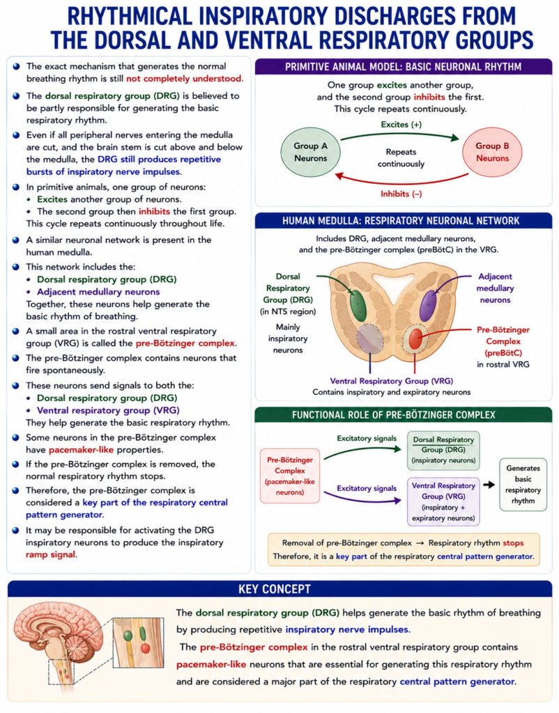
INSPIRATORY “RAMP” SIGNAL
- The nerve signal sent to the inspiratory muscles, mainly the diaphragm, is not a sudden burst of impulses.
- Instead, the signal:
- Starts weakly
- Gradually increases in strength in a ramp-like pattern.
- During normal breathing, this ramp signal lasts for about 2 seconds.
- After that, the signal stops suddenly for about 3 seconds.
- When the signal stops:
- The diaphragm relaxes.
- Elastic recoil of the lungs and chest wall causes expiration.
- After expiration, the ramp signal starts again, and the breathing cycle repeats continuously.
- Therefore, the inspiratory nerve signal is called the inspiratory ramp signal.
- The main advantage of the ramp signal is that it causes a smooth and gradual increase in lung volume during inspiration, instead of sudden inspiratory gasps.
- Two features of the inspiratory ramp are controlled:
- Rate of rise of the ramp
- The speed at which the ramp increases can be adjusted.
- During heavy breathing, the ramp rises more rapidly.
- This allows the lungs to fill more quickly.
- Point at which the ramp stops
- The point where the ramp suddenly ends is controlled.
- This is the main way the body regulates the breathing rate.
- If the ramp stops earlier:
- Inspiration becomes shorter.
- Expiration also becomes shorter.
- Therefore, the breathing frequency increases.
Easy Concept
Think of the inspiratory ramp like slowly pressing the accelerator of a car.
Normal Inspiration
Weak signal
     ↓
▁▂▃▄▅▆▇█
(Gradually increases for ~2 sec)

↓↓

Diaphragm contracts gradually

↓↓

Lungs fill smoothly
Expiration
Signal suddenly stops

█████│

↓↓

Diaphragm relaxes

↓↓

Lungs recoil

↓↓

Expiration occurs
Then the cycle starts again.
Easy Memory Trick
Ramp = Rise Slowly
Ramp ON
↓↓
Gradual diaphragm contraction
↓↓
Smooth inspiration

Ramp OFF
↓↓
Diaphragm relaxes
↓↓
Expiration
KEY CONCEPT
- The inspiratory ramp signal is a gradually increasing nerve signal sent mainly to the diaphragm. It lasts about 2 seconds, then stops for about 3 seconds, allowing expiration. The ramp produces smooth inspiration, while controlling its rate of rise and stopping point helps adjust the depth and frequency of breathing.
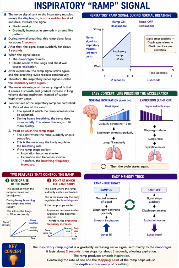
PNEUMOTAXIC CENTER LIMITS DURATION OF INSPIRATION AND INCREASES RESPIRATORY RATE
- The pneumotaxic center is located dorsally in the nucleus parabrachialis of the upper pons.
- It sends signals to the inspiratory center.
- Its main function is to control the “switch-off” point of the inspiratory ramp signal.
- By controlling when the ramp signal stops, it controls the duration of inspiration (lung filling phase).
- When the pneumotaxic signal is strong:
- Inspiration may last only about 0.5 second.
- The lungs fill with a smaller amount of air.
- When the pneumotaxic signal is weak:
- Inspiration may continue for 5 seconds or more.
- The lungs fill with a much larger amount of air.
- Therefore, the main function of the pneumotaxic center is to limit inspiration.
- Limiting inspiration also shortens expiration.
- As a result, the entire breathing cycle becomes shorter.
- This increases the respiratory rate.
- A strong pneumotaxic signal can increase the breathing rate to about 30–40 breaths/min.
- A weak pneumotaxic signal can decrease the breathing rate to about 3–5 breaths/min.
Easy Concept
Think of the pneumotaxic center as a timer for inspiration.
Strong Pneumotaxic Signal ⏱️
Ramp starts
      ↓
Timer stops it quickly
      ↓
Short inspiration (≈0.5 sec)
      ↓
Less air enters lungs
      ↓
More breaths per minute
(30–40 breaths/min)
Weak Pneumotaxic Signal ⏱️
Ramp starts
      ↓
Timer stops it late
      ↓
Long inspiration (≈5 sec or more)
      ↓
More air enters lungs
      ↓
Fewer breaths per minute
(3–5 breaths/min)
Easy Memory Trick
Strong Pneumotaxic
↓↓
Stops inspiration Early
↓↓
Short breaths
↓↓
Fast breathing
Weak Pneumotaxic
↓↓
Stops inspiration Late
↓↓
Deep breaths
↓↓
Slow breathingKEY CONCEPT
- The pneumotaxic center, located in the upper pons, controls the switch-off point of the inspiratory ramp signal. A strong pneumotaxic signal shortens inspiration, reduces lung filling, and increases the respiratory rate, whereas a weak signal prolongs inspiration, increases lung filling, and decreases the respiratory rate.
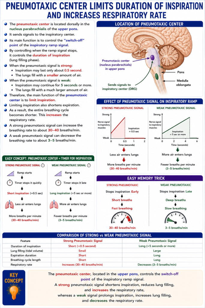
VENTRAL RESPIRATORY GROUP OF NEURONS—FUNCTIONS IN INSPIRATION AND EXPIRATION
- The ventral respiratory group (VRG) is located on each side of the medulla, about 5 mm anterior and lateral to the dorsal respiratory group (DRG).
- It is located in:
- Nucleus ambiguus (rostrally)
- Nucleus retroambiguus (caudally)
- The functions of the VRG differ from those of the DRG in several important ways.
1. Function in Inspiration and Expiration
- The VRG helps control both inspiration and expiration.
- During normal quiet breathing (eupnea):
- Most VRG neurons remain inactive.
- Repetitive inspiratory signals from the pre-Bötzinger complex and the DRG are transmitted to the diaphragm to maintain normal quiet breathing.
2. Switching From Inspiration to Expiration
- The Bötzinger complex, located in front of the pre-Bötzinger complex, sends inhibitory signals to stop inspiration.
- This helps switch breathing from inspiration to expiration.
- After inspiration stops:
- Normal expiration occurs passively because of elastic recoil of the lungs and thoracic cage.
- Active expiration is controlled by a separate central pattern generator located in the retrotrapezoid nucleus of the rostral medulla.
- The retrotrapezoid nucleus:
- Responds strongly to increased PCO₂ and H⁺.
- Receives chemosensory signals from the carotid bodies.
- Helps regulate respiration according to CO₂ and H⁺ levels.
3. Function During Heavy Breathing
- The VRG becomes especially active during heavy breathing.
- It sends strong expiratory signals to the abdominal muscles during forceful expiration.
- Therefore, the VRG acts as an “overdrive” center when the body needs high levels of ventilation, especially during heavy exercise.
Easy Concept
Think of the DRG as the normal breathing driver and the VRG as the turbo engine.
Normal Quiet Breathing
DRG ✅ Active
VRG 😴 Mostly inactive- Normal inspiration occurs.
- Expiration is passive.
Heavy Exercise
DRG ✅

+

VRG 🚀 Activated- Strong inspiration.
- Strong active expiration.
- Abdominal muscles contract.
- Breathing becomes faster and more forceful.
Easy Memory Trick
DRG
↓↓
Normal quiet inspiration
VRG
↓↓
Heavy inspiration
+
Forceful expirationBötzinger Complex
↓↓
Stops inspirationRetrotrapezoid Nucleus
↓↓
Responds to ↑ CO₂ and ↑ H⁺
↓↓
Controls active expirationKEY CONCEPT
- The ventral respiratory group (VRG), located in the medulla, controls both inspiration and expiration. It is mostly inactive during quiet breathing but becomes active during heavy breathing, providing powerful inspiratory and expiratory signals. The Bötzinger complex helps stop inspiration, while the retrotrapezoid nucleus generates active expiration and responds to increased CO₂ and H⁺ levels.
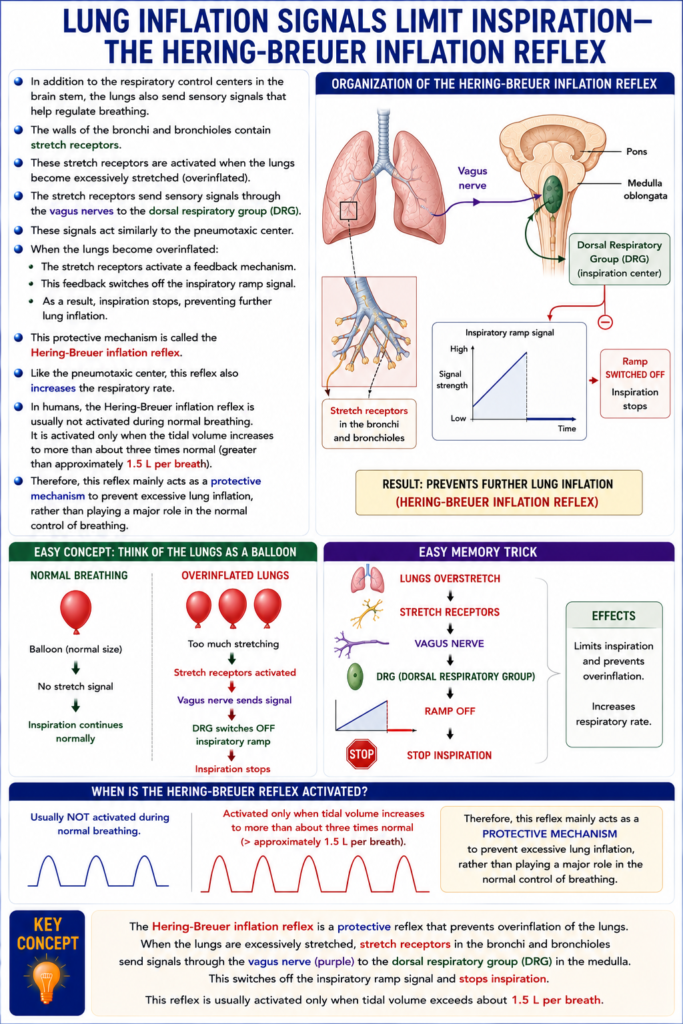
LUNG INFLATION SIGNALS LIMIT INSPIRATION—THE HERING-BREUER INFLATION REFLEX
- In addition to the respiratory control centers in the brain stem, the lungs also send sensory signals that help regulate breathing.
- Fig. 42.1 shows the organization of the respiratory center.
- The walls of the bronchi and bronchioles contain stretch receptors.
- These stretch receptors are activated when the lungs become excessively stretched (overinflated).
- The stretch receptors send sensory signals through the vagus nerves to the dorsal respiratory group (DRG).
- These signals act similarly to the pneumotaxic center.
- When the lungs become overinflated:
- The stretch receptors activate a feedback mechanism.
- This feedback switches off the inspiratory ramp signal.
- As a result, inspiration stops, preventing further lung inflation.
- This protective mechanism is called the Hering-Breuer inflation reflex.
- Like the pneumotaxic center, this reflex also increases the respiratory rate.
- In humans, the Hering-Breuer inflation reflex is usually not activated during normal breathing.
- It is activated only when the tidal volume increases to more than about three times normal (greater than approximately 1.5 L per breath).
- Therefore, this reflex mainly acts as a protective mechanism to prevent excessive lung inflation, rather than playing a major role in the normal control of breathing.
Fig. 42.1
- Stretch receptors are located in the bronchi and bronchioles.
- Vagus nerve carries stretch signals to the dorsal respiratory group (DRG).
- The inspiratory ramp is switched off when the lungs are overinflated.
- This produces the Hering-Breuer inflation reflex.
Easy Concept
Imagine the lungs are a balloon.
Normal Breathing
Balloon 🎈

Normal size

↓

No stretch signal

↓

Inspiration continues normally
Overinflated Lungs
Balloon 🎈🎈🎈

Too much stretching

↓

Stretch receptors activated

↓

Vagus nerve sends signal

↓

DRG switches OFF inspiratory ramp

↓

Inspiration stops
Easy Memory Trick
Lungs Overstretch
        ↓
Stretch Receptors
        ↓
Vagus Nerve
        ↓
DRG
        ↓
Ramp OFF
        ↓
Stop Inspiration
KEY CONCEPT
- The Hering-Breuer inflation reflex is a protective reflex that prevents overinflation of the lungs. When the lungs are excessively stretched, stretch receptors in the bronchi and bronchioles send signals through the vagus nerve to the dorsal respiratory group, which switches off the inspiratory ramp signal and stops inspiration. This reflex is usually activated only when tidal volume exceeds about 1.5 L per breath.
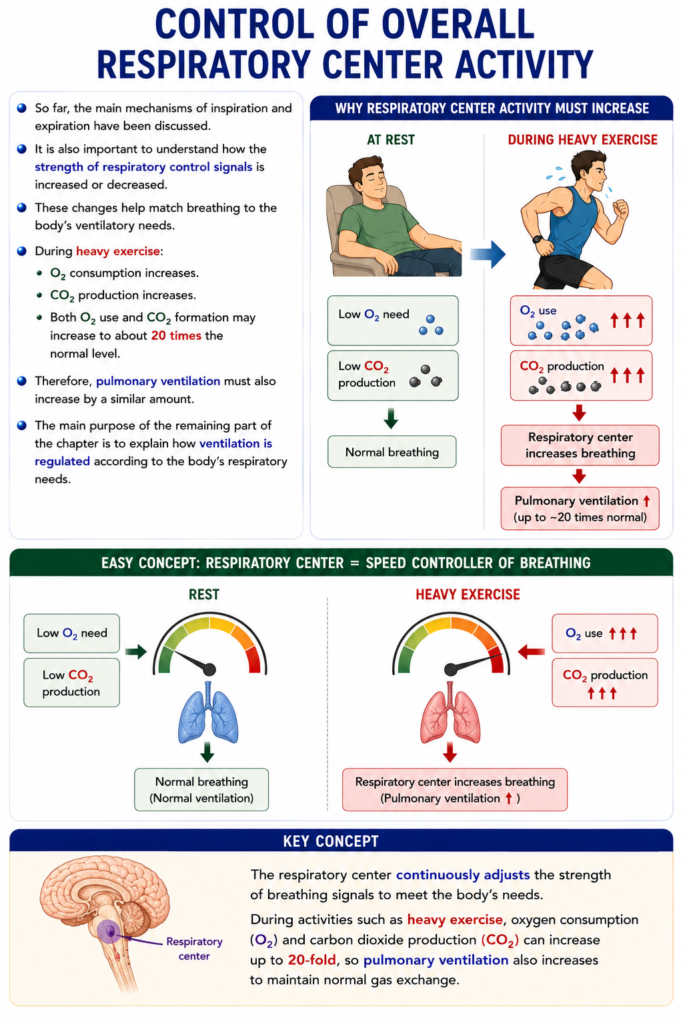
CONTROL OF OVERALL RESPIRATORY CENTER ACTIVITY
- So far, the main mechanisms of inspiration and expiration have been discussed.
- It is also important to understand how the strength of respiratory control signals is increased or decreased.
- These changes help match breathing to the body’s ventilatory needs.
- During heavy exercise:
- O₂ consumption increases.
- CO₂ production increases.
- Both O₂ use and CO₂ formation may increase to about 20 times the normal level.
- Therefore, pulmonary ventilation must also increase by a similar amount.
- The main purpose of the remaining part of the chapter is to explain how ventilation is regulated according to the body’s respiratory needs.
Easy Concept
Imagine the respiratory center is the speed controller of breathing.
Rest
Low O₂ need
Low CO₂ production

↓

Normal breathing
Heavy Exercise
O₂ use ↑↑↑
CO₂ production ↑↑↑

↓

Respiratory center increases breathing

↓

Pulmonary ventilation ↑
KEY CONCEPT
- The respiratory center continuously adjusts the strength of breathing signals to meet the body’s needs. During activities such as heavy exercise, oxygen consumption and carbon dioxide production can increase up to 20-fold, so pulmonary ventilation also increases to maintain normal gas exchange.
MADE BY SELF LEARNING CEO AND FOUNDER DR SHEEN
