


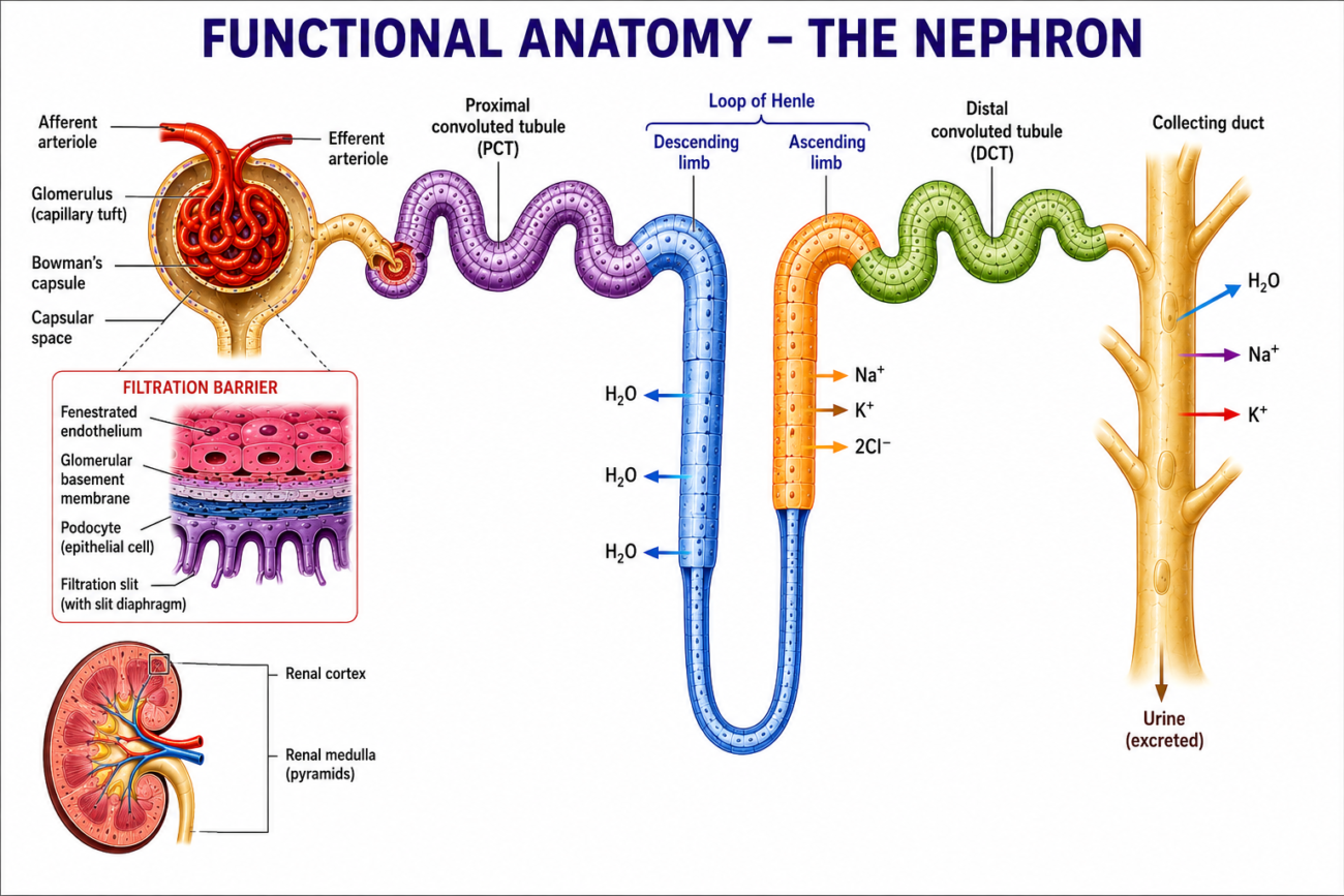
- The nephron is the functional unit of the kidney.
- Each nephron consists of:
- A renal tubule
- Its glomerulus
- Each human kidney contains about 1 million nephrons.
- Figure 37–1 shows the structure of the nephron.
- The glomerulus is about 200 µm in diameter.
- It is formed by a tuft of capillaries inside the blind, dilated end of the nephron called Bowman’s capsule.
- The glomerulus receives blood through the afferent arteriole.
- Blood leaves through the efferent arteriole.
- Glomerular filtrate is formed in the glomerulus.
- The afferent arteriole has a larger diameter than the efferent arteriole.
- Two cellular layers separate the blood from the glomerular filtrate:
- Fenestrated capillary endothelium
- Specialized epithelium of Bowman’s capsule
- The glomerular capillary endothelium contains fenestrations (pores) measuring 70–90 nm.
- The capillaries are surrounded by the glomerular basement membrane.
- Specialized epithelial cells called podocytes cover the basement membrane.
- Podocytes have many foot processes (pseudopodia) that interlock.
- These form filtration slits along the capillary wall.
- Each filtration slit is about 25 nm wide.
- Each slit is covered by a thin membrane.
- The glomerular basement membrane does not contain visible pores or gaps.
- Mesangial cells are located between the capillary endothelium and the basement membrane.
- They are similar to pericytes found around other capillaries.
- Mesangial cells are especially common between adjacent capillaries.
- They are contractile and help regulate glomerular filtration.
- They also:
- Secrete the extracellular matrix
- Remove immune complexes
- Participate in the progression of glomerular disease
- The glomerular filtration membrane:
- Allows free passage of neutral molecules up to 4 nm in diameter
- Almost completely blocks molecules larger than 8 nm
- Both molecular size and electrical charge influence filtration.
- The total glomerular filtration surface area in humans is about 0.8 m².
- The renal tubules are lined by different types of epithelial cells.
- Their structural differences are related to their specific functions.
- The proximal convoluted tubule (PCT):
- Is about 15 mm long
- Has a diameter of about 55 µm
- Is lined by a single layer of epithelial cells
- Cells are joined by tight junctions
- Contains lateral intercellular spaces
- Has a brush border made of numerous microvilli
- After the PCT, the tubule continues as the Loop of Henle.
- The descending limb and thin ascending limb:
- Contain thin, permeable cells
- The thick ascending limb:
- Contains large cells rich in mitochondria
- There are two types of nephrons:
- Cortical nephrons
- Have short loops of Henle
- Juxtamedullary nephrons
- Have long loops extending deep into the medulla
- Cortical nephrons
- Only about 15% of human nephrons are juxtamedullary nephrons.
- The thick ascending limb returns close to its own glomerulus.
- Specialized cells here form the macula densa.
- The macula densa, lacis cells, and renin-secreting granular cells together form the juxtaglomerular apparatus.
- The distal convoluted tubule (DCT):
- Begins at the macula densa
- Is about 5 mm long
- Has lower epithelial cells
- Contains few microvilli
- Has no brush border
- The distal tubules join to form collecting ducts.
- Collecting ducts are about 20 mm long.
- They pass through the renal cortex and medulla.
- They open into the renal pelvis at the apex of the medullary pyramids.
- Collecting ducts contain two main cell types:
- Principal (P) cells
- More numerous
- Relatively tall
- Contain few organelles
- Responsible for:
- Na⁺ reabsorption
- Vasopressin (ADH)-stimulated water reabsorption
- Intercalated (I) cells
- Fewer in number
- Also present in the DCT
- Contain more:
- Microvilli
- Cytoplasmic vesicles
- Mitochondria
- Responsible for:
- Acid (H⁺) secretion
- HCO₃⁻ transport
- Principal (P) cells
- The total length of a nephron, including the collecting duct, is about 45–65 mm.
- The kidney also contains secretory cells:
- Granular (juxtaglomerular) cells
- Renal medullary interstitial cells (RMICs)
- RMICs:
- Are specialized fibroblast-like cells
- Contain lipid droplets
- Express COX-2 and prostaglandin E synthase (PGES)
- RMICs produce prostaglandin E₂ (PGE₂).
- PGE₂ is an important local (paracrine) regulator of salt and water balance.
- PGE₂ is secreted by:
- RMICs
- Macula densa
- Collecting duct cells
- Prostacyclin (PGI₂) and other prostaglandins are secreted by the glomeruli and renal arterioles.
Figure 37–1
- Shows the overall structure of the nephron.
- Includes:
- Glomerulus
- Bowman’s capsule
- Proximal convoluted tubule
- Loop of Henle
- Distal convoluted tubule
- Collecting duct
Figure 37–2
- Shows:
- Afferent arteriole
- Efferent arteriole
- Podocytes
- Filtration slits
- Glomerular basement membrane
- Mesangial cells
- Macula densa
- Juxtaglomerular apparatus
KEY CONCEPT
- The nephron is the functional unit of the kidney. It consists of the glomerulus and renal tubule, where filtration, reabsorption, secretion, and urine formation occur. Specialized structures such as podocytes, mesangial cells, the macula densa, and the juxtaglomerular apparatus regulate filtration, while different tubular segments perform specific functions in maintaining fluid, electrolyte, and acid-base balance.
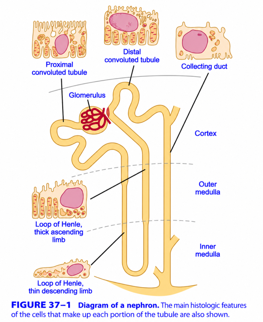
Nephron (Ganong Fig. 37-1) – Easiest & Most Conceptual Explanation
🎯 One-Line Concept
The nephron is the structural and functional unit of the kidney.
Its job is to filter blood, remove waste, and produce urine while conserving water and useful substances.
Think of a nephron as a water purification plant.
- 🩸 Blood enters dirty
- 🧹 Useful substances are recovered
- 🚽 Waste leaves as urine
Complete Nephron Flow
Blood
   ↓
Glomerulus
   ↓
Proximal Convoluted Tubule (PCT)
   ↓
Loop of Henle
   ↓
Distal Convoluted Tubule (DCT)
   ↓
Collecting Duct
   ↓
Urine
Big Picture
The nephron has 5 major parts.
| Step | Structure | Main Job |
|---|---|---|
| 1 | Glomerulus | Filters blood |
| 2 | PCT | Reabsorbs most useful substances |
| 3 | Loop of Henle | Concentrates urine |
| 4 | DCT | Fine adjustment of salts and pH |
| 5 | Collecting Duct | Final concentration of urine |
1. Glomerulus – The Filter
📍 First part of the nephron
The glomerulus is a ball of tiny capillaries.
Its function is simple:
Filter blood.
Think of it like a kitchen sieve.
Blood
 ↓
Glomerulus
 ↓
Filtrate
What passes through?
✅ Water
✅ Sodium
✅ Glucose
✅ Amino acids
✅ Urea
What does NOT pass?
❌ Red blood cells
❌ White blood cells
❌ Platelets
❌ Large proteins
These remain in the blood.
Easy Memory
Glomerulus = Blood Filter
2. Proximal Convoluted Tubule (PCT)
📍 The Recovery Center
After filtration,
almost everything useful is recovered here.
Approximately
65–70% of filtered fluid is reabsorbed.
PCT Reabsorbs
✅ Water
✅ Sodium
✅ Chloride
✅ Potassium
✅ Bicarbonate
✅ Glucose
✅ Amino acids
Think of PCT as
The “Saving Department.”
It asks,
“Is this useful?”
If YES,
it sends it back into the blood.
Why does PCT have a Brush Border?
Look at the figure.
The PCT cells have many tiny finger-like projections.
These are
Microvilli (Brush Border).
Purpose:
Increase surface area
↓
More absorption
Think of a sponge.
More surface area
↓
Absorbs more water.
3. Loop of Henle
The Loop of Henle has
Two limbs
Descending Limb
        ↓
Hairpin Turn
        ↑
Ascending Limb
Its main function is
Concentrating urine.
A. Thin Descending Limb
Main Rule
Water leaves.
Salt stays.
The descending limb is
Permeable to water
Not permeable to salt.
Therefore
Water OUT

Salt stays
Result
Tubular fluid becomes
More concentrated.
Easy Memory
Descending = Water Down
B. Thick Ascending Limb
Here,
the opposite happens.
Water cannot leave.
Instead,
Salt is pumped out.
Main ions transported
✅ Sodium
✅ Potassium
✅ Chloride
This is called
NKCC2 transporter.
Result
The tubular fluid becomes
Dilute.
Easy Memory
Ascending = Salt Up
Why Is the Loop of Henle Important?
It creates the
Medullary Osmotic Gradient
This gradient allows the kidneys to produce
- Concentrated urine (during dehydration)
- Dilute urine (when excess water is present)
4. Distal Convoluted Tubule (DCT)
The Fine-Tuning Department
Most work has already been done.
Now the kidney makes
small adjustments.
DCT controls
Sodium
Potassium
Calcium
Hydrogen ions
Water
Hormones Acting Here
Aldosterone
↑ Sodium reabsorption
↓
Potassium secretion
Parathyroid Hormone (PTH)
↑ Calcium reabsorption
Think of DCT as
Final Editing Before Printing
5. Collecting Duct
Final Decision Point
This is where the kidney decides
How much water should be saved?
The hormone
ADH (Antidiuretic Hormone)
controls this.
When ADH is High
Water is reabsorbed.
Urine becomes
Small volume
Dark
Concentrated
When ADH is Low
Water remains in urine.
Urine becomes
Large volume
Pale
Dilute
Easy Memory
Collecting Duct = Water Saver
Cortex vs Medulla
The figure also shows three kidney regions.
Cortex
Contains
✅ Glomerulus
✅ PCT
✅ DCT
This is where filtration begins.
Outer Medulla
Contains
Loop of Henle
Collecting ducts
Inner Medulla
Contains
Long loops of Henle
Collecting ducts
This region creates the highest osmotic concentration, allowing maximum water reabsorption.
Why Do the Cells Look Different?
The figure also shows the microscopic appearance (histology) of each nephron segment.
Proximal Convoluted Tubule (PCT)
- Tall cuboidal cells
- Dense brush border (microvilli)
- Many mitochondria
- Built for maximum reabsorption
Thin Descending Limb
- Thin, flat cells
- Few mitochondria
- Specialized for water movement
Thick Ascending Limb
- Cuboidal cells
- Many mitochondria
- No brush border
- Built for active salt transport
Distal Convoluted Tubule (DCT)
- Cuboidal cells
- Few microvilli
- Fine-tunes electrolyte balance
Collecting Duct
- Larger cells with clear borders
- Responds to ADH
- Determines the final concentration of urine
The Entire Nephron Story
Blood
   │
   ▼
Glomerulus
(Filters Blood)
   │
   ▼
PCT
(Reabsorbs most water,
glucose, amino acids,
and electrolytes)
   │
   ▼
Descending Limb
(Water leaves)
   │
   ▼
Ascending Limb
(Salt leaves,
water stays)
   │
   ▼
DCT
(Fine adjustment
under hormones)
   │
   ▼
Collecting Duct
(ADH controls
final water reabsorption)
   │
   ▼
Urine
Easy Analogy
Imagine a water purification factory:
- 🧺 Glomerulus = Entrance filter (separates fluid from blood).
- ♻️ PCT = Recovery department (returns valuable items like glucose and most water).
- ⚖️ Loop of Henle = Concentration machine (adjusts water and salt to create a concentration gradient).
- 🔧 DCT = Quality control section (fine-tunes electrolytes and pH under hormone control).
- 🚰 Collecting duct = Final packaging (decides how much water to keep before urine leaves).
MBBS High-Yield Summary
| Part | Main Function | Memory Keyword |
|---|---|---|
| Glomerulus | Filters blood | Filter |
| PCT | Reabsorbs ~65–70% of filtrate | Recover |
| Descending Limb | Water leaves | Water Down |
| Ascending Limb | Salt leaves, water cannot | Salt Up |
| DCT | Hormonal fine-tuning | Adjust |
| Collecting Duct | ADH controls final water reabsorption | Save Water |
🌟 Golden Flow to Remember
FILTER
   ↓
RECOVER
   ↓
CONCENTRATE
   ↓
ADJUST
   ↓
SAVE WATER
   ↓
URINE
💡 Golden Rule
The nephron filters blood in the glomerulus, reabsorbs useful substances in the tubules, secretes selected wastes into the tubular fluid, and finally produces urine in the collecting duct. Every segment has a specialized role, working together to maintain the body’s fluid, electrolyte, and acid–base balance.
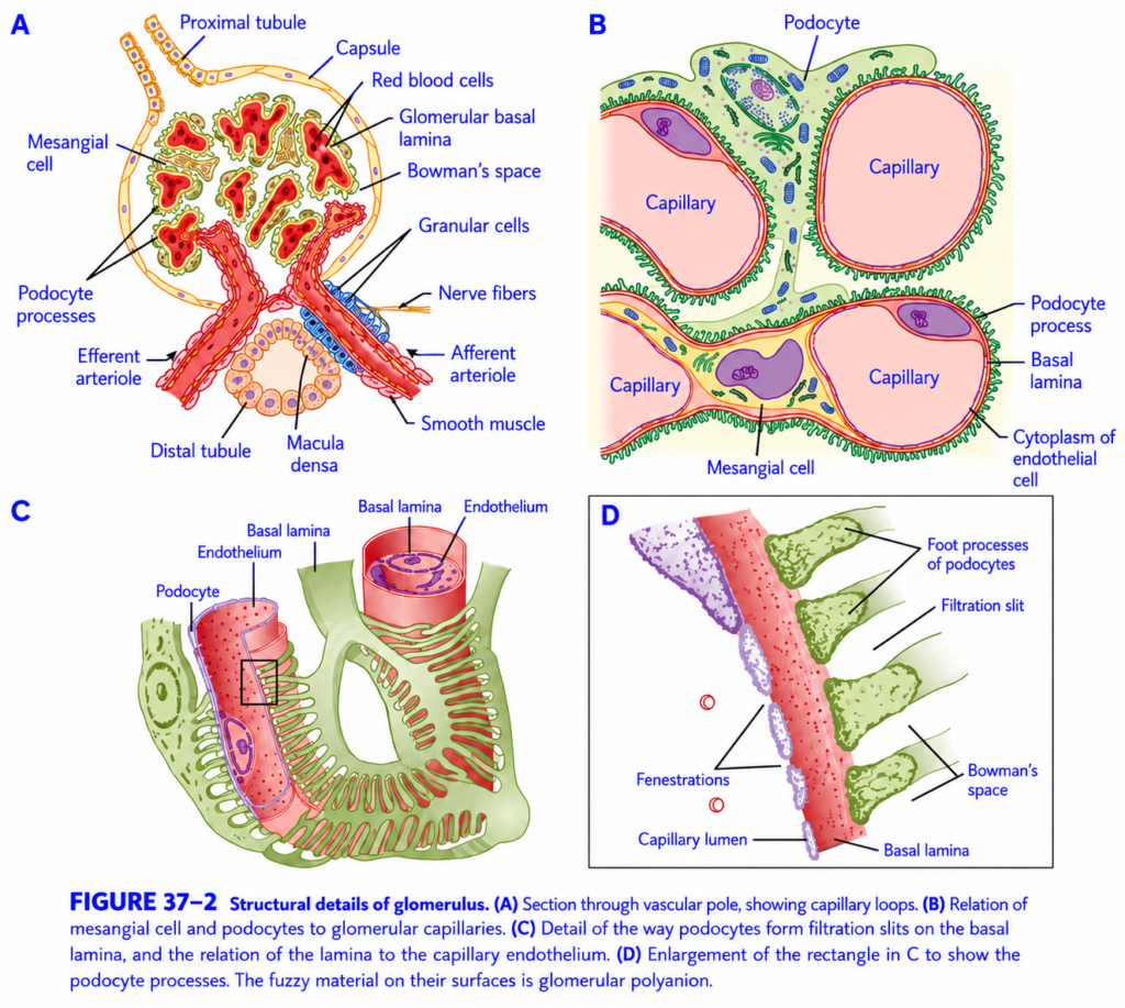
Structural Details of the Glomerulus (Ganong Fig. 37-2) – Easiest & Most Conceptual Explanation
🎯 One-Line Concept
The glomerulus is the kidney’s microscopic filter that allows water and small molecules to pass into Bowman’s capsule while preventing blood cells and proteins from leaving the blood.
Think of it as a high-security water filter.
- ✅ Water and small molecules pass.
- ❌ Blood cells and large proteins are blocked.
Big Picture
The figure has 4 parts (A–D), each showing the glomerulus in greater detail.
A → Complete glomerulus
        ↓
B → Relationship of cells
        ↓
C → Filtration barrier
        ↓
D → Magnified filtration slit
PART A – Whole Glomerulus
This is the overall structure of the renal corpuscle.
Components
Afferent Arteriole
        ↓
   Glomerulus
        ↓
Bowman's Capsule
        ↓
Proximal Tubule
1. Afferent Arteriole
Function
Brings blood into the glomerulus.
Think of it as
🚰 The inlet pipe.
2. Glomerular Capillaries
These are tiny capillary loops where blood filtration occurs.
Blood pressure inside these capillaries forces fluid outward.
3. Efferent Arteriole
Carries blood away from the glomerulus.
Think of it as
🚰 The outlet pipe.
Easy Memory
Afferent = Arrives
Efferent = Exits
4. Bowman’s Capsule
A cup-shaped structure surrounding the glomerulus.
It collects the filtered fluid.
Think of it as
🥣 A collecting bowl placed under a filter.
5. Bowman’s Space
The small gap between
- Glomerular capillaries
- Bowman’s capsule
This is where the filtrate first collects.
6. Proximal Tubule
The filtrate enters here after leaving Bowman’s space.
This is the beginning of tubular reabsorption.
7. Distal Tubule & Macula Densa
The distal tubule passes close to the glomerulus.
Here lies the
Macula Densa
Its function:
Measures sodium chloride in tubular fluid.
If sodium delivery changes,
the macula densa helps adjust the glomerular filtration rate (GFR) through tubuloglomerular feedback.
Think of it as the kidney’s quality-control sensor.
8. Granular (Juxtaglomerular) Cells
Located in the wall of the afferent arteriole.
They secrete
Renin
Renin is released when:
- Blood pressure falls.
- Renal blood flow decreases.
- Sodium delivery to the macula densa falls.
Renin activates the Renin–Angiotensin–Aldosterone System (RAAS) to help restore blood pressure.
9. Mesangial Cells
These cells lie between the capillary loops.
Functions
- Provide structural support.
- Remove debris by phagocytosis.
- Contract to alter the surface area available for filtration, helping regulate GFR.
Think of them as the support and cleaning staff of the glomerulus.
PART B – Relationship Between Cells
This panel shows the close arrangement of the filtration barrier.
Main structures
- Glomerular capillary
- Endothelial cell
- Basement membrane (basal lamina)
- Podocyte
- Mesangial cell
Together, they form the filtration apparatus.
What is a Podocyte?
Podocytes are specialized epithelial cells covering the outer surface of the glomerular capillaries.
They have many finger-like extensions called
Foot Processes (Pedicels)
These wrap around the capillaries.
Think of them as
🖐 Hands gently holding a tube.
PART C – Filtration Barrier
This panel shows how the filtration barrier is organized.
The filter has three layers.
Layer 1 – Fenestrated Endothelium
The capillary wall contains many tiny pores called
Fenestrations
These allow
✅ Water
✅ Electrolytes
✅ Glucose
to pass easily.
However,
blood cells are too large to pass.
Layer 2 – Glomerular Basement Membrane (GBM)
This is the main filter.
It acts as both
- A size barrier
- A charge barrier
It prevents
❌ Large proteins (e.g., albumin)
from passing into the filtrate.
Layer 3 – Podocyte Foot Processes
The podocyte processes interlock, leaving narrow gaps called
Filtration Slits
These slits provide the final level of filtration before fluid enters Bowman’s space.
PART D – Magnified Filtration Slit
This is a close-up of the filtration barrier.
Fluid must pass through three filters in sequence:
Blood
   │
   ▼
Fenestrated Endothelium
   │
   ▼
Glomerular Basement Membrane
   │
   ▼
Filtration Slit (Between Podocyte Foot Processes)
   │
   ▼
Bowman's Space
Only after crossing all three layers does the fluid become glomerular filtrate.
What Passes Through the Filter?
✅ Can Pass
- Water
- Sodium
- Potassium
- Chloride
- Glucose
- Amino acids
- Urea
- Creatinine
- Bicarbonate
❌ Cannot Pass
- Red blood cells
- White blood cells
- Platelets
- Most plasma proteins (especially albumin)
Easy Analogy
Imagine making tea with a tea strainer.
- 🍵 Water and dissolved sugar pass through.
- 🍃 Tea leaves stay behind.
Similarly:
- Water and small molecules pass.
- Blood cells and proteins remain in the circulation.
The Complete Filtration Journey
Blood enters
(Afferent arteriole)
        │
        ▼
Glomerular Capillaries
        │
        ▼
Fenestrated Endothelium
        │
        ▼
Glomerular Basement Membrane
        │
        ▼
Filtration Slits
(Podocyte Foot Processes)
        │
        ▼
Bowman's Space
        │
        ▼
Proximal Tubule
High-Yield Functions of the Main Cells
| Structure | Main Function | Easy Memory |
|---|---|---|
| Afferent arteriole | Brings blood to the glomerulus | Arrives |
| Efferent arteriole | Carries blood away | Exits |
| Mesangial cells | Support capillaries, remove debris, regulate filtration surface area | Support & Clean |
| Podocytes | Form filtration slits | Final Filter |
| Basement membrane | Main size and charge barrier | Main Filter |
| Macula densa | Senses tubular NaCl and helps regulate GFR | Salt Sensor |
| Granular (JG) cells | Release renin | Blood Pressure Controller |
MBBS High-Yield Points
- ✅ Glomerular filtration barrier has three layers: fenestrated endothelium, glomerular basement membrane, and podocyte filtration slits.
- ✅ Podocyte foot processes create the filtration slits.
- ✅ Mesangial cells provide support, clear debris, and help regulate filtration.
- ✅ Macula densa + juxtaglomerular (granular) cells form part of the juxtaglomerular apparatus, which regulates GFR and blood pressure.
- ✅ Albumin and blood cells normally do not appear in urine because the filtration barrier prevents their passage.
🌟 Super Memory Summary
Blood
   │
Afferent Arteriole
   │
Glomerular Capillaries
   │
Fenestrated Endothelium
   │
Basement Membrane
   │
Podocyte Filtration Slits
   │
Bowman's Space
   │
Proximal Tubule
Golden Rule
The glomerulus acts as a highly selective three-layer filter. It allows water and small solutes to enter Bowman’s space while retaining blood cells and most plasma proteins within the bloodstream, ensuring efficient filtration without losing essential blood components.
BLOOD VESSELS
- The renal blood circulation is shown in Figure 37–3.
- The afferent arterioles arise from the interlobular arteries.
- They are short and straight blood vessels.
- Each afferent arteriole divides into many capillaries to form the glomerulus.
- The glomerular capillaries then join together to form the efferent arteriole.
- The efferent arteriole divides again into peritubular capillaries, which surround the renal tubules.
- These capillaries finally drain into the interlobular veins.
- The blood vessels between the glomerulus and renal tubules form a portal system.
- The glomerular capillaries are unique because they are the only capillaries in the body that drain into an arteriole (the efferent arteriole).
- The efferent arteriole contains relatively little smooth muscle.
- In cortical nephrons:
- The efferent arteriole forms a peritubular capillary network.
- In juxtamedullary nephrons:
- The efferent arteriole forms:
- Peritubular capillaries
- Vasa recta
- The efferent arteriole forms:
- The vasa recta are hairpin-shaped blood vessels.
- They descend into the renal medulla alongside the loops of Henle.
- The descending vasa recta:
- Have nonfenestrated endothelium
- Contain facilitated urea transporters
- The ascending vasa recta:
- Have fenestrated endothelium
- Help conserve solutes
- One efferent arteriole supplies capillaries around several different nephrons.
- Therefore, a nephron does not always receive blood only from its own efferent arteriole.
- In humans:
- The total surface area of the renal capillaries is about 12 m².
- The total surface area of the renal tubules is also about 12 m².
- At any moment, the renal capillaries contain about 30–40 mL of blood.
Figure 37–3
- Shows the pathway of renal blood flow:
- Interlobular artery
- Afferent arteriole
- Glomerulus
- Efferent arteriole
- Peritubular capillaries
- Vasa recta (juxtamedullary nephrons)
- Interlobular vein
KEY CONCEPT
- Blood enters the glomerulus through the afferent arteriole, is filtered, and leaves through the efferent arteriole. The efferent arteriole forms peritubular capillaries in cortical nephrons and vasa recta in juxtamedullary nephrons. The glomerular capillaries are unique because they drain into an arteriole, creating a portal system that supports filtration and tubular exchange.
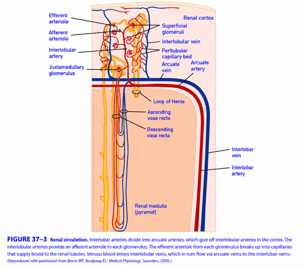
Renal Circulation (GANONG Fig. 37-3) – Easiest & Most Conceptual Explanation
🎯 One-Line Concept
Renal circulation is the pathway by which blood enters the kidney, gets filtered in the glomerulus, nourishes the kidney tubules, and then returns to the heart.
Think of it as a two-step blood circulation:
- Blood is filtered.
- Filtered blood then nourishes the kidney tissue.
This is unique because the kidney has two capillary beds connected in series.
Big Picture
Heart
   ↓
Renal Artery
   ↓
Interlobar Artery
   ↓
Arcuate Artery
   ↓
Interlobular Artery
   ↓
Afferent Arteriole
   ↓
Glomerulus ⭐
   ↓
Efferent Arteriole
   ↓
Peritubular Capillaries / Vasa Recta
   ↓
Interlobular Vein
   ↓
Arcuate Vein
   ↓
Interlobar Vein
   ↓
Renal Vein
   ↓
Heart
Step 1. Blood Enters the Kidney
Blood enters through the
Renal Artery
This artery brings oxygen-rich blood to the kidney.
It divides into smaller branches.
Renal artery
      ↓
Interlobar arteries
Step 2. Interlobar Arteries
These arteries run between the renal pyramids.
Think of them as
🛣️ Main highways inside the kidney.
They then divide into
Arcuate Arteries
Step 3. Arcuate Arteries
These arteries run along the boundary between
- Cortex
- Medulla
Think of them as
🌉 Border roads separating the cortex and medulla.
They give rise to
Interlobular Arteries
Step 4. Interlobular Arteries
These arteries extend upward into the cortex.
Their job is to supply every nephron.
Each interlobular artery gives rise to
Afferent Arterioles
Step 5. Afferent Arteriole
Function
Brings blood into the glomerulus.
Easy Memory
Afferent = Arrives
Think of it as
🚰 The inlet pipe of the filter.
Step 6. Glomerulus ⭐
This is the first capillary bed.
Its function is
Filter blood.
Blood pressure forces water and small molecules into Bowman’s capsule.
What is filtered?
✅ Water
✅ Sodium
✅ Glucose
✅ Amino acids
✅ Urea
What stays in blood?
❌ RBCs
❌ WBCs
❌ Platelets
❌ Large proteins
Step 7. Efferent Arteriole
After filtration,
blood leaves through the
Efferent Arteriole
Easy Memory
Efferent = Exits
Unlike most organs,
the glomerular capillaries drain into another arteriole, not a venule.
This helps maintain pressure for efficient filtration.
Why Is This Unique?
Most body organs have:
Artery
   ↓
Capillary
   ↓
Vein
But the kidney has:
Artery
   ↓
Capillary (Glomerulus)
   ↓
Arteriole
   ↓
Second Capillary Bed
   ↓
Vein
This arrangement is unique.
Step 8. Second Capillary Bed
After leaving the efferent arteriole,
blood enters one of two capillary networks.
A. Peritubular Capillaries
These surround the renal tubules in the cortex.
Function
- Reabsorb water
- Reabsorb electrolytes
- Reabsorb nutrients
- Secrete some substances into the tubules
Think of them as
♻️ The recovery network.
B. Vasa Recta
These are present mainly in juxtamedullary nephrons.
They run alongside the Loop of Henle.
Function
- Supply oxygen to the medulla
- Maintain the medullary osmotic gradient by acting as a countercurrent exchanger
- Help produce concentrated urine
Think of them as
💧 The water-saving blood vessels.
Two Types of Nephrons in the Figure
1. Superficial (Cortical) Nephrons
Located in the outer cortex.
Features
- Short loops of Henle
- Mainly associated with peritubular capillaries
- Responsible for most filtration and reabsorption
About 85% of nephrons are cortical.
2. Juxtamedullary Nephrons
Located near the corticomedullary junction.
Features
- Long loops of Henle extending deep into the medulla
- Closely associated with the vasa recta
- Essential for producing concentrated urine
About 15% of nephrons are juxtamedullary.
Step 9. Venous Drainage
After exchange in the capillaries,
blood returns through the veins.
Peritubular capillaries
        ↓
Interlobular vein
        ↓
Arcuate vein
        ↓
Interlobar vein
        ↓
Renal vein
        ↓
Heart
Cortex vs Medulla
Renal Cortex
Contains
- Glomeruli
- PCT
- DCT
- Peritubular capillaries
This is where filtration begins.
Renal Medulla
Contains
- Loop of Henle
- Collecting ducts
- Vasa recta
This region is responsible for urine concentration.
Why Are There Two Capillary Beds?
This is the most important concept.
First Capillary Bed (Glomerulus)
Purpose
➡ Filtration
Blood pressure is high, allowing fluid to be filtered into Bowman’s capsule.
Second Capillary Bed (Peritubular Capillaries/Vasa Recta)
Purpose
➡ Reabsorption and secretion
Blood pressure is low, making it easier to reabsorb water and solutes back into the blood.
Easy Analogy
Imagine a water treatment plant.
High-Yield Flow Chart
Renal Artery
      │
      ▼
Interlobar Artery
      │
      ▼
Arcuate Artery
      │
      ▼
Interlobular Artery
      │
      ▼
Afferent Arteriole
      │
      ▼
Glomerulus
(Filtration)
      │
      ▼
Efferent Arteriole
      │
      ├──────────────► Peritubular Capillaries
      │                 (Reabsorption & Secretion)
      │
      └──────────────► Vasa Recta
                        (Maintains Medullary Gradient)
      │
      ▼
Interlobular Vein
      │
      ▼
Arcuate Vein
      │
      ▼
Interlobar Vein
      │
      ▼
Renal Vein
MBBS High-Yield Summary Table
| Structure | Main Function | Memory Keyword |
|---|---|---|
| Renal artery | Brings blood to kidney | Entry |
| Interlobar artery | Travels between pyramids | Main Highway |
| Arcuate artery | Runs along corticomedullary junction | Border Road |
| Interlobular artery | Supplies nephrons | Branch Road |
| Afferent arteriole | Delivers blood to glomerulus | Arrives |
| Glomerulus | Filters blood | Filter |
| Efferent arteriole | Carries blood away | Exits |
| Peritubular capillaries | Reabsorption and secretion | Recovery Network |
| Vasa recta | Maintains medullary osmotic gradient | Water Saver |
| Renal vein | Returns blood to circulation | Exit |
⭐ Super Memory Summary
Blood Enters Kidney
        │
Renal Artery
        │
Interlobar Artery
        │
Arcuate Artery
        │
Interlobular Artery
        │
Afferent Arteriole
        │
Glomerulus
⭐ FILTERS BLOOD
        │
Efferent Arteriole
        │
Peritubular Capillaries
(Reabsorb Useful Substances)
        │
OR
        ▼
Vasa Recta
(Maintains Medullary Gradient)
        │
Veins
        │
Renal Vein
        │
Heart
💡 Golden Rule
The kidney is unique because blood passes through two capillary beds in series. The first capillary bed (glomerulus) filters plasma under high pressure, while the second capillary bed (peritubular capillaries or vasa recta) operates under lower pressure to reabsorb water and solutes, nourish the kidney tissue, and help concentrate urine.
LYMPHATICS
- The kidneys have a rich (abundant) lymphatic supply.
- The renal lymphatic vessels drain into the thoracic duct.
- The thoracic duct carries the lymph back into the venous circulation in the thorax.
KEY CONCEPT
- The kidneys have an abundant lymphatic network. Renal lymph drains through the thoracic duct and finally returns to the venous circulation in the thorax.
CAPSULE
- The renal capsule is thin but strong (tough).
- If the kidney becomes edematous (swollen with fluid):
- The renal capsule limits the swelling because it cannot stretch much.
- As a result:
- Renal interstitial pressure increases.
- The increased renal interstitial pressure decreases the glomerular filtration rate (GFR).
- A reduced GFR may enhance and prolong anuria in acute kidney injury (AKI).
Easy Concept
Imagine the kidney is a balloon inside a tight plastic cover.
Normal Kidney
Kidney 😊
Inside a loose capsule

↓

Normal GFR
Swollen Kidney (Edema)
Kidney 💧 Swells

↓

Tight capsule prevents expansion

↓

Pressure inside kidney ↑

↓

GFR ↓

↓

Less urine (Anuria)
KEY CONCEPT
- The renal capsule is a tough covering that limits kidney swelling. During kidney edema, this increases renal interstitial pressure, reduces the glomerular filtration rate (GFR), and can worsen or prolong anuria in acute kidney injury (AKI).
INNERVATION OF THE RENAL VESSELS
- The renal nerves travel along the renal blood vessels as they enter the kidney.
- These nerves contain:
- Many postganglionic sympathetic efferent fibers
- A few afferent (sensory) fibers
- The kidneys may also receive cholinergic nerve fibers through the vagus nerve.
- However, the function of the vagal innervation is still uncertain.
- The sympathetic preganglionic fibers arise mainly from the:
- Lower thoracic spinal cord
- Upper lumbar spinal cord
- The cell bodies of the postganglionic sympathetic neurons are located in:
- Sympathetic chain ganglia
- Superior mesenteric ganglion
- Ganglia along the renal artery
- Sympathetic nerve fibers are mainly distributed to:
- Afferent arterioles
- Efferent arterioles
- Proximal tubules
- Distal tubules
- Juxtaglomerular apparatus
- There is also a dense noradrenergic nerve supply to the:
- Thick ascending limb of the Loop of Henle
- Pain (nociceptive) afferent fibers from the kidney:
- Carry pain signals during kidney disease
- Travel alongside the sympathetic nerves
- Enter the spinal cord through the:
- Thoracic dorsal roots
- Upper lumbar dorsal roots
- Other renal afferent fibers are involved in the renorenal reflex.
- In this reflex:
- Increased ureteral pressure in one kidney
- Causes decreased sympathetic efferent activity to the opposite kidney
- This allows the opposite kidney to:
- Increase sodium (Na⁺) excretion
- Increase water excretion
Figure 37–2
- Shows:
- Afferent arteriole
- Efferent arteriole
- Juxtaglomerular apparatus
- Macula densa
- Podocytes
- Mesangial cells
- Glomerular filtration barrier
Easy Concept
Think of the kidney nerves as communication wires.
Sympathetic Nerves
Brain & Spinal Cord
        │
        ▼
Kidney
They mainly control:
- Blood vessels
- Renal tubules
- Juxtaglomerular apparatus
Pain Pathway
Kidney pain
      ↓
Sensory nerves
      ↓
Thoracic & Upper Lumbar Spinal Cord
Renorenal Reflex
Kidney 1

↑ Ureter Pressure

↓

Signal sent

↓

Kidney 2

↓

↓ Sympathetic activity

↓

↑ Na⁺ excretion

↑ Water excretion
KEY CONCEPT
- The kidneys receive mainly sympathetic nerve supply, which regulates the renal blood vessels, renal tubules, and juxtaglomerular apparatus. Pain fibers travel with sympathetic nerves to the spinal cord, while renal sensory fibers also participate in the renorenal reflex, helping the opposite kidney increase sodium and water excretion when ureteral pressure rises in one kidney.
MADE BY SELF LEARNING CEO AND FOUNDER DR SHEEN
