


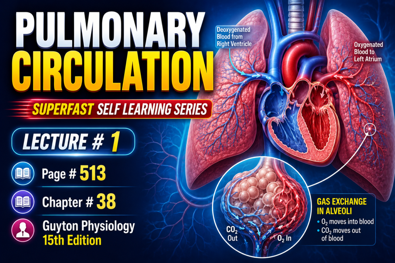
- The lung has two separate circulations.
- These are:
- High-pressure, low-flow circulation
- Low-pressure, high-flow circulation
High-Pressure, Low-Flow Circulation
- The high-pressure, low-flow circulation supplies systemic arterial blood to:
- Trachea
- Bronchial tree (including terminal bronchioles)
- Supporting tissues of the lung
- Outer coats (adventitia) of the pulmonary arteries and veins
- The bronchial arteries supply most of this systemic arterial blood.
- The bronchial arteries are branches of the thoracic aorta.
- The blood pressure in the bronchial arteries is only slightly lower than the aortic pressure.
Low-Pressure, High-Flow Circulation
- The low-pressure, high-flow circulation carries venous blood from all parts of the body to the lungs.
- The blood reaches the alveolar capillaries.
- In the alveolar capillaries:
- Oxygen (O₂) is added to the blood.
- Carbon dioxide (CO₂) is removed from the blood.
Pulmonary Artery
- The pulmonary artery receives blood from the right ventricle.
- The pulmonary artery and its branches carry blood to the alveolar capillaries for gas exchange.
Pulmonary Veins
- The pulmonary veins return the oxygenated blood to the left atrium.
- The left ventricle then pumps this blood into the systemic circulation.
Focus of This Chapter
- This chapter discusses the special features of the pulmonary circulation that are important for gas exchange in the lungs.
Key Concept
- The lungs have two circulations:
- High-pressure, low-flow circulation → Supplies the trachea, bronchial tree, supporting lung tissues, and the outer coats of pulmonary vessels through the bronchial arteries, which arise from the thoracic aorta.
- Low-pressure, high-flow circulation → Carries venous blood from the right ventricle to the alveolar capillaries, where O₂ is added and CO₂ is removed, and returns oxygenated blood through the pulmonary veins to the left atrium for distribution by the left ventricle.
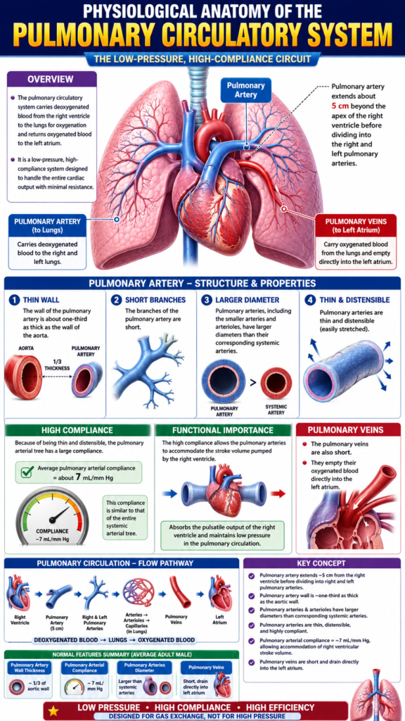
PHYSIOLOGICAL ANATOMY OF THE PULMONARY CIRCULATORY SYSTEM
Pulmonary Blood Vessels
- The pulmonary artery extends about 5 cm beyond the apex of the right ventricle.
- It then divides into the right and left main pulmonary arteries.
- These two branches carry blood to the right and left lungs.
Pulmonary Artery
- The wall of the pulmonary artery is about one-third as thick as the wall of the aorta.
- The branches of the pulmonary artery are short.
- The pulmonary arteries, including the smaller arteries and arterioles, have larger diameters than their corresponding systemic arteries.
- The pulmonary arteries are:
- Thin
- Distensible (easily stretched)
- Because of these features, the pulmonary arterial tree has a large compliance.
- The average pulmonary arterial compliance is about 7 mL/mm Hg.
- This compliance is similar to that of the entire systemic arterial tree.
- The high compliance allows the pulmonary arteries to accommodate the stroke volume pumped by the right ventricle.
Pulmonary Veins
- The pulmonary veins are also short.
- They empty their oxygenated blood directly into the left atrium.
Key Concept
- The pulmonary artery extends about 5 cm from the right ventricle before dividing into the right and left pulmonary arteries.
- The pulmonary artery wall is about one-third as thick as the aortic wall.
- Pulmonary arteries and arterioles have larger diameters than corresponding systemic arteries.
- The pulmonary arteries are thin, distensible, and highly compliant.
- Pulmonary arterial compliance = about 7 mL/mm Hg, allowing the vessels to accommodate the right ventricular stroke volume.
- Pulmonary veins are short and drain directly into the left atrium.
Bronchial Blood Vessels
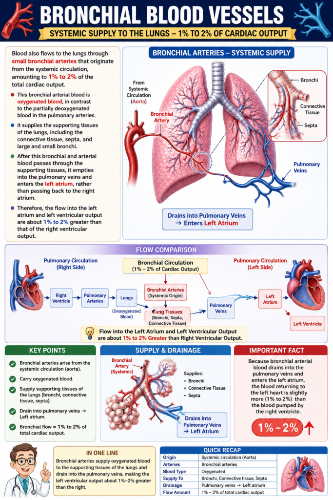
- Blood also reaches the lungs through small bronchial arteries.
- These bronchial arteries originate from the systemic circulation.
- The blood flowing through the bronchial arteries is about 1%–2% of the total cardiac output.
Bronchial Arterial Blood
- The blood in the bronchial arteries is oxygenated.
- This is different from the blood in the pulmonary arteries, which is partially deoxygenated.
Structures Supplied by the Bronchial Arteries
- Bronchial arterial blood supplies the supporting tissues of the lungs, including:
- Connective tissue
- Septa
- Large bronchi
- Small bronchi
Drainage of Bronchial Blood
- After passing through the supporting tissues, the bronchial arterial blood enters the pulmonary veins.
- It then flows into the left atrium.
- It does not return to the right atrium.
Effect on Cardiac Output
- Because bronchial blood enters the left atrium, the blood flow into the left atrium increases.
- Therefore, the output of the left ventricle is about 1%–2% greater than the output of the right ventricle.
Key Concept
- Bronchial arteries arise from the systemic circulation.
- They carry oxygenated blood equal to 1%–2% of the total cardiac output.
- They supply the connective tissue, septa, and large and small bronchi.
- After supplying these tissues, the blood drains into the pulmonary veins and then into the left atrium, not the right atrium.
- As a result, the left ventricular output is about 1%–2% greater than the right ventricular output.
Lymphatics
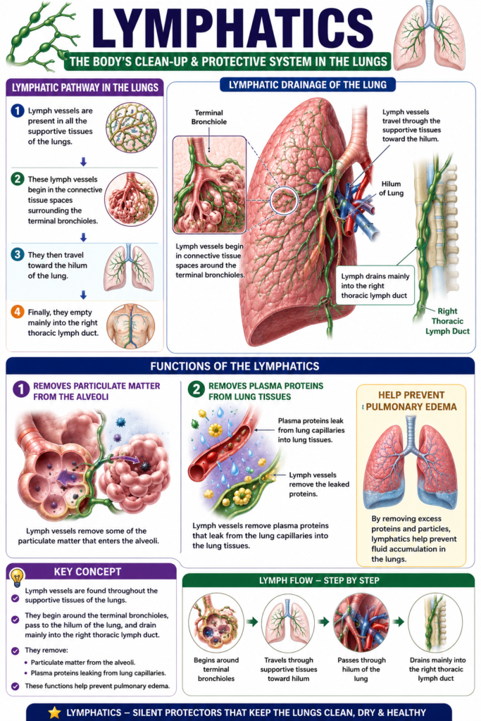
- Lymph vessels are present in all the supportive tissues of the lungs.
- These lymph vessels begin in the connective tissue spaces surrounding the terminal bronchioles.
- They then travel toward the hilum of the lung.
- Finally, they empty mainly into the right thoracic lymph duct.
Functions of the Lymphatics
- Lymph vessels remove some of the particulate matter that enters the alveoli.
- They also remove plasma proteins that leak from the lung capillaries into the lung tissues.
- By removing these proteins and particles, the lymphatics help prevent pulmonary edema.
Key Concept
- Lymph vessels are found throughout the supportive tissues of the lungs.
- They begin around the terminal bronchioles, pass to the hilum of the lung, and drain mainly into the right thoracic lymph duct.
- They remove:
- Particulate matter from the alveoli.
- Plasma proteins leaking from lung capillaries.
- These functions help prevent pulmonary edema.
PRESSURES IN THE PULMONARY CIRCULATORY SYSTEM
Figure: Fig. 39.1
Pressures in the Right Ventricle
- Fig. 39.1 shows the pressure pulse curves of the right ventricle and the pulmonary artery.
- These pressure curves are shown in the lower part of the figure.
- The upper part of the figure shows the aortic pressure curve.
- The aortic pressure is much higher than the pressure in the right ventricle and pulmonary artery.
- The normal systolic pressure of the right ventricle is about 25 mm Hg.
- This pressure is only one-fifth of the systolic pressure of the left ventricle.
- The normal diastolic pressure of the right ventricle is about 0–1 mm Hg.
Easy Concept
Think of the two ventricles as pumping blood to two different circulations:
- Right ventricle → Pumps blood to the lungs
- Short distance
- Low resistance
- Needs low pressure (25 mm Hg)
- Left ventricle → Pumps blood to the entire body
- Long distance
- High resistance
- Needs much higher pressure
Therefore,
Right ventricular pressure is much lower than left ventricular pressure.
Key Concept
- Fig. 39.1 compares the pressure curves of the right ventricle, pulmonary artery, and aorta.
- The right ventricular and pulmonary artery pressure curves are shown in the lower part of the figure.
- The aortic pressure curve is shown in the upper part of the figure.
- Normal right ventricular systolic pressure = about 25 mm Hg.
- Normal right ventricular diastolic pressure = about 0–1 mm Hg.
- The right ventricular systolic pressure is only about one-fifth of the left ventricular systolic pressure because the pulmonary circulation is a low-pressure system.
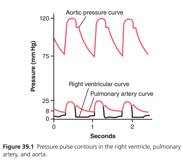
This figure compares the pressure changes in three different parts of the circulation during each heartbeat:
- Aorta (Systemic circulation)
- Right Ventricle
- Pulmonary Artery (Pulmonary circulation)
The most important message of this graph is:
The right side of the heart pumps blood at much lower pressure than the left side, but the pressure patterns are very similar.
Let’s understand every line, every peak, every dip, and every label in the simplest MBBS way.
Big Concept (One-Line Summary)
Think of blood traveling like this:
Right Ventricle → Pulmonary Artery → Lungs → Left Heart → Aorta → Body
Each chamber or vessel has its own pressure pattern.
This graph records those pressures over time.
Step 1: Understanding the Axes
Y-axis (Vertical)
Pressure (mmHg)
This tells us:
How much force the blood is pushing with.
Higher on the graph = Higher blood pressure.
Lower on the graph = Lower blood pressure.
Notice the markings:
- 0 mmHg
- 8 mmHg
- 25 mmHg
- 75 mmHg
- 120 mmHg
These values are important because they represent normal physiological pressures.
X-axis (Horizontal)
Time (Seconds)
This shows how pressure changes during each heartbeat.
Each repeated wave = One cardiac cycle.
There Are Three Curves
1. Top Red Curve
Aortic Pressure Curve
This represents blood pressure inside the aorta.
2. Middle Red Curve
Pulmonary Artery Pressure Curve
This represents pressure inside the pulmonary artery.
3. Black Curve
Right Ventricular Pressure Curve
This represents pressure inside the right ventricle.
PART 1 — Aortic Pressure Curve (Top Red Wave)
This is the pressure inside the aorta.
Normal values are approximately:
- Systolic = 120 mmHg
- Diastolic = 80 mmHg (about 75–80 mmHg in the graph)
Beginning of the Wave
The pressure is about 75–80 mmHg.
Why doesn’t it fall to zero?
Because:
The aorta is elastic.
Even when the heart relaxes, the stretched aortic wall recoils and continues pushing blood forward.
So pressure never falls to zero.
Rapid Rise
The pressure suddenly increases to 120 mmHg.
Why?
The left ventricle contracts (ventricular systole) and ejects blood into the aorta.
More blood enters than leaves.
Pressure rises rapidly.
This is called:
Systolic pressure
Rounded Peak
Notice the top is smooth, not pointed.
Why?
Blood continues to be ejected for a short period.
Pressure stays high before it starts falling.
Small Notch
After the peak there is a tiny dip.
This is called the:
Dicrotic notch (Incisura)
Why does it occur?
The left ventricle finishes ejecting blood.
The aortic valve closes.
A small amount of blood briefly flows backward toward the closed valve.
The valve snaps shut.
This creates the notch.
Gradual Fall
Pressure slowly decreases from 120 → 80 mmHg.
Why?
No more blood is entering the aorta.
Blood continues flowing into the body’s arteries.
As blood leaves the aorta, pressure gradually falls until the next heartbeat.
PART 2 — Right Ventricular Curve (Black Wave)
This is the pressure inside the right ventricle.
Normal pressure:
- Systolic ≈ 25 mmHg
- Diastolic ≈ 0–5 mmHg
Notice how much lower it is than the aorta.
Before Contraction
Pressure is almost zero.
Why?
The ventricle is relaxed and filling with blood from the right atrium.
Only a small pressure is needed.
Sudden Vertical Rise
The right ventricle contracts.
Pressure rapidly increases.
This is ventricular systole.
Maximum pressure reaches about 25 mmHg.
Flat Top
The ventricle continues ejecting blood into the pulmonary artery.
Pressure remains high briefly.
Sudden Fall
The ventricle relaxes.
Pressure quickly returns to almost zero.
This is ventricular diastole.
Why Does It Drop Almost to Zero?
Unlike arteries,
the ventricle is a muscular chamber.
When it relaxes,
it no longer maintains pressure.
Therefore pressure becomes very low.
PART 3 — Pulmonary Artery Curve (Middle Red Wave)
This represents pressure inside the pulmonary artery.
Normal pressure:
- Systolic ≈ 25 mmHg
- Diastolic ≈ 8 mmHg
Notice something interesting.
Its systolic pressure is almost the same as the right ventricle.
But its diastolic pressure is higher.
Let’s understand why.
Rapid Rise
When the right ventricle contracts,
blood enters the pulmonary artery.
Pressure rises rapidly to 25 mmHg.
Small Notch
Like the aorta,
the pulmonary artery also has a dicrotic notch.
Why?
The pulmonary valve closes.
A tiny backward movement of blood causes a brief dip.
Slow Decline
Pressure decreases slowly to 8 mmHg.
Why doesn’t it fall to zero?
Because:
The pulmonary artery is elastic.
It stretches during systole and recoils during diastole.
This recoil keeps pressure above zero, even when the right ventricle relaxes.
Why Is Pulmonary Pressure Much Lower Than Aortic Pressure?
This is one of the highest-yield concepts.
The lungs are:
- Very close to the heart.
- Highly compliant (easy to expand).
- Low resistance to blood flow.
So the right ventricle does not need to generate high pressure.
By contrast, the left ventricle must pump blood through the entire body, which has much higher resistance.
Therefore:
- Aortic pressure ≈ 120/80 mmHg
- Pulmonary artery pressure ≈ 25/8 mmHg
Comparing the Three Curves
| Feature | Right Ventricle | Pulmonary Artery | Aorta |
|---|---|---|---|
| Peak pressure | 25 mmHg | 25 mmHg | 120 mmHg |
| Lowest pressure | 0–5 mmHg | ≈8 mmHg | ≈75–80 mmHg |
| Falls almost to zero? | ✅ Yes | ❌ No | ❌ No |
| Valve closure notch | ❌ Not seen | ✅ Present | ✅ Present |
| Main valve involved | Pulmonary valve opens/closes | Pulmonary valve closure produces notch | Aortic valve closure produces notch |
Why Does the Right Ventricular Pressure Fall to Nearly Zero, but Arterial Pressures Do Not?
This is a favorite MBBS viva question.
Right Ventricle
- Muscular pumping chamber.
- Generates pressure only during contraction.
- When relaxed, pressure becomes almost zero.
Pulmonary Artery and Aorta
- Elastic arteries.
- Store energy during systole.
- Release that energy during diastole (elastic recoil).
- Maintain blood flow and pressure even after the ventricles relax.
Sequence During One Heartbeat
- Right ventricle contracts → pressure rises to 25 mmHg.
- Pulmonary valve opens → blood enters the pulmonary artery.
- Pulmonary artery pressure rises to 25 mmHg.
- Pulmonary valve closes → dicrotic notch appears.
- Pulmonary artery pressure falls slowly to 8 mmHg due to elastic recoil.
- Left ventricle contracts → aortic pressure rises from 80 to 120 mmHg.
- Aortic valve closes → dicrotic notch appears.
- Aortic pressure falls gradually to 80 mmHg because of elastic recoil.
Easy Memory Trick
Imagine two water pumps:
- Small pump → Right ventricle → Lungs
- Needs only gentle pressure (25/8 mmHg) because the lungs are close and offer low resistance.
- Powerful pump → Left ventricle → Whole body
- Must generate much higher pressure (120/80 mmHg) to overcome the body’s greater resistance.
High-Yield MBBS Points
- Aortic pressure: Approximately 120/80 mmHg; maintained during diastole by elastic recoil of the aorta.
- Right ventricular pressure: Approximately 25/0–5 mmHg; falls almost to zero during relaxation because the ventricle is a muscular chamber.
- Pulmonary artery pressure: Approximately 25/8 mmHg; systolic pressure matches the right ventricle, while diastolic pressure stays above zero due to arterial elasticity.
- Dicrotic notch: Seen in the aortic and pulmonary artery curves after closure of the aortic and pulmonary valves, respectively.
- Key concept: The right side of the heart pumps at low pressure into the low-resistance pulmonary circulation, whereas the left side pumps at high pressure into the high-resistance systemic circulation.
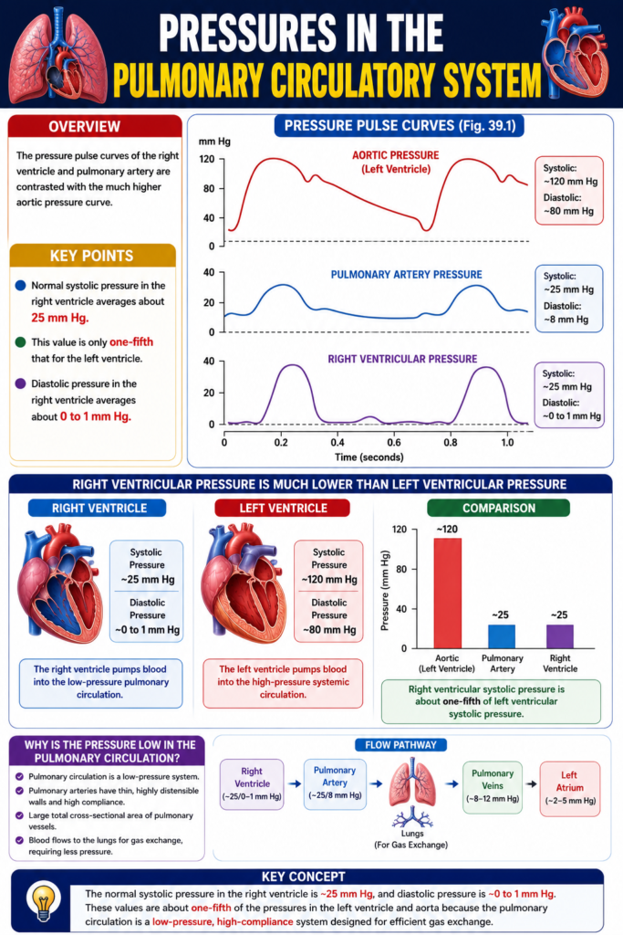
PRESSURES IN THE PULMONARY ARTERY
Figure: Fig. 39.1 and Fig. 39.2
Pressure During Systole
- During systole, the pressure in the pulmonary artery is almost the same as the pressure in the right ventricle.
- This relationship is shown in Fig. 39.1.
Pressure After Systole
- At the end of systole, the pulmonary valve closes.
- After the valve closes, the pressure in the right ventricle falls rapidly.
- However, the pressure in the pulmonary artery falls more slowly.
- This slower fall occurs because blood continues to flow through the lungs.
Normal Pulmonary Artery Pressures
Figure: Fig. 39.2
- The normal systolic pulmonary arterial pressure is about 25 mm Hg.
- The normal diastolic pulmonary arterial pressure is about 8 mm Hg.
- The normal mean pulmonary arterial pressure is 15 mm Hg.
Easy Concept
Think of the right ventricle and pulmonary artery as two connected chambers.
During Systole
Right Ventricle Contracts
          │
          ▼
Pulmonary Valve Opens
          │
          ▼
Blood Flows Into Pulmonary Artery
          │
          ▼
Both Have Nearly the Same Pressure
(≈25 mm Hg)
After Systole
Pulmonary Valve Closes
          │
          ▼
Right Ventricle Relaxes
          │
          ▼
Right Ventricular Pressure Falls Quickly
          │
          ▼
Pulmonary Artery Still Contains Blood
          │
          ▼
Pressure Falls Slowly
Why Does the Pulmonary Artery Pressure Fall Slowly?
- The right ventricle stops pumping after systole.
- However, the pulmonary artery still contains blood.
- This blood continues flowing through the lungs.
- Therefore, the pulmonary artery pressure decreases gradually, not suddenly.
Key Concept
- Fig. 39.1 shows that during systole, the pulmonary artery pressure is almost equal to the right ventricular pressure.
- After the pulmonary valve closes, the right ventricular pressure falls rapidly, while the pulmonary artery pressure falls gradually because blood continues to flow through the lungs.
- Fig. 39.2 shows the normal pulmonary artery pressures:
- Systolic pressure = 25 mm Hg
- Diastolic pressure = 8 mm Hg
- Mean pressure = 15 mm Hg
PRESSURES IN THE PULMONARY ARTERY
Figure: Fig. 39.1 and Fig. 39.2
Pressure During Systole
- During systole, the pressure in the pulmonary artery is almost the same as the pressure in the right ventricle.
- This relationship is shown in Fig. 39.1.
Pressure After Systole
- At the end of systole, the pulmonary valve closes.
- After the valve closes, the pressure in the right ventricle falls rapidly.
- However, the pressure in the pulmonary artery falls more slowly.
- This slower fall occurs because blood continues to flow through the lungs.
Normal Pulmonary Artery Pressures
Figure: Fig. 39.2
- Systolic pulmonary arterial pressure = 25 mm Hg
- Diastolic pulmonary arterial pressure = 8 mm Hg
- Mean pulmonary arterial pressure = 15 mm Hg
Easy Concept
Think of the right ventricle as a pump and the pulmonary artery as a pipe filled with blood.
During Systole
Right Ventricle Contracts
          │
          ▼
Pulmonary Valve Opens
          │
          ▼
Blood Enters Pulmonary Artery
          │
          ▼
Right Ventricle and Pulmonary Artery
Have Almost the Same Pressure
(≈25 mm Hg)
Concept:
- The pulmonary valve is open.
- Blood flows freely from the right ventricle into the pulmonary artery.
- Therefore, both chambers have nearly the same pressure.
After Systole
Right Ventricle Stops Contracting
          │
          ▼
Pulmonary Valve Closes
          │
          ▼
Right Ventricular Pressure Falls Quickly
          │
          ▼
Blood Already Inside Pulmonary Artery
Continues Flowing Through the Lungs
          │
          ▼
Pulmonary Artery Pressure Falls Slowly
Concept:
- The right ventricle relaxes immediately.
- The pulmonary artery still contains blood.
- This blood continues moving through the lungs.
- Therefore, pulmonary artery pressure decreases gradually, not suddenly.
Why Does the Pulmonary Artery Pressure Fall Slowly?
- The right ventricle has stopped pumping.
- But the pulmonary artery still contains blood under pressure.
- This blood continues flowing through the lungs.
- Therefore, the pulmonary artery pressure falls slowly, while the right ventricular pressure falls rapidly.
Key Concept
- Fig. 39.1 shows that during systole, the right ventricle and pulmonary artery have almost the same pressure because the pulmonary valve is open.
- After systole, the pulmonary valve closes.
- The right ventricle relaxes immediately, so its pressure falls rapidly.
- The pulmonary artery still contains blood, so its pressure falls gradually as blood continues to flow through the lungs.
- Fig. 39.2 shows the normal pulmonary artery pressures:
- Systolic = 25 mm Hg
- Diastolic = 8 mm Hg
- Mean = 15 mm Hg
This is the style I’ll continue using: sentence-by-sentence conversion, followed by a conceptual flow diagram, a “Why?” explanation for difficult physiology, and a concise Key Concept section for revision. I think this approach will help MBBS students understand the concepts much more quickly than memorizing the textbook alone.
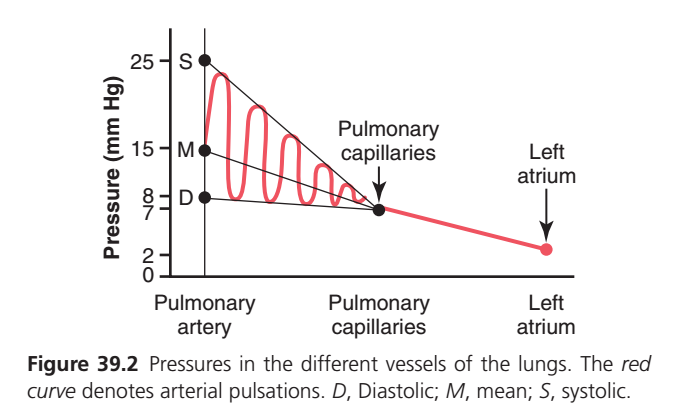
This figure explains how blood pressure changes as blood flows through the pulmonary circulation, from the pulmonary artery → pulmonary capillaries → left atrium.
The most important concept is:
As blood moves through the lungs, its pressure gradually falls because energy is lost due to resistance in the pulmonary blood vessels.
Let’s understand every line, every point, every wave, and every label in the easiest conceptual way for MBBS students.
Big Picture (One-Line Story)
Blood follows this path:
Right Ventricle → Pulmonary Artery → Pulmonary Capillaries → Pulmonary Veins → Left Atrium
As blood moves forward,
Pressure continuously decreases.
This graph shows exactly how much it decreases.
Step 1: Understanding the Axes
Y-axis (Vertical)
Pressure (mmHg)
This tells us
How much force blood is pushing against the vessel wall.
Higher on the graph = Higher pressure
Lower on the graph = Lower pressure
Important values shown are:
- 25 mmHg
- 15 mmHg
- 8 mmHg
- 7 mmHg
- 2 mmHg
X-axis (Horizontal)
This is not time.
Instead, it shows where the blood is.
Blood moves from left to right.
Pulmonary Artery
        ↓
Pulmonary Capillaries
        ↓
Left Atrium
Three Main Regions
The graph is divided into three parts.
Pulmonary Artery
        ↓
Pulmonary Capillaries
        ↓
Left Atrium
Each part has a different pressure.ART 1 — Pulmonary Artery
Look at the left side.
Three letters are marked.
S
M
D
These represent three different pressures.
S = Systolic Pressure
Approximately
25 mmHg
This is the highest pressure.
Why?
The right ventricle contracts.
Blood is forcefully pushed into the pulmonary artery.
Pressure rises to about 25 mmHg.
M = Mean Pressure
Approximately
15 mmHg
This is the average pressure during the whole cardiac cycle.
It is not the highest and not the lowest.
It is simply the average pressure in the pulmonary artery.
D = Diastolic Pressure
Approximately
8 mmHg
This is the pressure after the right ventricle relaxes.
Even though the ventricle has stopped pumping,
the pulmonary artery still has pressure because it is elastic.
So the Pulmonary Artery Pressure is:
| Pressure | Value |
|---|---|
| Systolic | 25 mmHg |
| Mean | 15 mmHg |
| Diastolic | 8 mmHg |
PART 2 — Red Pulsating Curve
This is the red wavy line.
Many students wonder:
Why are there waves?
Because every heartbeat produces one pressure pulse.
One heartbeat
↓
One pressure wave
↓
Another heartbeat
↓
Another pressure wave
So each wave represents one cardiac cycle.
Why Are the Waves Tall at First?
Near the pulmonary artery,
blood is very close to the right ventricle.
The pumping force is still strong.
Therefore,
the pressure pulses are large.
Why Do the Waves Become Smaller?
As blood travels farther,
it passes through many branching arteries and arterioles.
These vessels absorb part of the pressure.
Energy is lost because of vascular resistance.
Therefore,
each pulse becomes smaller and smaller.
This process is called:
Damping (attenuation) of the pressure pulse.
Think of dropping a stone into water.
Near the stone,
waves are big.
Far away,
waves become tiny.
Exactly the same happens to blood pressure waves.
PART 3 — Sloping Black Lines
There are two black sloping lines.
One starts from 25 mmHg.
Another starts from 15 mmHg.
These connect to the pulmonary capillaries.
What do they show?
They illustrate that
both
- Systolic pressure
- Mean pressure
gradually decrease as blood moves through the pulmonary circulation.
The fall is continuous.
PART 4 — Pulmonary Capillaries
This point is marked around
7 mmHg
Why has the pressure become so low?
Because blood has already passed through many pulmonary vessels.
Resistance has reduced the pressure.
Why is this low pressure important?
The lungs contain extremely delicate air sacs (alveoli).
If capillary pressure were very high,
fluid would leak into the alveoli,
causing pulmonary edema.
A low capillary pressure helps keep the alveoli dry and efficient for gas exchange.
Why Is Pulmonary Capillary Pressure About 7 mmHg?
This value is ideal because:
- It is high enough to maintain blood flow.
- It is low enough to prevent fluid leakage into the alveoli.
This balance allows efficient gas exchange without flooding the lungs.PART 5 — Straight Red Line After Capillaries
After the capillaries,
pressure continues to decrease.
There are no more pressure waves.
Why?
Because the capillaries and small veins absorb almost all of the pulsations.
By the time blood reaches the pulmonary veins,
the flow is nearly steady.
PART 6 — Left Atrium
The pressure finally reaches about
2 mmHg
This is the normal left atrial pressure.
Why is it so low?
The left atrium is relaxed and continuously receiving oxygenated blood from the lungs.
Only a small pressure is needed for blood to flow into the left ventricle.
Why Does Pressure Continuously Fall?
Imagine water flowing through a long garden hose.
At the tap,
pressure is high.
As water travels,
friction with the hose walls reduces pressure.
By the end,
pressure is lower.
Blood behaves in the same way.
Every vessel offers some resistance.
As blood moves forward,
pressure is gradually lost.
Why Do Pressure Pulses Disappear?
Initially,
the pulmonary artery receives blood directly from the pumping right ventricle.
So pressure rises and falls with every heartbeat.
As blood passes through many elastic arteries, arterioles, and capillaries,
these vessels stretch and absorb the pulsations.
By the time blood reaches the capillaries and veins,
the flow becomes smooth and almost non-pulsatile.
Clinical Importance
1. Prevents Pulmonary Edema
Low pulmonary capillary pressure (~7 mmHg) minimizes filtration of fluid into the alveoli, keeping them dry for efficient gas exchange.
2. Protects the Alveoli
The thin alveolar-capillary membrane could be damaged by high pressures.
The low-pressure pulmonary circulation protects this delicate structure.
3. Reduces the Workload of the Right Ventricle
Because the lungs have low vascular resistance, the right ventricle only needs to generate about 25 mmHg during systole, much less than the left ventricle.
Easy Story to Remember
Imagine blood as a cyclist going downhill.
At the pulmonary artery, the cyclist starts with a lot of energy (high pressure and strong pulses).
As the cyclist rides through many small roads (arterioles and capillaries), friction slows them down, and the bumps become smoother.
By the time they reach the left atrium, they are moving gently with low, steady pressure.
High-Yield MBBS Points
- Pulmonary artery pressure: 25/8 mmHg, with a mean pressure of ~15 mmHg.
- S (25 mmHg): Systolic pressure during right ventricular contraction.
- M (15 mmHg): Mean pulmonary artery pressure.
- D (8 mmHg): Diastolic pulmonary artery pressure.
- Pulmonary capillary pressure: Approximately 7 mmHg, ideal for gas exchange while preventing pulmonary edema.
- Left atrial pressure: Approximately 2 mmHg, allowing easy filling of the left heart.
- Pressure pulses decrease (damping): As blood moves away from the right ventricle, resistance and vessel elasticity absorb the pulsations.
- Key concept: Blood pressure falls progressively from the pulmonary artery to the left atrium because of vascular resistance, while the pulsatile flow gradually becomes smooth before reaching the pulmonary veins and left atrium.
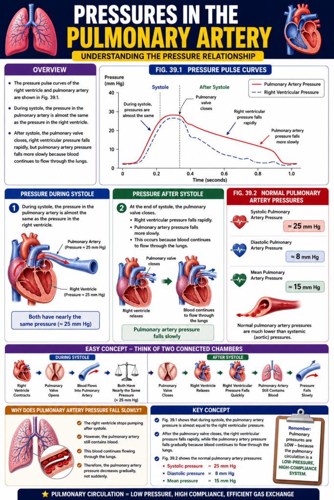
Pulmonary Capillary Pressure
Figure: Fig. 39.2
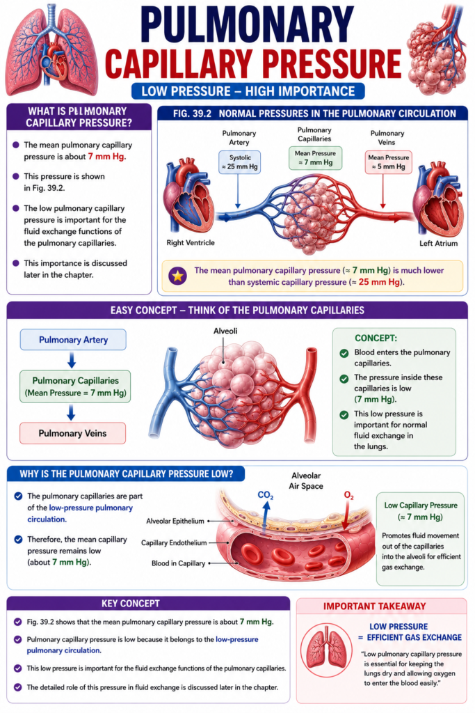
- The mean pulmonary capillary pressure is about 7 mm Hg.
- This pressure is shown in Fig. 39.2.
- The low pulmonary capillary pressure is important for the fluid exchange functions of the pulmonary capillaries.
- This importance is discussed later in the chapter.
Easy Concept
Think of the pulmonary capillaries as tiny blood vessels surrounding the alveoli.
Pulmonary Artery
      │
      ▼
Pulmonary Capillaries
(Mean Pressure = 7 mm Hg)
      │
      ▼
Pulmonary Veins
Concept:
- Blood enters the pulmonary capillaries.
- The pressure inside these capillaries is low (7 mm Hg).
- This low pressure is important for normal fluid exchange in the lungs.
Why Is the Pulmonary Capillary Pressure Low?
- The pulmonary capillaries are part of the low-pressure pulmonary circulation.
- Therefore, the mean capillary pressure remains low (about 7 mm Hg).
Key Concept
- Fig. 39.2 shows that the mean pulmonary capillary pressure is about 7 mm Hg.
- Pulmonary capillary pressure is low because it belongs to the low-pressure pulmonary circulation.
- This low pressure is important for the fluid exchange functions of the pulmonary capillaries.
- The detailed role of this pressure in fluid exchange is discussed later in the chapter.
Left Atrial and Pulmonary Venous Pressures
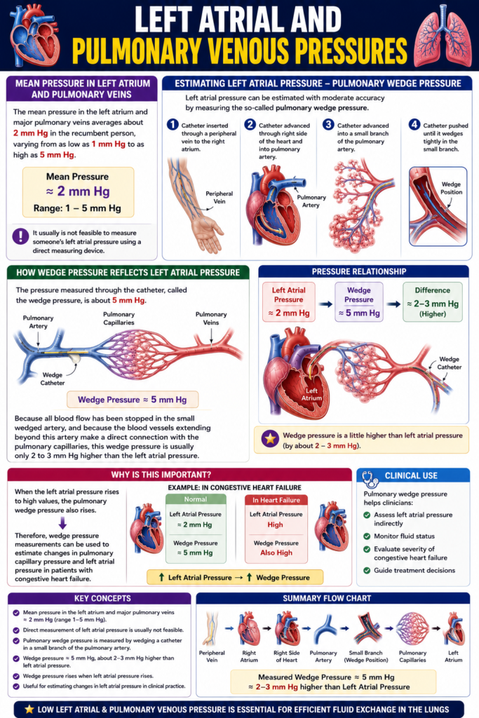
- The mean pressure in the left atrium and major pulmonary veins is about 2 mm Hg in a recumbent (lying) person.
- This pressure normally ranges from 1 mm Hg to 5 mm Hg.
Direct Measurement of Left Atrial Pressure
- Direct measurement of left atrial pressure is usually not feasible.
- This is because it is difficult to pass a catheter through the heart chambers into the left atrium.
Pulmonary Wedge Pressure
- The left atrial pressure can be estimated by measuring the pulmonary wedge pressure.
- This method provides moderately accurate estimation of left atrial pressure.
How Pulmonary Wedge Pressure Is Measured
Easy Concept
Think of the catheter as traveling step by step through the circulation.
Peripheral Vein
      │
      ▼
Right Atrium
      │
      ▼
Right Ventricle
      │
      ▼
Pulmonary Artery
      │
      ▼
Small Pulmonary Artery Branch
      │
      ▼
Catheter Becomes Wedged
Concept:
- The catheter blocks the small pulmonary artery branch.
- Beyond the blockage, the blood vessels are directly connected to the pulmonary capillaries.
- Therefore, the measured pressure closely reflects the pressure beyond the blockage, including the left atrial pressure.
Wedge Pressure
- The normal pulmonary wedge pressure is about 5 mm Hg.
- Blood flow stops in the small wedged artery.
- The blood vessels beyond the wedged artery are directly connected to the pulmonary capillaries.
- Therefore, the pulmonary wedge pressure is usually only 2–3 mm Hg higher than the left atrial pressure.
Easy Concept
Left Atrial Pressure
      │
      ▼
About 2 mm Hg
      │
      ▼
Pulmonary Wedge Pressure
      │
      ▼
About 5 mm Hg
Concept:
- The catheter cannot enter the left atrium directly.
- Instead, the wedge pressure acts as an indirect estimate of left atrial pressure.
- Since the difference is usually only 2–3 mm Hg, the wedge pressure provides a good estimate of left atrial pressure.
Clinical Importance
- If the left atrial pressure increases, the pulmonary wedge pressure also increases.
- Therefore, pulmonary wedge pressure can be used to estimate changes in:
- Pulmonary capillary pressure
- Left atrial pressure
- This measurement is useful in patients with congestive heart failure.
Key Concept
- Mean left atrial and pulmonary venous pressure = about 2 mm Hg (normal range 1–5 mm Hg).
- Direct measurement of left atrial pressure is difficult, so it is usually estimated using pulmonary wedge pressure.
- The catheter passes through:
- Peripheral vein → Right atrium → Right ventricle → Pulmonary artery → Small pulmonary artery branch (wedged).
- Normal pulmonary wedge pressure = about 5 mm Hg.
- Pulmonary wedge pressure is usually only 2–3 mm Hg higher than the left atrial pressure because the vessels beyond the wedged artery are directly connected to the pulmonary capillaries.
- An increase in left atrial pressure causes an increase in pulmonary wedge pressure.
- Pulmonary wedge pressure is used to estimate pulmonary capillary pressure and left atrial pressure in patients with congestive heart failure.
MADE BY SELF LEARNING CEO AND FOUNDER DR SHEEN
