



- Gas exchange between the alveolar air and the pulmonary capillary blood is discussed in the next chapter.
- Here, an important feature of the pulmonary capillaries is explained.
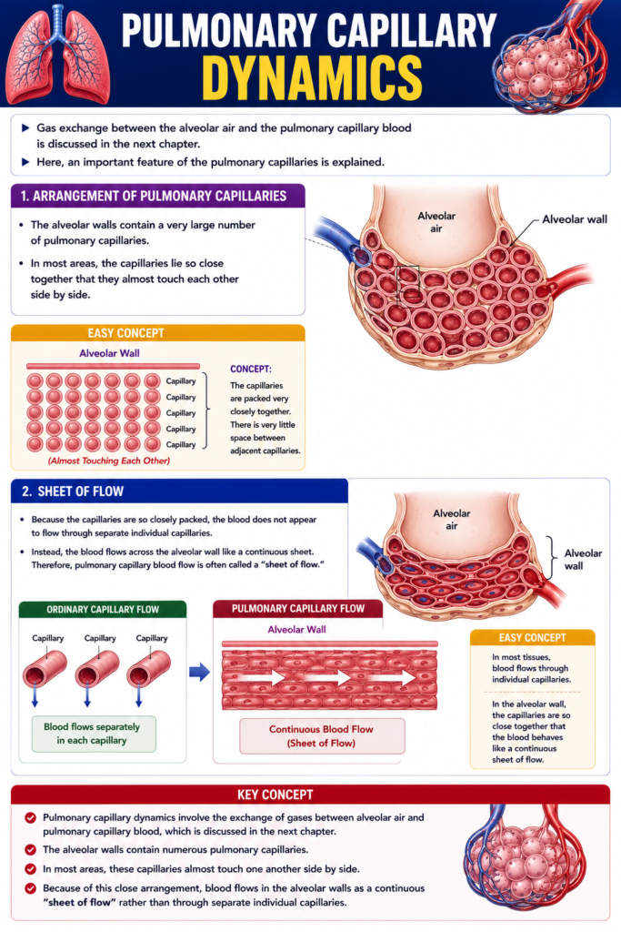
Arrangement of Pulmonary Capillaries
- The alveolar walls contain a very large number of pulmonary capillaries.
- In most areas, the capillaries lie so close together that they almost touch each other side by side.
Easy Concept
Think of the alveolar wall as being covered by a dense network of capillaries.
Alveolar Wall

══════════════════════════

||||||||||||||||||||||||||
Capillary Capillary Capillary
Capillary Capillary Capillary
Capillary Capillary Capillary

(Almost Touching Each Other)
Concept:
- The capillaries are packed very closely together.
- There is very little space between adjacent capillaries.
Sheet of Flow
- Because the capillaries are so closely packed, the blood does not appear to flow through separate individual capillaries.
- Instead, the blood flows across the alveolar wall like a continuous sheet.
- Therefore, pulmonary capillary blood flow is often called a “sheet of flow.”
Easy Concept
Ordinary Capillary Flow
Capillary     Capillary     Capillary

   │             │             │
   ▼             ▼             ▼

Blood flows separately
in each capillary
Pulmonary Capillary Flow
══════════════════════════
Alveolar Wall

██████████████████████████
Continuous Blood Flow
(Sheet of Flow)
Concept:
- In most tissues, blood flows through individual capillaries.
- In the alveolar wall, the capillaries are so close together that the blood behaves like a continuous sheet of flow.
Key Concept
- Pulmonary capillary dynamics involve the exchange of gases between alveolar air and pulmonary capillary blood, which is discussed in the next chapter.
- The alveolar walls contain numerous pulmonary capillaries.
- In most areas, these capillaries almost touch one another side by side.
- Because of this close arrangement, blood flows in the alveolar walls as a continuous “sheet of flow” rather than through separate individual capillaries.
Pulmonary Capillary Pressure
- Direct measurement of pulmonary capillary pressure has not been reported.
- Therefore, the pulmonary capillary pressure is estimated indirectly.
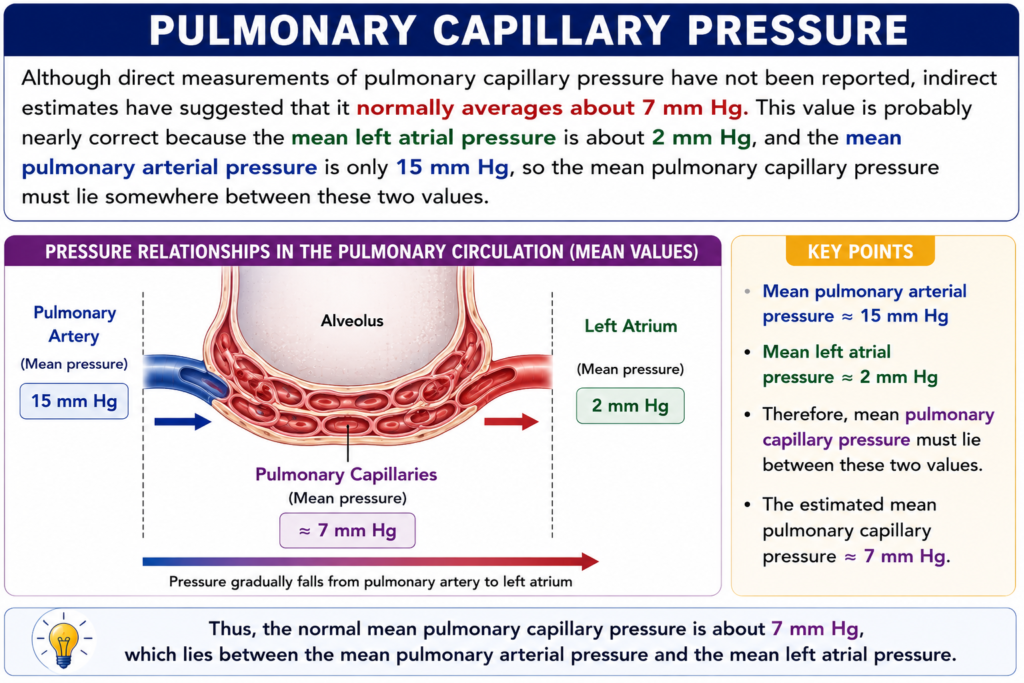
- These indirect estimates suggest that the mean pulmonary capillary pressure is about 7 mm Hg.
Why Is the Pulmonary Capillary Pressure About 7 mm Hg?
- The mean left atrial pressure is about 2 mm Hg.
- The mean pulmonary arterial pressure is about 15 mm Hg.
- The pulmonary capillaries lie between the pulmonary artery and the left atrium.
- Therefore, the pulmonary capillary pressure must lie somewhere between these two pressures.
- For this reason, the mean pulmonary capillary pressure is about 7 mm Hg.
Easy Concept
Think of the pulmonary capillaries as being between the pulmonary artery and the left atrium.
Pulmonary Artery
(15 mm Hg)
        │
        ▼
Pulmonary Capillaries
(?)
        │
        ▼
Left Atrium
(2 mm Hg)
Concept:
- Blood flows from the pulmonary artery to the left atrium.
- Therefore, the capillary pressure must be between 15 mm Hg and 2 mm Hg.
Step-by-Step Understanding
Step 1
Mean Pulmonary Arterial Pressure = 15 mm Hg
↓
Step 2
Mean Left Atrial Pressure = 2 mm Hg
↓
Step 3
Pulmonary capillaries are located between these two structures.
↓
Step 4
Therefore,
Pulmonary Capillary Pressure must lie between 15 mm Hg and 2 mm Hg.
↓
Final Value
Mean Pulmonary Capillary Pressure ≈ 7 mm Hg
Easy Memory Trick
Pulmonary Artery
15 mm Hg
      │
      ▼
Pulmonary Capillary
7 mm Hg
      │
      ▼
Left Atrium
2 mm Hg
Remember:
15 → 7 → 2
(Artery → Capillary → Left Atrium)
Key Concept
- Pulmonary capillary pressure is not measured directly.
- It is estimated indirectly.
- The mean pulmonary capillary pressure is about 7 mm Hg.
- This value is reasonable because:
- Mean pulmonary arterial pressure = 15 mm Hg
- Mean left atrial pressure = 2 mm Hg
- Since the pulmonary capillaries are located between the pulmonary artery and the left atrium, their pressure must lie between these two values, giving an average of about 7 mm Hg.
Length of Time Blood Stays in the Pulmonary Capillaries
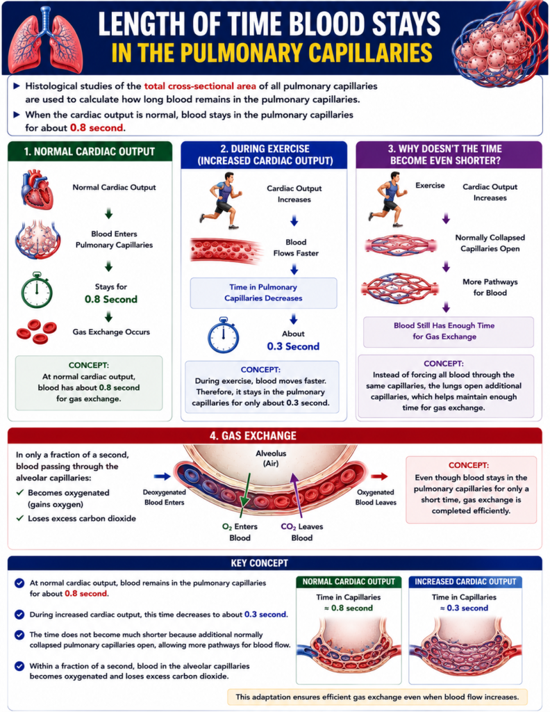
- Histological studies of the total cross-sectional area of all pulmonary capillaries are used to calculate how long blood remains in the pulmonary capillaries.
- When the cardiac output is normal, blood stays in the pulmonary capillaries for about 0.8 second.
During Increased Cardiac Output
- When the cardiac output increases, the time blood stays in the pulmonary capillaries decreases.
- It may shorten to about 0.3 second.
Easy Concept
Normal Cardiac Output
Normal Cardiac Output
        │
        ▼
Blood Enters
Pulmonary Capillaries
        │
        ▼
Stays for
0.8 Second
        │
        ▼
Gas Exchange Occurs
Concept:
- At normal cardiac output, blood has about 0.8 second for gas exchange.
During Exercise (Increased Cardiac Output)
Cardiac Output Increases
        │
        ▼
Blood Flows Faster
        │
        ▼
Time in Pulmonary Capillaries
Decreases
        │
        ▼
About 0.3 Second
Concept:
- During exercise, blood moves faster.
- Therefore, it stays in the pulmonary capillaries for only about 0.3 second.
Why Doesn’t the Time Become Even Shorter?
- The time does not become much shorter because additional pulmonary capillaries open.
- These capillaries are normally collapsed.
- When they open, they accommodate the increased blood flow.
Easy Concept
Exercise
        │
        ▼
Cardiac Output Increases
        │
        ▼
Normally Collapsed
Capillaries Open
        │
        ▼
More Pathways for Blood
        │
        ▼
Blood Still Has Enough Time
for Gas Exchange
Concept:
- Instead of forcing all blood through the same capillaries, the lungs open additional capillaries.
- This helps maintain enough time for gas exchange, even when blood flow increases.
Gas Exchange
- In only a fraction of a second, blood passing through the alveolar capillaries:
- Becomes oxygenated (gains oxygen)
- Loses excess carbon dioxide
Easy Concept
Blood Enters
Pulmonary Capillary
        │
        ▼
O₂ Enters Blood
        │
        ▼
CO₂ Leaves Blood
        │
        ▼
Blood Leaves Oxygenated
Concept:
- Even though blood stays in the pulmonary capillaries for only a short time, gas exchange is completed efficiently.
Key Concept
- At normal cardiac output, blood remains in the pulmonary capillaries for about 0.8 second.
- During increased cardiac output, this time decreases to about 0.3 second.
- The time does not become much shorter because additional normally collapsed pulmonary capillaries open, allowing more pathways for blood flow.
- Within a fraction of a second, blood in the alveolar capillaries becomes oxygenated and loses excess carbon dioxide.
Capillary Exchange of Fluid in the Lungs and Pulmonary Interstitial Fluid Dynamics
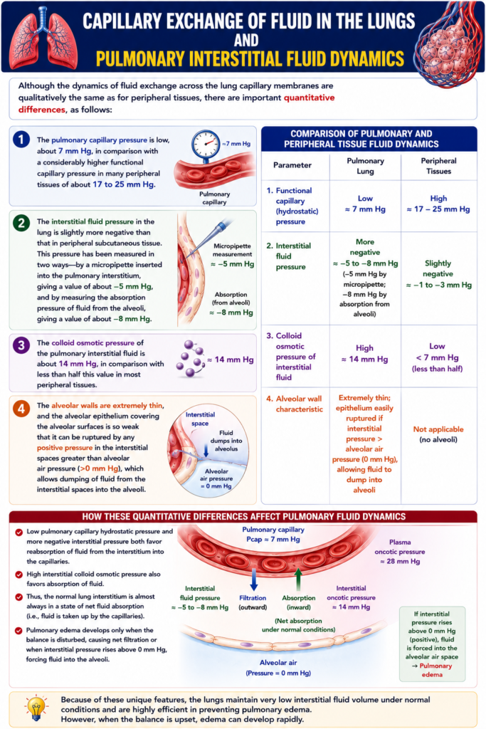
- Fluid exchange across the pulmonary capillary membrane is qualitatively similar to fluid exchange in peripheral tissues.
- However, there are important quantitative differences between the lungs and peripheral tissues.
1. Pulmonary Capillary Pressure
- The pulmonary capillary pressure is low, about 7 mm Hg.
- In comparison, the functional capillary pressure in many peripheral tissues is about 17–25 mm Hg.
Easy Concept
Pulmonary Capillary
Pressure
= 7 mm Hg

↓

Peripheral Tissue
Capillary Pressure
= 17–25 mm Hg
Concept:
- The lungs have much lower capillary pressure than most peripheral tissues.
2. Pulmonary Interstitial Fluid Pressure
- The interstitial fluid pressure in the lungs is slightly more negative than in peripheral subcutaneous tissue.
- This pressure has been measured in two ways.
Method 1: Micropipette Measurement
- A micropipette inserted into the pulmonary interstitium measures a pressure of about −5 mm Hg.
Method 2: Alveolar Fluid Absorption Measurement
- Measuring the absorption pressure of fluid from the alveoli gives a value of about −8 mm Hg.
Easy Concept
Pulmonary Interstitium

Method 1
Micropipette
        │
        ▼
−5 mm Hg

Method 2
Fluid Absorption
from Alveoli
        │
        ▼
−8 mm Hg
Concept:
- Both methods show that the interstitial pressure in the lungs is negative.
3. Colloid Osmotic Pressure of Pulmonary Interstitial Fluid
- The colloid osmotic pressure of the pulmonary interstitial fluid is about 14 mm Hg.
- In most peripheral tissues, the colloid osmotic pressure is less than half of this value.
Easy Concept
Pulmonary Interstitial
Colloid Osmotic Pressure

= 14 mm Hg

↓

Peripheral Tissues

Less Than Half
of 14 mm Hg
Concept:
4. Thin Alveolar Walls
- The alveolar walls are extremely thin.
- The alveolar epithelium covering the alveolar surface is very weak.
- If the interstitial pressure becomes greater than the alveolar air pressure (>0 mm Hg), the alveolar epithelium can rupture.
- This allows fluid to move from the interstitial space into the alveoli.
Easy Concept
Interstitial Pressure
>0 mm Hg
        │
        ▼
Alveolar Epithelium
Ruptures
        │
        ▼
Fluid Moves
Into Alveoli
Concept:
- Positive interstitial pressure can break the thin alveolar wall.
- As a result, fluid enters the alveoli.
Step-by-Step Understanding
Step 1
Normal Pulmonary Capillary Pressure = 7 mm Hg
↓
Step 2
Pulmonary Interstitial Pressure = −5 to −8 mm Hg
↓
Step 3
Pulmonary Interstitial Colloid Osmotic Pressure = 14 mm Hg
↓
Step 4
If Interstitial Pressure > 0 mm Hg
↓
Alveolar Epithelium Ruptures
↓
Fluid Moves Into the Alveoli
Key Concept
- Pulmonary capillary fluid exchange is qualitatively similar to peripheral tissues, but important quantitative differences exist.
- Pulmonary capillary pressure = about 7 mm Hg, compared with 17–25 mm Hg in many peripheral tissues.
- Pulmonary interstitial fluid pressure is negative:
- Micropipette measurement = about −5 mm Hg
- Alveolar fluid absorption measurement = about −8 mm Hg
- Pulmonary interstitial colloid osmotic pressure = about 14 mm Hg, which is higher than in most peripheral tissues.
- The alveolar walls are extremely thin, and the alveolar epithelium is weak.
- If interstitial pressure rises above alveolar air pressure (>0 mm Hg), the alveolar epithelium can rupture, allowing fluid to move from the interstitial space into the alveoli.
Interrelationships Between Interstitial Fluid Pressure and Other Pressures in the Lung
Figure: Fig. 39.7
- Fig. 39.7 shows:
- A pulmonary capillary
- An alveolus
- A lymphatic capillary
- The lymphatic capillary drains the interstitial space between the pulmonary capillary and the alveolus.
- Fig. 39.7 also shows the balance of forces acting across the pulmonary capillary membrane.
Forces Causing Fluid to Move Out of the Capillary
1. Capillary Hydrostatic Pressure
- Capillary hydrostatic pressure = 7 mm Hg
- This pressure pushes fluid out of the pulmonary capillary into the interstitial space.
2. Interstitial Fluid Colloid Osmotic Pressure
- Interstitial fluid colloid osmotic pressure = 14 mm Hg
- This pressure pulls fluid out of the pulmonary capillary into the interstitial space.
3. Negative Interstitial Fluid Hydrostatic Pressure
- Negative interstitial fluid hydrostatic pressure = 8 mm Hg
- This negative pressure pulls fluid outward from the capillary into the interstitial space.
Step-by-Step Calculation of Total Outward Force
Formula
Total Outward Force

= Capillary Hydrostatic Pressure
+ Interstitial Fluid Colloid Osmotic Pressure
+ Negative Interstitial Fluid Hydrostatic Pressure
Substitute the Values
= 7 + 14 + 8Solve
= 29 mm HgFinal Answer
Total Outward Force = 29 mm Hg
Easy Concept
Fluid Inside Capillary

        │
        ▼

Capillary Hydrostatic Pressure
        7 mm Hg

+

Interstitial Colloid Osmotic Pressure
       14 mm Hg

+

Negative Interstitial Pressure
        8 mm Hg

        │
        ▼

Total Outward Force

29 mm Hg
Concept:
- These three forces work together.
- They move fluid from the pulmonary capillary into the interstitial space.
Force Causing Fluid to Move Into the Capillary
Plasma Colloid Osmotic Pressure
- Plasma colloid osmotic pressure = 28 mm Hg
- This pressure pulls fluid back into the pulmonary capillary.
Easy Concept
Interstitial Space

        │
        ▼

Plasma Colloid Osmotic Pressure

28 mm Hg

        │
        ▼

Fluid Pulled Back
Into the Capillary
Concept:
- Plasma proteins attract water back into the blood.
Step-by-Step Calculation of Net Filtration Pressure
Formula
Net Filtration Pressure

= Total Outward Force
− Total Inward Force
Substitute the Values
= 29 − 28Solve
= +1 mm HgFinal Answer
Net Filtration Pressure = +1 mm Hg
Easy Concept
Outward Force
29 mm Hg

        │
        ▼

Inward Force
28 mm Hg

        │
        ▼

Net Filtration Pressure

+1 mm Hg
Concept:
- The outward force is only slightly greater than the inward force.
- Therefore, only a small amount of fluid leaves the pulmonary capillary.
Effect of the +1 mm Hg Filtration Pressure
- The +1 mm Hg filtration pressure causes a small continuous movement of fluid from the pulmonary capillaries into the interstitial spaces.
- A small amount of this fluid evaporates into the alveoli.
- Most of the filtered fluid is returned to the circulation through the pulmonary lymphatic system.
Easy Concept
Pulmonary Capillary
        │
        ▼
Small Amount of Fluid
Leaves the Capillary
(+1 mm Hg)
        │
        ▼
Interstitial Space
        │
        ├──────────────┐
        ▼              ▼
Small Amount      Pulmonary
Evaporates        Lymphatics
Into Alveoli      Return Fluid
                  to Blood
Concept:
- A tiny amount of fluid normally leaves the capillary.
- The lymphatic vessels continuously remove this fluid, preventing fluid accumulation.
Key Concept
- Fig. 39.7 shows a pulmonary capillary, alveolus, and lymphatic capillary, along with the forces controlling fluid movement.
- Outward forces:
- Capillary hydrostatic pressure = 7 mm Hg
- Interstitial fluid colloid osmotic pressure = 14 mm Hg
- Negative interstitial fluid hydrostatic pressure = 8 mm Hg
- Total outward force = 7 + 14 + 8 = 29 mm Hg
- Inward force:
- Plasma colloid osmotic pressure = 28 mm Hg
- Net filtration pressure = 29 − 28 = +1 mm Hg
- This +1 mm Hg pressure causes a small continuous filtration of fluid into the pulmonary interstitium.
- Most of this fluid is returned to the circulation by the pulmonary lymphatic system, while a small amount evaporates into the alveoli.
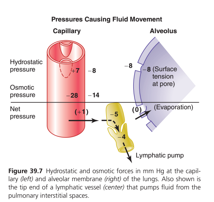
Figure 39.7: Pressures Causing Fluid Movement Across Pulmonary Capillaries (Guyton Physiology 15th Edition)
🎯 One-Line Core Concept
Normally, only a very tiny amount of fluid filters out of pulmonary capillaries into the lung interstitium (+1 mmHg), and this fluid is immediately removed by the lymphatic system. This keeps the alveoli dry and prevents pulmonary edema.
🧠 First Understand the Big Picture
Imagine an alveolus (air sac) beside a pulmonary capillary (blood vessel).
Pulmonary Capillary
        │
        │
 Interstitial Space
        │
        │
     Alveolus
Fluid is continuously trying to move from the blood into the lung tissue.
At the same time,
other forces pull the fluid back.
The balance between these forces determines whether:
- Fluid stays inside blood vessels ✅
- Fluid enters lung tissue ❌
- Pulmonary edema develops ❌
This figure explains all these forces.
First Learn Four Important Pressures
There are 4 Starling forces acting across the pulmonary capillary.
Think of them as two forces pushing fluid out and two forces pulling fluid back in.
Force 1
Pulmonary Capillary Hydrostatic Pressure
Value = +7 mmHg
Shown in the capillary.
What is Hydrostatic Pressure?
This is simply
Blood pressure inside the pulmonary capillary.
Blood pushes against the capillary wall.
It tries to push fluid outward.
Imagine squeezing a water-filled balloon.
Water wants to escape.
Exactly the same thing happens here.
Direction
Capillary
   │
   │
Fluid pushed OUT
Therefore
Hydrostatic pressure
➡ Pushes fluid OUT
Force 2
Interstitial Hydrostatic Pressure
Value = −8 mmHg
This is written outside the capillary.
Many students find the negative sign confusing.
Why is it Negative?
The pressure in the lung interstitium is actually slightly below atmospheric pressure.
Think of it as a gentle vacuum.
Instead of pushing fluid back,
it pulls fluid outward.
Imagine gently sucking on a straw.
Fluid is pulled toward the suction.
The negative interstitial pressure does the same.
Direction
Interstitial Space
      ↑
Fluid pulled outward
So,
although it is called hydrostatic pressure,
because it is negative, it helps pull fluid out of the capillary.
Total Outward Hydrostatic Force
There are now two outward forces.
Capillary Hydrostatic = +7

Interstitial Pressure = -8
Total outward force
7 + 8 = 15 mmHg outwardForce 3
Plasma Colloid Osmotic Pressure
Value = 28 mmHg
This is shown as
−28
What is this?
Blood proteins (especially albumin) cannot leave the capillary.
They attract water.
Think of albumin as a sponge inside the capillary.
It continuously pulls water back.
Direction
Fluid
← Back into capillary
Therefore,
Plasma proteins
➡ Pull fluid INTO the capillary.
Force 4
Interstitial Colloid Osmotic Pressure
Value = 14 mmHg
Shown as
−14
Why does it exist?
A small amount of protein is present in the lung interstitium.
These proteins also attract water.
Therefore,
they pull fluid out of the capillary.
Direction
Capillary
      │
Fluid pulled outward
Calculate the Net Pressure (Step by Step)
This is the easiest way to remember.
Step 1
Outward Forces
Capillary hydrostatic
+7Plus
Negative interstitial pressure
+8Total
15 mmHg outwardStep 2
Inward Force
Plasma oncotic pressure
28 mmHg inwardStep 3
Outward Osmotic Force
Interstitial oncotic pressure
14 mmHg outwardFinal Equation
Outward
15 + 14 = 29Inward
28Net movement
29 − 28 = +1 mmHg outwardThis is exactly why the figure shows
Net Pressure = +1 mmHg
What Does +1 mmHg Mean?
It means
Only a very tiny amount of fluid leaves the pulmonary capillary.
This is completely normal.
Without this tiny filtration,
lung tissues would not receive nutrients.
Where Does This Fluid Go?
Look at the yellow structure.
This is the
Lymphatic Vessel
Numbers
Outside the lymphatic
−5 mmHgInside the lymphatic
−4 mmHgThese negative pressures create a suction effect.
The lymphatic pump continuously removes filtered fluid.
Why Is the Lymphatic System So Important?
Imagine a floor where someone spills a few drops of water every minute.
A vacuum cleaner immediately removes those drops.
The floor stays dry.
The lymphatic system acts as that vacuum cleaner.
It prevents fluid from accumulating around the alveoli.
Now Look at the Alveolus
On the right side is the alveolus.
Notice several numbers.
Alveolar Pressure
Approximately
−8 mmHg
This negative pressure is mainly due to:
- Surface tension
- Interstitial forces around the alveolus
It helps keep the interstitial space under slight suction.
Surface Tension at the Pore
Surface tension also contributes to maintaining a negative pressure around the alveolus.
This assists in preventing fluid from flooding the alveolar air space.
Evaporation (0 mmHg)
Inside the alveolus,
water continuously evaporates into inspired air.
This evaporation helps keep the alveolar surface almost dry.
The figure marks this as approximately 0 mmHg because evaporation itself is not a major Starling pressure, but it contributes to keeping alveolar fluid minimal.
Why Don’t Alveoli Fill With Water?
This is the most important concept.
Three protective mechanisms keep alveoli dry:
① Very Low Net Filtration
Only
+1 mmHg
So only a tiny amount of fluid leaves capillaries.
② Lymphatic Drainage
Any filtered fluid is immediately removed.
③ Continuous Evaporation
Tiny amounts of fluid on the alveolar surface evaporate into inspired air.
What Happens in Left Heart Failure?
Suppose pulmonary capillary pressure rises.
Instead of
7 mmHgit becomes
20 mmHgNow,
Outward force becomes much greater.
Large amounts of fluid leave the capillary.
The lymphatics cannot remove all of it.
Fluid accumulates.
↓
Pulmonary edema develops.
↓
Alveoli fill with fluid.
↓
Severe breathlessness occurs.
Easy Flowchart
Normal Lung
        ↓
Only +1 mmHg filtration
        ↓
Small amount of fluid enters interstitium
        ↓
Lymphatic pump removes it
        ↓
Alveoli remain dry
        ↓
Normal gas exchange
Memory Trick
Think of a leaking water tank.
- 🚰 Capillary = Water tank
- 💧 Small leak = +1 mmHg filtration
- 🪣 Lymphatic = Drain removing leaked water
- 🌬️ Alveolus = Floor kept dry by evaporation
As long as the drain works, the floor stays dry.
If the leak becomes too large (e.g., increased pulmonary capillary pressure), the drain is overwhelmed, water accumulates, and the floor floods—just like pulmonary edema.
📚 High-Yield MBBS Points
Forces pushing fluid OUT of the pulmonary capillary
- Pulmonary capillary hydrostatic pressure = +7 mmHg
- Negative interstitial hydrostatic pressure = −8 mmHg (acts as an outward suction)
Forces opposing or modifying filtration
- Plasma colloid osmotic pressure = 28 mmHg → pulls fluid into the capillary.
- Interstitial colloid osmotic pressure = 14 mmHg → pulls fluid out into the interstitium.
Net filtration pressure
- Outward forces = 7 + 8 + 14 = 29 mmHg
- Inward force = 28 mmHg
- Net = +1 mmHg outward, producing only minimal filtration.
Why alveoli stay dry
- Very low net filtration pressure (+1 mmHg).
- Pulmonary lymphatics continuously remove filtered fluid.
- Evaporation from the alveolar surface limits fluid accumulation.
Clinical significance
An increase in pulmonary capillary hydrostatic pressure (e.g., left-sided heart failure or mitral valve disease) increases filtration, overwhelms lymphatic drainage, and leads to pulmonary edema, impairing gas exchange.
Negative Pulmonary Interstitial Pressure and Mechanism for Keeping Alveoli Dry
- Under normal conditions, the alveoli do not fill with fluid.
What Keeps the Alveoli Dry?
- The pulmonary capillaries and the pulmonary lymphatic system maintain a slight negative pressure in the pulmonary interstitial spaces.
Effect of Negative Interstitial Pressure
- Whenever extra fluid appears in the alveoli, the negative interstitial pressure mechanically pulls (sucks) the fluid into the lung interstitium.
- The fluid passes through the small openings between the alveolar epithelial cells.
Easy Concept
Extra Fluid
Inside Alveolus
        │
        ▼
Negative Interstitial
Pressure
        │
        ▼
Fluid Is Pulled Into
Lung Interstitium
Concept:
- The negative pressure acts like a gentle suction.
- It pulls extra fluid out of the alveoli, helping to keep them dry.
Removal of the Excess Fluid
- The excess fluid in the lung interstitium is carried away by the pulmonary lymphatic vessels.
Easy Concept
Alveolus
        │
Extra Fluid
        │
        ▼
Lung Interstitium
        │
        ▼
Pulmonary Lymphatics
        │
        ▼
Fluid Returned
to the Circulation
Concept:
- After fluid leaves the alveoli, the lymphatic system removes it.
- This prevents fluid accumulation in the lungs.
Normal Condition of the Alveoli
Easy Concept
Normal Alveolus
        │
        ▼
Mostly Dry
        │
        ▼
Small Amount of Fluid
Covers the Surface
        │
        ▼
Keeps the Alveoli Moist
Concept:
- The alveoli are not completely dry.
- A thin layer of fluid is normally present to keep the alveolar surface moist.
Step-by-Step Understanding
Step 1
Extra fluid enters the alveoli.
↓
Step 2
Negative interstitial pressure pulls the fluid into the lung interstitium.
↓
Step 3
Pulmonary lymphatics remove the excess fluid.
↓
Step 4
Only a thin moist layer remains on the alveolar surface.
↓
Final Result
Alveoli remain dry and ready for efficient gas exchange.
Key Concept
- Pulmonary capillaries and the pulmonary lymphatic system maintain a slight negative pressure in the pulmonary interstitium.
- This negative pressure pulls extra fluid from the alveoli into the lung interstitium through small openings between the alveolar epithelial cells.
- The pulmonary lymphatics remove the excess interstitial fluid.
- Under normal conditions, the alveoli remain dry, except for a thin layer of fluid that keeps the alveolar surface moist.
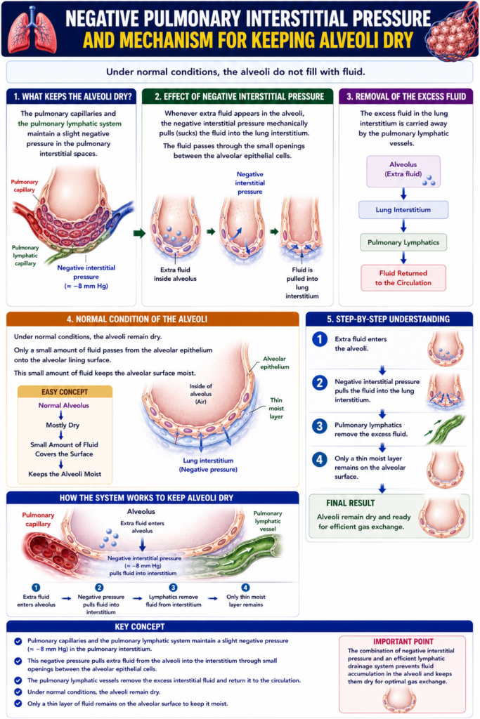
MADE BY SELF LEARNING CEO AND FOUNDER DR SHEEN
