


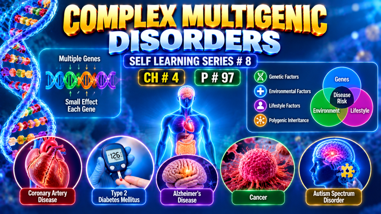
- Complex multigenic disorders are also called multifactorial or polygenic disorders.
- They result from interaction between:
- multiple genetic variants
- environmental factors
- A genetic variant present in at least 1% of the population is called a polymorphism.
- According to the common disease–common variant hypothesis:
- many common polymorphisms are inherited together
- each has only a small effect
- each usually has low penetrance
- together they increase disease risk
- Different polymorphisms do not contribute equally.
- Example: in type 1 diabetes, many genes are involved, but only a few contribute most of the risk.
- Certain HLA alleles account for more than 50% of the genetic risk.
- Some polymorphisms:
- are shared by several related diseases
- while others are specific to one disease
- Many disease-associated polymorphisms occur in noncoding DNA.
- Therefore, they may alter epigenetic regulation and gene expression, rather than directly changing protein structure.
- Multigenic inheritance also controls many normal traits, including:
- hair color
- eye color
- skin color
- height
- intelligence
- These traits show continuous variation among individuals.
- Environmental factors strongly modify how multigenic traits are expressed.
- Example:
- Type 2 diabetes has a strong genetic component.
- Weight gain and obesity may reveal or worsen the underlying genetic tendency.
- Thus:
genetic susceptibility + obesity/environment → clinical diabetes
- A disorder should be called multigenic only after careful evaluation.
- Important clues include:
- familial clustering
- absence of a clear Mendelian inheritance pattern
- absence of a chromosomal abnormality
- variable severity among affected individuals
- However, variable severity alone is not enough, because reduced penetrance and variable expressivity can also occur in single-gene disorders.
KEY CONCEPT
- Multigenic disease = many genes + environment working together.
- Polymorphism = common genetic variant present in ≥1% of the population.
- Each variant usually has a small effect, but many together can produce significant disease risk.
- Environment can unmask genetic susceptibility.
CONCEPTUAL EXAMPLES
- Many diabetes-risk genes + obesity → type 2 diabetes becomes clinically apparent.
- Several HLA-related variants together → increased risk of type 1 diabetes.
- Multiple genes + environment → continuous traits such as height or skin color.
CYTOGENETIC DISORDERS
- Cytogenetic disorders result from abnormalities in the number or structure of chromosomes.
- They may involve:
- autosomes
- sex chromosomes
- Chromosomal abnormalities are relatively common:
- about 1 in 200 newborns has some chromosomal abnormality
- frequency is much higher in fetuses that do not survive to term
- up to 50% of first-trimester spontaneous abortions may involve a chromosomal abnormality
- Karyotyping is a basic method used to study chromosomes.
- A karyotype = digital arrangement of stained chromosomes from a metaphase cell, organized from largest to smallest.
- Chromosomes can be stained using different techniques.
- The commonly used Giemsa stain (G-banding) produces a characteristic pattern of:
- alternating dark bands
- alternating light bands
on each chromosome (Fig. 4.17).
- Each chromosome has its own characteristic banding pattern.
- Therefore, G-banding helps:
- identify individual chromosomes
- detect structural chromosome abnormalities
- locate abnormalities that are large enough to alter the normal banding pattern
KEY CONCEPT
- Cytogenetic disorder = abnormal chromosome number or structure.
- Karyotype = metaphase chromosomes arranged by decreasing size.
- G-banding = characteristic light/dark chromosome pattern used for identification and detection of large structural abnormalities.
CONCEPTUAL EXAMPLES
- Extra or missing chromosome → numerical chromosomal abnormality.
- Missing or rearranged chromosome segment → structural abnormality.
- Abnormal G-band pattern on karyotype → helps identify the affected chromosome and region.
Numeric Abnormalities
- Normal human diploid cells contain 46 chromosomes:
- 22 pairs of autosomes
- 1 pair of sex chromosomes
- Written as 2n = 46
- Haploid number (n) = one complete chromosome set.
- Any exact multiple of n is called euploid.
- More than two complete chromosome sets are called polyploid:
- 3n = triploidy
- 4n = tetraploidy
- In fetuses, polyploidy usually leads to spontaneous abortion.
- Aneuploidy = chromosome number that is not an exact multiple of n.
- The major cause of aneuploidy is nondisjunction → chromosomes fail to separate normally.
- Nondisjunction may occur during:
- Meiosis I → homologous chromosomes fail to separate.
- Meiosis II → sister chromatids fail to separate.
- Mitosis → sister chromatids fail to separate in somatic cells.
- Anaphase lag can also cause aneuploidy:
- one chromosome fails to move properly during cell division
- → it is lost from one daughter cell.
- If nondisjunction occurs during meiosis, abnormal gametes may contain:
- n + 1 → one extra chromosome
- n − 1 → one chromosome missing
- After fertilization with a normal gamete:
- (n + 1) + n = 2n + 1 → trisomy
- one extra chromosome
- (n − 1) + n = 2n − 1 → monosomy
- one chromosome missing
- (n + 1) + n = 2n + 1 → trisomy
- Autosomal monosomy is incompatible with life.
- Some autosomal trisomies can survive to birth.
- Trisomy 21 may allow survival into adulthood.
- Monosomy involving a sex chromosome can also be compatible with survival to birth.
- Mosaicism = one individual has two or more cell populations with different chromosome complements.
- Mosaicism can result from postzygotic mitotic nondisjunction:
- one daughter cell becomes trisomic
- another becomes monosomic
- their descendants form different cell populations in the same person.
- Mosaicism involving sex chromosomes is relatively common, whereas autosomal mosaicism is less common.
KEY CONCEPT
- Euploid = complete chromosome sets.
- Polyploid = extra complete sets, such as 3n or 4n.
- Aneuploid = extra or missing individual chromosome.
- Nondisjunction → abnormal chromosome separation → trisomy or monosomy.
- Mosaicism = genetically different chromosome populations within one individual.
CONCEPTUAL EXAMPLES
- Normal cell → 2n = 46.
- Gamete receives one extra chromosome → n + 1 → after fertilization → 2n + 1 = trisomy.
- Gamete loses one chromosome → n − 1 → after fertilization → 2n − 1 = monosomy.
- Nondisjunction after fertilization → some cells normal and others abnormal → mosaicism.
Structural Abnormalities
- Structural chromosome abnormalities usually result from:
chromosome breakage → loss or rearrangement of genetic material. - Cytogenetic shorthand:
- p = short arm of chromosome
- q = long arm
- Regions and bands are numbered from the centromere outward (Fig. 4.17).
- Example: 2q34 = chromosome 2 → long arm (q) → region 3 → band 4.
- Major chromosome rearrangements after breakage are shown in Fig. 4.18.
- Translocation:
- Part of one chromosome is transferred to another chromosome.
- Usually reciprocal → two chromosomes exchange fragments.
- Example: 46,XX,t(2;5)(q31;p14) means:
- female with 46 chromosomes
- reciprocal translocation between chromosomes 2 and 5
- chromosome 2 breaks at q31
- chromosome 5 breaks at p14
- Balanced reciprocal translocation:
- all genetic material is still present
- carrier is usually normal unless a breakpoint disrupts an important gene
- during gamete formation → unbalanced gametes may form → abnormal offspring
- Robertsonian translocation:
- occurs between two acrocentric chromosomes
- breaks occur near centromeres
- produces one large chromosome + one very small chromosome
- small chromosome is lost → carrier has 45 chromosomes
- loss is usually tolerated because acrocentric short arms contain highly repetitive genes
- however, abnormal gametes may form → affected offspring
- Isochromosome:
- abnormal chromosome ends up with two identical arms:
- two p arms, or
- two q arms
- The acentric abnormal chromosome is lost.
- Most common live-born example → i(Xq).
- If fertilization also provides a normal X:
- genes on Xp → monosomy
- genes on Xq → trisomy
- abnormal chromosome ends up with two identical arms:
- Deletion:
- loss of part of a chromosome.
- One break → loss of a terminal segment.
- Two breaks → loss of an internal segment.
- Lost fragment usually lacks a centromere → disappears → genes are lost.
- Inversion:
- two chromosome breaks occur
- released segment turns around
- rejoins in the opposite orientation.
- Ring chromosome:
- a form of deletion
- chromosome ends are lost
- remaining ends join together → ring-shaped chromosome.
KEY CONCEPT
- Translocation = chromosome material moves/exchanges.
- Robertsonian = fusion of two acrocentric chromosomes → usually 45 chromosomes in carrier.
- Isochromosome = two identical arms.
- Deletion = chromosome segment lost.
- Inversion = segment reversed.
- Ring chromosome = both ends lost → chromosome forms a ring.
CONCEPTUAL EXAMPLES
- Two chromosomes exchange pieces but no DNA is lost → balanced translocation.
- Balanced carrier makes an abnormal gamete → child may receive too much or too little chromosome material.
- Chromosome loses an internal piece → deletion.
- Broken segment turns 180° and rejoins → inversion.
- Chromosome loses both ends and joins into a circle → ring chromosome.
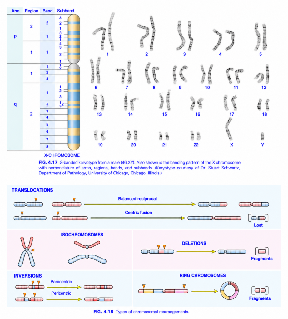
FIG. 4.17 & 4.18 — CHROMOSOME BANDING + CHROMOSOMAL REARRANGEMENTS
🧠 Simplest idea
Chromosome breaks → pieces may be exchanged, lost, reversed, duplicated, or joined abnormally.
FIG. 4.17 — KARYOTYPE + X-CHROMOSOME BANDING
1️⃣ Karyotype
The chromosomes are arranged as:
1 → 22 + X/Y
This image shows a male karyotype = 46,XY.
- Chromosomes 1–22 = autosomes
- X and Y = sex chromosomes
2️⃣ X chromosome: p arm and q arm
- p = short arm
- q = long arm
- Gray constricted center = centromere
🧠 Memory:
p = petit = short
3️⃣ Region → Band → Subband
The chromosome is divided from the centromere outward.
Example:
Xq23
means:
X chromosome
→ q arm
→ region 2
→ band 3
If another decimal/subband is given:
Xq23.1
→ subband 1 within band 3.
Easy idea:
Chromosome → arm → region → band → subband
FIG. 4.18 — TYPES OF CHROMOSOMAL REARRANGEMENTS
1️⃣ TRANSLOCATION
A. Balanced reciprocal translocation
🔵 chromosome + 🩷 chromosome break.
Pieces are exchanged.
Chromosome A gives piece to B
↔
Chromosome B gives piece to A
Result:
Usually no net DNA loss or gain.
➡️ Called balanced reciprocal translocation.
🧠 Memory:
Balanced = exchange, but total material preserved
B. Centric fusion
Two chromosomes fuse around the centromeric region.
Small fragments may be:
lost
➡️ This is the basic idea of a Robertsonian-type translocation.
2️⃣ ISOCHROMOSOME
Normally a chromosome has:
one p arm + one q arm
In an isochromosome, abnormal centromere division produces:
- two identical p arms
or - two identical q arms
Result:
One arm is duplicated and the other is lost.
🧠 Memory:
Iso = same arms
3️⃣ DELETION
A chromosome breaks at two points.
The intervening piece is removed.
Result:
DNA fragment lost
➡️ chromosome becomes shorter.
🧠 Memory:
Deletion = DNA missing
4️⃣ INVERSION
A chromosome segment breaks off, turns 180°, and reinserts.
A. Paracentric inversion
The inverted segment does NOT include the centromere.
B. Pericentric inversion
The inverted segment DOES include the centromere.
🧠 Memory:
PARA = away from centromere
PERI = around centromere
5️⃣ RING CHROMOSOME
Both ends of a chromosome break.
The terminal fragments are lost.
The remaining ends join together.
⬇️
Ring chromosome
🧠 Easy flow:
Break both ends → lose tips → ends fuse → RING
🎨 COLOR / ARROW GUIDE
- 🔵 Blue chromosome = one chromosome
- 🩷 Pink chromosome = another chromosome
- 🟨 Arrows = direction of rearrangement
- 🔺 Orange triangles = break points
- Bracketed small pieces = lost fragments
- Gray circle = centromere
⭐ Fastest exam recall
Translocation = exchange
Deletion = loss
Inversion = segment flipped
Isochromosome = two same arms
Ring = chromosome ends join
🧠 One-line memory
BREAK → exchange, lose, flip, duplicate same arm, or make a ring.
General Features of Chromosomal Disorders
- Chromosomal disorders may result from:
- loss of genetic material → deletion or monosomy
- gain of genetic material → trisomy
- rearrangement → translocation
- In general, loss of chromosomal material causes more severe abnormalities than gain.
- Extra chromosomal material may involve:
- a whole chromosome → trisomy
- part of a chromosome → e.g., Robertsonian translocation
- Abnormalities of sex chromosomes are usually better tolerated than similar abnormalities of autosomes.
- Sex chromosome disorders may cause only subtle changes and may not be recognized at birth.
- Infertility may become apparent only during adolescence.
- Most chromosomal disorders arise de novo:
- parents are usually unaffected
- recurrence risk in siblings is usually low
- An important exception is translocation Down syndrome, which may be inherited.
Cytogenetic Disorders Involving Autosomes
- Important autosomal abnormalities include:
- trisomy 21
- trisomy 18
- trisomy 13
- deletion involving 22q
Trisomy 21 (Down Syndrome)
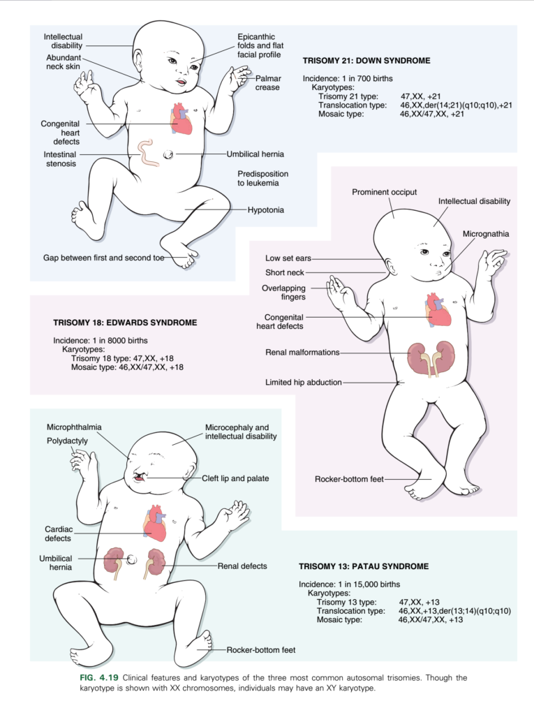
- Down syndrome results from an extra copy of chromosome 21 and is the most common chromosomal disorder (Fig. 4.19).
- About 95% of affected individuals have complete trisomy 21:
- chromosome number = 47
- most commonly caused by meiotic nondisjunction
- In these cases, the parents are usually unaffected.
- Maternal age strongly increases risk:
- younger than 20 years → about 1 in 1550 live births
- older than 45 years → about 1 in 25
- In about 95% of cases, the extra chromosome 21 is of maternal origin.
- The reason aging increases meiotic nondisjunction in the ovum is not fully understood.
- About 4% of cases result from translocation of chromosome 21 material to chromosome 14 or 22.
- These cases may be familial.
- A parent may be a healthy carrier of a Robertsonian translocation.
- About 1% of patients are mosaics:
- mixture of 46-chromosome and 47-chromosome cells
- caused by mitotic nondisjunction early in embryonic development
- manifestations are often milder and depend on the proportion of abnormal cells
- Typical physical features are usually recognizable at birth:
- flat facial profile
- oblique palpebral fissures
- epicanthic folds (Fig. 4.19)
- Down syndrome is an important cause of intellectual disability.
- About 80% have IQ values of approximately 25–50.
- Some mosaic patients may have much milder changes and near-average intelligence.
- About 40% have congenital heart disease, especially:
- endocardial cushion defects
- atrial septal defects
- atrioventricular valve abnormalities
- ventricular septal defects
- Cardiac abnormalities are a major cause of death in infancy and early childhood.
- Other congenital abnormalities include:
- esophageal atresia
- small-bowel atresia
- Children with trisomy 21 have a 10–20 times greater risk of acute leukemia:
- acute lymphoblastic leukemia
- acute myeloid leukemia
- Nearly all patients older than 40 years develop neuropathologic changes characteristic of Alzheimer disease.
- Abnormal immune responses increase susceptibility to:
- serious infections, especially pulmonary infections
- thyroid autoimmunity
- Other increased abnormalities include:
- intestinal stenosis
- Hirschsprung disease
- cataracts and refractive errors
- hearing loss
- slow growth
- cryptorchidism
- hypospadias
- Improved medical care has increased survival.
- The source text gives a median age at death of about 60 years.
- The exact molecular mechanism remains uncertain.
- Increased dosage of chromosome 21 genes may contribute.
- One implicated gene is the amyloid-β precursor gene, related to Alzheimer disease.
- microRNAs and long noncoding RNAs may also contribute.
- Prenatal screening may include:
- ↑ β-hCG
- ↓ pregnancy-associated plasma protein A (PAPP-A)
- ultrasound assessment of nuchal folds
- PAPP-A is produced by the syncytiotrophoblast, and a low level suggests poor placental function.
- Cell-free fetal DNA can also be detected in maternal blood.
- About 5%–10% of total cell-free DNA in maternal blood is fetal in origin.
- Next-generation sequencing can measure the amount of chromosome 21 DNA → provides a sensitive, specific noninvasive prenatal screening method.
- In the source text, positive screening results are confirmed by conventional cytogenetic analysis of fetal cells obtained by amniocentesis.
KEY CONCEPT
- Down syndrome = extra chromosome 21.
- Main mechanisms:
95% meiotic nondisjunction + 4% translocation + 1% mosaicism. - Increasing maternal age → increasing risk.
- Major associations:
intellectual disability + congenital heart disease + leukemia + early Alzheimer-type changes + infections. - Mosaic Down syndrome is generally milder.
- Prenatal screening → β-hCG, PAPP-A, nuchal assessment, and cell-free fetal DNA.
CONCEPTUAL EXAMPLES
- Chromosome 21 fails to separate during maternal meiosis → ovum receives an extra chromosome 21 → fertilization → trisomy 21.
- Healthy parent carries a Robertsonian translocation → abnormal chromosome 21 material passed to child → translocation Down syndrome.
- Nondisjunction occurs after fertilization → some cells have 46 chromosomes and others 47 → mosaic Down syndrome.
- Extra chromosome 21 gene dosage → contributes to the characteristic multisystem abnormalities of Down syndrome.
22q11.2 Deletion Syndrome
- 22q11.2 deletion syndrome results from a small interstitial deletion of chromosome 22 at band q11.2.
- It produces a variable combination of:
- congenital heart disease, especially outflow-tract defects
- palatal abnormalities
- characteristic facial abnormalities
- developmental delay
- thymic hypoplasia → impaired T-cell immunity
- parathyroid hypoplasia → hypocalcemia
- These manifestations were previously separated into:
- DiGeorge syndrome
- velocardiofacial syndrome
- Both are now recognized as part of the 22q11.2 deletion syndrome.
- Different sizes and positions of the deletion help explain the variable clinical presentation.
- When T-cell immunodeficiency + hypocalcemia dominate → DiGeorge syndrome.
- When facial abnormalities + cardiac defects dominate, with milder immunodeficiency → velocardiofacial syndrome.
- Patients also have an increased risk of psychiatric disorders:
- schizophrenia
- bipolar disorder
- About 25% of affected adults develop schizophrenia.
- Conversely, 22q11.2 deletions occur in about 2%–3% of individuals with childhood-onset schizophrenia.
- The exact molecular mechanism is not fully understood because the deleted region contains many genes, including:
- protein-coding genes
- noncoding regulatory RNAs
- Clinical findings may suggest the diagnosis, but confirmation requires detection of the deletion, typically by fluorescence in situ hybridization (FISH).
KEY CONCEPT
- 22q11.2 deletion → heart + palate + face + thymus + parathyroids + developmental abnormalities.
- DiGeorge pattern → thymic hypoplasia → ↓ T cells + parathyroid hypoplasia → hypocalcemia.
- Velocardiofacial pattern → prominent facial/palatal + cardiac abnormalities with milder immune deficiency.
- Diagnosis → FISH detection of the deletion.
CONCEPTUAL EXAMPLES
- Thymus underdeveloped → fewer functional T cells → impaired cellular immunity.
- Parathyroids underdeveloped → hypocalcemia.
- Same 22q11.2 deletion with different extent → one patient may mainly show DiGeorge features, while another mainly shows velocardiofacial features.
Cytogenetic Disorders Involving Sex Chromosomes
- Sex chromosome abnormalities are better tolerated than autosomal abnormalities.
- Karyotypes ranging from 45,X to 49,XXXXY may be compatible with life.
- Even males with two or three Y chromosomes have been identified.
- This tolerance mainly occurs because of:
- X-chromosome inactivation (lyonization)
- the relatively small amount of genetic information on the Y chromosome
- Lyonization means that in females, only one X chromosome remains genetically active in each cell.
- X inactivation begins early in fetal life, about 16 days after conception.
- In each embryonic cell:
- either the maternal X or
- the paternal X
is randomly inactivated.
- Once an X chromosome is inactivated:
same X remains inactive in all daughter cells. - All X chromosomes except one are inactivated.
- Therefore, even a female with 48,XXXX has only one active X chromosome.
- This prevents females from receiving a double dose of X-linked gene products compared with males.
- Because X inactivation is random, females are mosaics:
- some cells have the maternal X active
- other cells have the paternal X active
- X inactivation is controlled by XIST, a gene producing a long noncoding RNA.
- XIST RNA:
coats the X chromosome from which it is produced → silences genes on that chromosome. - On the active X chromosome:
- the XIST allele is switched off
- genes remain active
- X inactivation is not complete.
- About:
- 30% of genes on Xp
- 3% of genes on Xq
escape inactivation.
- These escaping genes are important in disorders such as Turner syndrome.
- Extra Y chromosomes are relatively well tolerated because the Y chromosome carries comparatively little genetic information.
- Most known Y-linked information is related to male differentiation.
- According to the source, the presence of a Y chromosome determines the male phenotype.
- The key gene is SRY (sex-determining region of Y), located on the short arm of the Y chromosome.
- Two important sex chromosome disorders discussed next are:
- Klinefelter syndrome
- Turner syndrome
KEY CONCEPT
- Sex chromosome abnormalities are better tolerated than autosomal abnormalities.
- Lyonization = all X chromosomes except one are inactivated.
- XIST RNA → coats and silences the inactive X chromosome.
- Females are cellular mosaics for maternal vs paternal X activity.
- Some X-linked genes escape inactivation, especially on Xp.
- SRY on Y chromosome → male differentiation.
CONCEPTUAL EXAMPLES
- Female cell randomly inactivates maternal X → all daughter cells from it keep the maternal X inactive.
- Another cell inactivates paternal X → its descendants keep the paternal X inactive.
- Thus one female contains two cell populations → X-chromosome mosaicism.
- Extra X chromosomes are mostly silenced → explains why multiple X chromosomes may still be compatible with life.
