


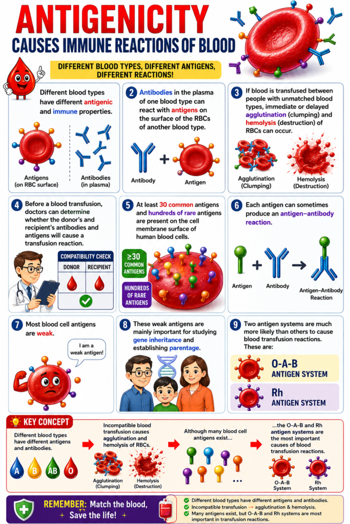
- Different blood types have different antigenic and immune properties.
- Antibodies in the plasma of one blood type can react with antigens on the surface of the red blood cells (RBCs) of another blood type.
- If blood is transfused between people with unmatched blood types, immediate or delayed agglutination (clumping) and hemolysis (destruction) of RBCs can occur.
- Before a blood transfusion, doctors can determine whether the donor’s and recipient’s antibodies and antigens will cause a transfusion reaction.
- At least 30 common antigens and hundreds of rare antigens are present on the cell membrane surface of human blood cells.
- Each antigen can sometimes produce an antigen–antibody reaction.
- Most blood cell antigens are weak.
- These weak antigens are mainly important for studying gene inheritance and establishing parentage.
- Two antigen systems are much more likely than others to cause blood transfusion reactions.
- These are:
- O-A-B antigen system
- Rh antigen system
Key Concept
- Different blood types have different antigens and antibodies.
- Incompatible blood transfusion causes agglutination and hemolysis of RBCs.
- Although many blood cell antigens exist, the O-A-B and Rh antigen systems are the most important causes of blood transfusion reactions.
O-A-B BLOOD TYPES
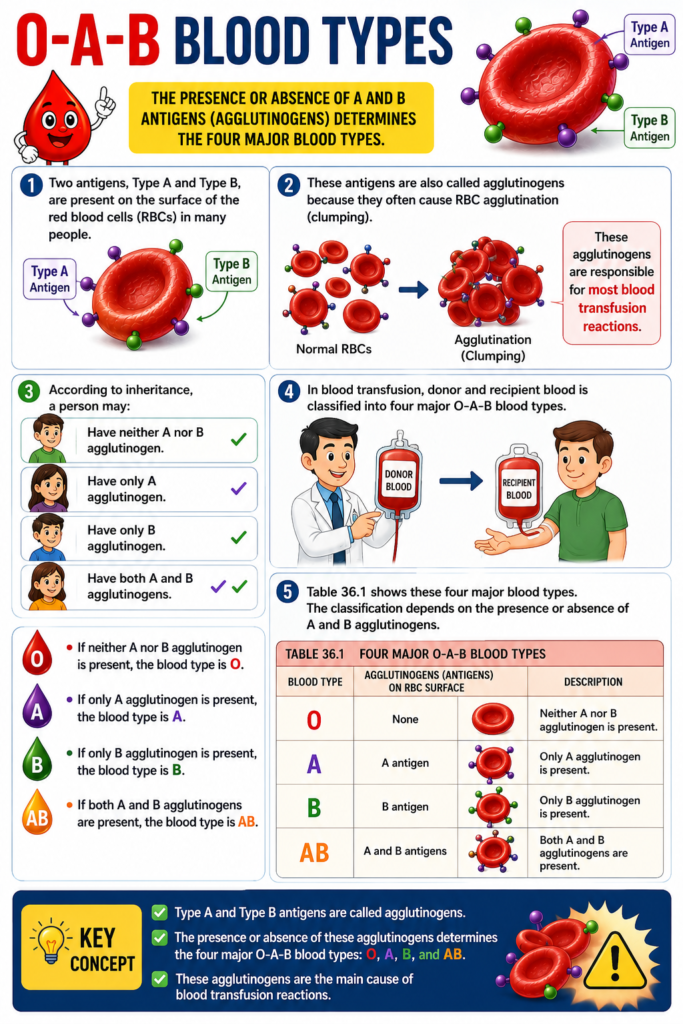
- Two antigens, Type A and Type B, are present on the surface of the red blood cells (RBCs) in many people.
- These antigens are also called agglutinogens because they often cause RBC agglutination (clumping).
- These agglutinogens are responsible for most blood transfusion reactions.
- According to inheritance, a person may:
- Have neither A nor B agglutinogen.
- Have only A agglutinogen.
- Have only B agglutinogen.
- Have both A and B agglutinogens.
- In blood transfusion, donor and recipient blood is classified into four major O-A-B blood types.
- Table 36.1 shows these four major blood types.
- The classification depends on the presence or absence of A and B agglutinogens.
- If neither A nor B agglutinogen is present, the blood type is O.
- If only A agglutinogen is present, the blood type is A.
- If only B agglutinogen is present, the blood type is B.
- If both A and B agglutinogens are present, the blood type is AB.
Key Concept
- Type A and Type B antigens are called agglutinogens.
- The presence or absence of these agglutinogens determines the four major O-A-B blood types: O, A, B, and AB.
- These agglutinogens are the main cause of blood transfusion reactions.
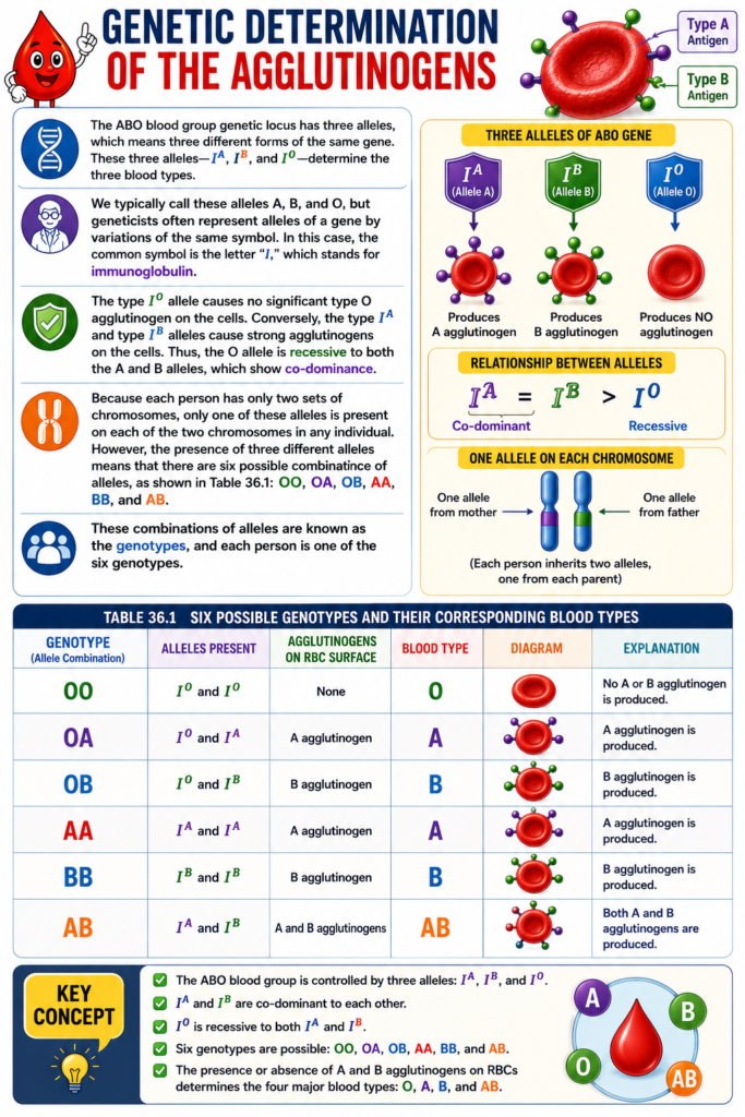
Genetic Determination of the Agglutinogens
- The ABO blood group genetic locus has three alleles.
- Three alleles mean three different forms of the same gene.
- These three alleles are IA, IB, and IO.
- These alleles determine the three blood group genes.
- These alleles are commonly called A, B, and O.
- Geneticists often represent these alleles using the letter “I”.
- The letter “I” stands for immunoglobulin.
- The O allele does not produce a significant type O agglutinogen on red blood cells.
- The A allele produces strong type A agglutinogens on red blood cells.
- The B allele produces strong type B agglutinogens on red blood cells.
- The O allele is recessive to both the A and B alleles.
- The A and B alleles show co-dominance.
- Each person has two sets of chromosomes.
- Therefore, each person has one ABO allele on each chromosome.
- Since there are three different alleles, there are six possible allele combinations.
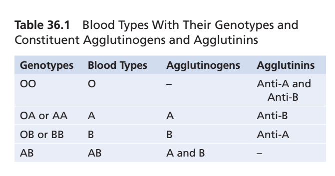
- Table 36.1 shows these six combinations.
- The six genotypes are:
- OO
- OA
- OB
- AA
- BB
- AB
- These allele combinations are called genotypes.
- Every person has one of these six genotypes.
- A person with genotype OO produces no agglutinogens.
- Therefore, the blood type is O.
- A person with genotype OA or AA produces type A agglutinogens.
- Therefore, the blood type is A.
- Genotypes OB and BB produce type B blood.
- Genotype AB produces type AB blood.
Key Concept
- The ABO blood group is controlled by three alleles: IA, IB, and IO.
- IO is recessive, while IA and IB are co-dominant.
- The six genotypes (OO, OA, OB, AA, BB, AB) determine the four blood types: O, A, B, and AB.
Relative Frequencies of Different Blood Types
- The frequency of blood types can vary among different populations.
- In one group of people, the approximate frequency of blood types was:
- O: 47%
- A: 41%
- B: 9%
- AB: 3%
- These percentages show that the O gene occurs more frequently than the B gene.
- They also show that the A gene occurs more frequently than the B gene.
Key Concept
- Blood type frequencies differ among populations.
- In the studied group:
- O = 47%
- A = 41%
- B = 9%
- AB = 3%
- The O and A genes are more common than the B gene.
Agglutinins
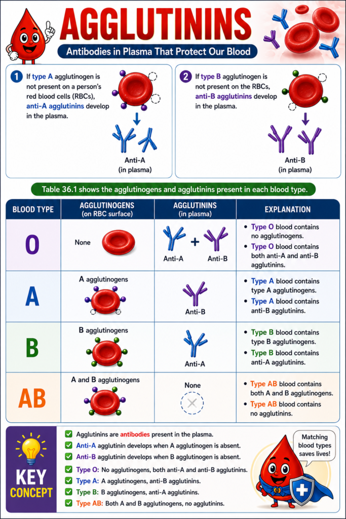
- If type A agglutinogen is not present on a person’s red blood cells (RBCs), anti-A agglutinins develop in the plasma.
- If type B agglutinogen is not present on the RBCs, anti-B agglutinins develop in the plasma.
- Table 36.1 shows the agglutinogens and agglutinins present in each blood type.
- Type O blood contains no agglutinogens.
- Type O blood contains both anti-A and anti-B agglutinins.
- Type A blood contains type A agglutinogens.
- Type A blood contains anti-B agglutinins.
- Type B blood contains type B agglutinogens.
- Type B blood contains anti-A agglutinins.
- Type AB blood contains both A and B agglutinogens.
- Type AB blood contains no agglutinins.
Key Concept
- Agglutinins are antibodies present in the plasma.
- Anti-A agglutinin develops when A agglutinogen is absent.
- Anti-B agglutinin develops when B agglutinogen is absent.
- Type O: No agglutinogens, both anti-A and anti-B agglutinins.
- Type A: A agglutinogens, anti-B agglutinins.
- Type B: B agglutinogens, anti-A agglutinins.
- Type AB: Both A and B agglutinogens, no agglutinins.
Titer of Agglutinins at Different Ages
- Immediately after birth, the amount of agglutinins in the plasma is almost zero.
- At 2 to 8 months after birth, an infant begins to produce agglutinins.
- Anti-A agglutinins are produced when type A agglutinogens are not present on the cells.
- Anti-B agglutinins are produced when type B agglutinogens are not present on the cells.
- Fig. 36.1 shows the changing titers of anti-A and anti-B agglutinins at different ages.
- The maximum agglutinin titer is usually reached at 8 to 10 years of age.
- After 8 to 10 years, the agglutinin titer gradually decreases throughout the rest of life.
Key Concept
- Agglutinins are almost absent at birth.
- Production begins at 2–8 months of age.
- Maximum titer occurs at 8–10 years of age.
- The agglutinin titer gradually declines with increasing age.
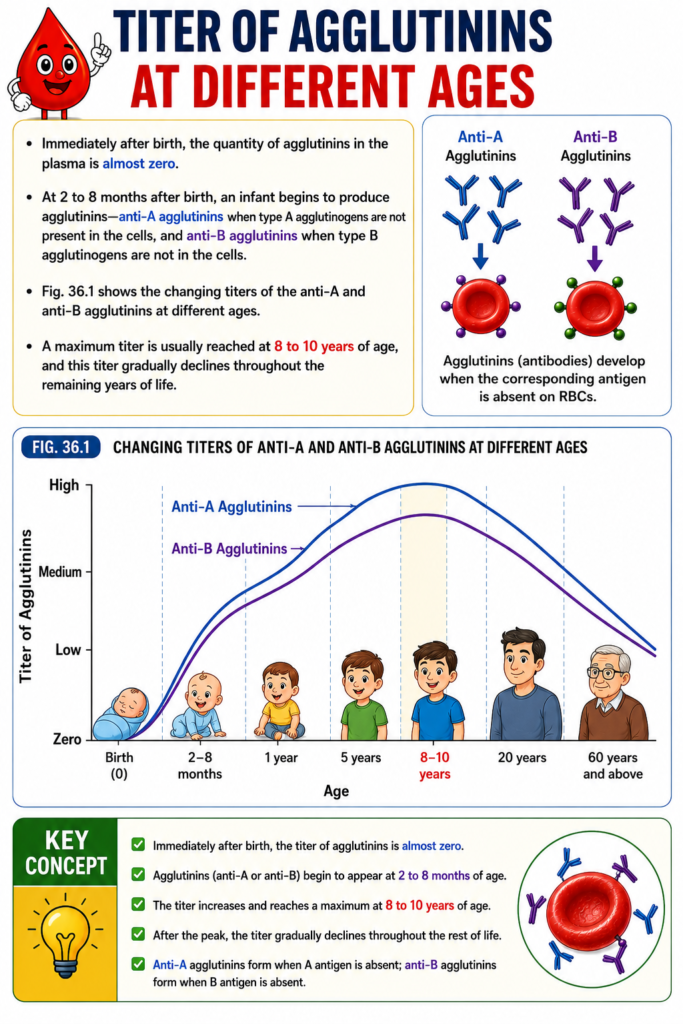
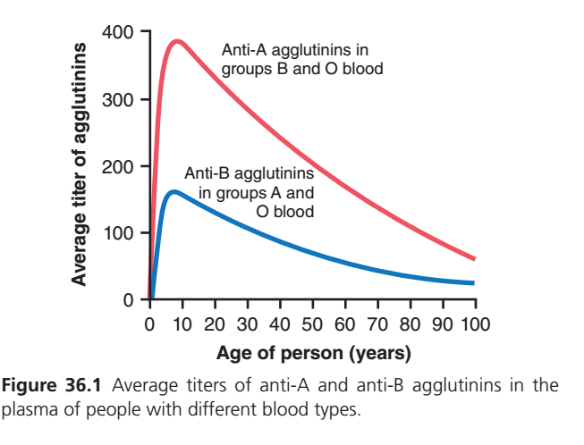
Figure 36.1: Average Titers of Anti-A and Anti-B Agglutinins at Different Ages
This graph explains how the amount (titer) of naturally occurring antibodies (agglutinins) changes throughout life.
- Red Line = Anti-A Agglutinins
- Blue Line = Anti-B Agglutinins
Step 1: Understand the Axes
X-Axis (Horizontal)
Age of person (Years)
- Starts from birth (0 years)
- Ends at 100 years
- Shows how antibody levels change as a person grows older.
Y-Axis (Vertical)
Average Titer of Agglutinins
“Titer” means:
The concentration (amount) of antibodies present in plasma.
Higher value = More antibodies
Lower value = Fewer antibodies
Step 2: Red Line (Anti-A Agglutinins)
What is Anti-A?
Anti-A antibodies attack A antigen.
They are present in:
- Blood Group B
- Blood Group O
They are NOT present in Group A or AB.
At Birth (0 years)
The red line starts almost at zero.
Meaning:
- Babies are born with almost no Anti-A antibodies.
Why?
Because the baby’s immune system is still immature.
During First Few Months
The red line rises very steeply.
Meaning:
The baby begins producing Anti-A antibodies rapidly.
Why?
Because after birth the baby is exposed to:
- Food
- Bacteria
- Environment
Some bacterial antigens resemble A antigen.
The immune system mistakenly learns to produce Anti-A antibodies.
Around 8–10 Years
The red line reaches its highest point.
This is called the Peak Titer.
Meaning:
This is the age when Anti-A antibody concentration is maximum.
The immune system is strongest during childhood.
Approximate value:
Nearly 380–390 units
After 10 Years
The red line starts going downward.
Meaning:
Anti-A antibodies gradually decrease with age.
Not because they disappear,
but because immune activity slowly declines.
Adult Age (20–50 Years)
The decrease becomes gradual.
Adults still have Anti-A antibodies,
but less than children.
Old Age (60–100 Years)
The red line continues falling.
Meaning:
Older people have much lower Anti-A antibody levels.
Reason:
Aging causes reduced antibody production.
This is called immune senescence.
Even at 90–100 years,
Anti-A antibodies are still present,
just in lower amounts.
Step 3: Blue Line (Anti-B Agglutinins)
What is Anti-B?
Anti-B antibodies attack B antigen.
They are present in:
- Blood Group A
- Blood Group O
At Birth
The blue line also starts at zero.
Meaning:
Newborn babies have almost no Anti-B antibodies.
During Infancy
The blue line rises quickly,
just like the red line,
but not as high.
Meaning:
Anti-B antibodies are produced after exposure to environmental antigens.
Peak Around 8–10 Years
The blue line reaches its highest level.
Approximate value:
Around 150–160 units
Notice:
This peak is much lower than the red line.Why is the Blue Peak Lower?
Normally,
the body produces
less Anti-B antibody than Anti-A antibody.
Therefore,
Anti-B titers are naturally lower.
This is an important physiological fact.
Adult Life
After childhood,
Anti-B antibodies slowly decrease.Old Age
The blue line continues declining.
At 90–100 years,
only a small amount remains.
Step 4: Compare Both Lines
| Feature | Red Line (Anti-A) | Blue Line (Anti-B) |
|---|---|---|
| Antibody | Anti-A | Anti-B |
| Present in | Group B & O | Group A & O |
| At birth | Almost zero | Almost zero |
| Peak age | Around 8–10 years | Around 8–10 years |
| Maximum level | Very high (~380) | Lower (~160) |
| Adult level | Decreases gradually | Decreases gradually |
| Old age | Low but present | Low but present |
Step 5: Why Do Both Lines Rise After Birth?
The baby is not born with these antibodies.
Instead,
after birth,
the immune system is exposed to:
- Intestinal bacteria
- Food proteins
- Environmental microorganisms
Many of these organisms contain molecules similar to A and B antigens.
The immune system responds by producing:
- Anti-A antibodies (if A antigen is absent)
- Anti-B antibodies (if B antigen is absent)
These are called naturally occurring ABO antibodies.
Step 6: Why Do They Fall in Old Age?
As people age:
- Bone marrow becomes less active.
- B-lymphocyte function decreases.
- Antibody production slows.
- Immune system becomes weaker.
Therefore,
both Anti-A and Anti-B titers gradually decline.
- Newborns have almost no ABO antibodies.
- ABO antibodies begin to appear at 2–8 months of age.
- Maximum antibody titer occurs around 8–10 years.
- Anti-A antibody titer is normally much higher than Anti-B titer.
- Both antibody titers gradually decrease with aging.
- Anti-A is present in Blood Groups B and O.
- Anti-B is present in Blood Groups A and O.
- Blood Group AB has neither Anti-A nor Anti-B antibodies.
- Blood Group O has both Anti-A and Anti-B antibodies.
- These antibodies are naturally acquired due to exposure to environmental and intestinal bacterial antigens after birth.
One-Minute Exam Summary
- Birth: Almost no Anti-A or Anti-B antibodies.
- 2–8 months: Antibody production begins.
- Childhood (8–10 years): Highest antibody levels.
- Red line (Anti-A): Higher than blue line throughout life.
- Blue line (Anti-B): Lower peak but follows the same pattern.
- Adulthood: Gradual decline.
- Old age: Lowest antibody titers due to decreased immune function.
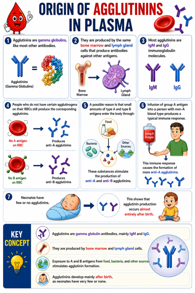
Origin of Agglutinins in Plasma
- Agglutinins are gamma globulins, like most other antibodies.
- They are produced by the same bone marrow and lymph gland cells that produce antibodies against other antigens.
- Most agglutinins are IgM and IgG immunoglobulin molecules.
- People who do not have certain agglutinogens on their RBCs still produce the corresponding agglutinins.
- A possible reason is that small amounts of type A and type B antigens enter the body through:
- Food
- Bacteria
- Other sources
- These substances stimulate the production of anti-A and anti-B agglutinins.
- Infusion of group A antigen into a person with non-A blood type produces a typical immune response.
- This immune response causes the formation of more anti-A agglutinins.
- Neonates have few or no agglutinins.
- This shows that agglutinin production occurs almost entirely after birth.
Key Concept
- Agglutinins are gamma globulin antibodies, mainly IgM and IgG.
- They are produced by bone marrow and lymph gland cells.
- Exposure to A and B antigens from food, bacteria, and other sources stimulates agglutinin formation.
- Agglutinins develop mainly after birth, as neonates have very few or none.
Agglutination Process in Transfusion Reactions
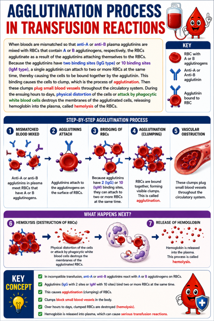
- When mismatched blood is transfused, anti-A or anti-B plasma agglutinins mix with RBCs that contain A or B agglutinogens, respectively.
- The RBCs agglutinate (clump together) because the agglutinins attach to the RBCs.
- IgG agglutinins have 2 binding sites.
- IgM agglutinins have 10 binding sites.
- One agglutinin can attach to two or more RBCs at the same time.
- This causes the RBCs to become linked together by the agglutinin.
- As a result, the RBCs form clumps.
- This clumping process is called agglutination.
- These RBC clumps can block small blood vessels throughout the circulatory system.
- Over the next hours to days, the agglutinated RBCs are destroyed.
- Destruction occurs because of:
- Physical distortion of the cells
- Attack by phagocytic white blood cells
- The RBC membranes break down and release hemoglobin into the plasma.
- This destruction of RBCs is called hemolysis.
Key Concept
- Mismatched blood transfusion causes agglutination when agglutinins bind to incompatible RBC agglutinogens.
- IgG has 2 binding sites, while IgM has 10 binding sites, allowing RBCs to clump together.
- Agglutination blocks small blood vessels.
- Hemolysis occurs when agglutinated RBCs are destroyed, releasing hemoglobin into the plasma.
Acute Hemolysis Occurs in Some Transfusion Reactions
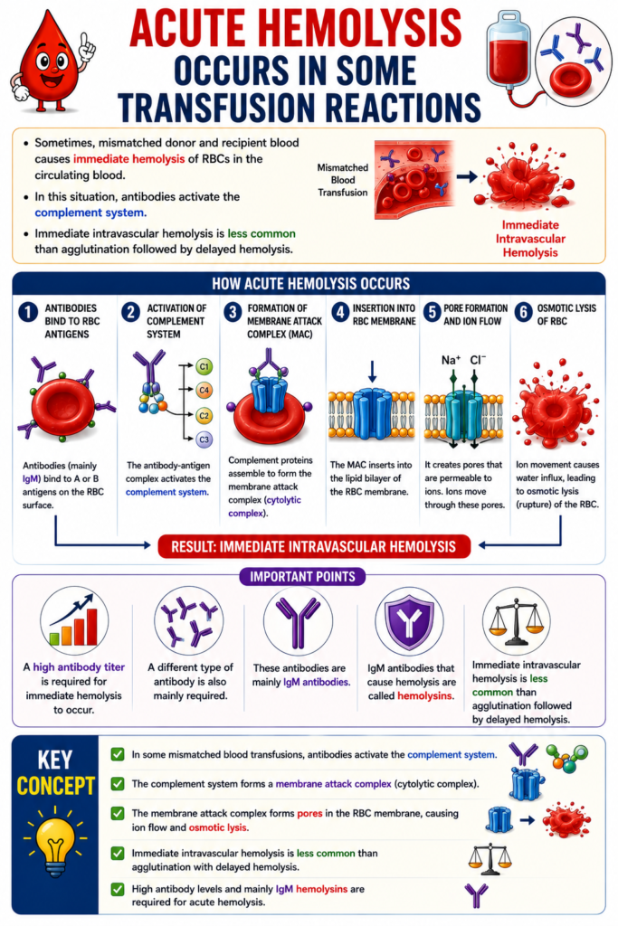
- Sometimes, mismatched donor and recipient blood causes immediate hemolysis of RBCs in the circulating blood.
- In this situation, antibodies activate the complement system.
- The complement system forms a membrane attack complex (cytolytic complex).
- This complex inserts into the lipid bilayer of the RBC membrane.
- It creates membrane pores that are permeable to ions.
- Ions move through these pores, causing osmotic lysis of the RBCs.
- This results in immediate intravascular hemolysis.
- Immediate intravascular hemolysis is less common than agglutination followed by delayed hemolysis.
- A high antibody titer is required for immediate hemolysis to occur.
- A different type of antibody is also mainly required.
- These antibodies are mainly IgM antibodies.
- IgM antibodies that cause hemolysis are called hemolysins.
Key Concept
- In some mismatched blood transfusions, antibodies activate the complement system.
- The membrane attack complex forms pores in the RBC membrane, causing osmotic lysis.
- Immediate intravascular hemolysis is less common than agglutination with delayed hemolysis.
- High antibody levels and mainly IgM hemolysins are required for acute hemolysis.
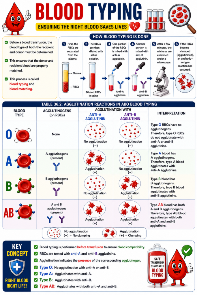
Blood Typing
- Before a blood transfusion, the blood type of both the recipient and donor must be determined.
- This ensures that the donor and recipient blood are properly matched.
- This process is called blood typing and blood matching.
- First, the RBCs are separated from the plasma.
- The RBCs are then diluted with saline solution.
- One portion of the RBCs is mixed with anti-A agglutinin.
- Another portion is mixed with anti-B agglutinin.
- After a few minutes, the mixtures are examined under a microscope.
- If the RBCs become clumped (agglutinated), an antibody–antigen reaction has occurred.
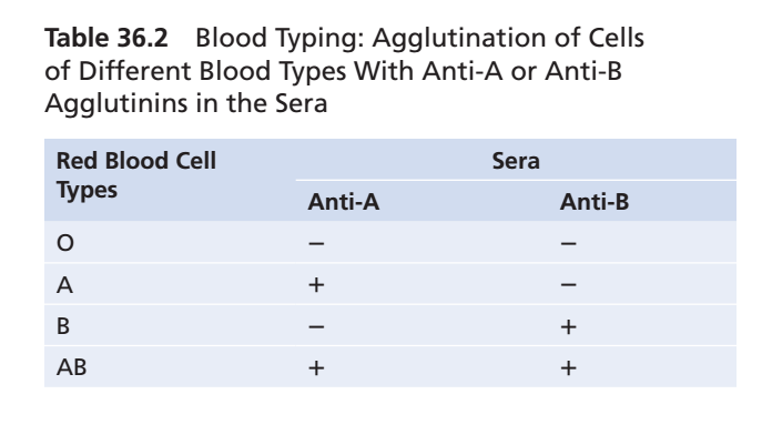
- Table 36.2 shows the presence (+) or absence (−) of agglutination in the four blood types.
- Type O RBCs have no agglutinogens.
- Therefore, type O RBCs do not agglutinate with anti-A or anti-B agglutinins.
- Type A blood has A agglutinogens.
- Therefore, type A blood agglutinates with anti-A agglutinins.
- Type B blood has B agglutinogens.
- Therefore, type B blood agglutinates with anti-B agglutinins.
- Type AB blood has both A and B agglutinogens.
- Therefore, type AB blood agglutinates with both anti-A and anti-B agglutinins.
Key Concept
- Blood typing is performed before transfusion to ensure blood compatibility.
- RBCs are tested with anti-A and anti-B agglutinins.
- Agglutination indicates the presence of the corresponding agglutinogen.
- Type O: No agglutination with anti-A or anti-B.
- Type A: Agglutinates with anti-A.
- Type B: Agglutinates with anti-B.
- Type AB: Agglutinates with both anti-A and anti-B.
MADE BY SELF LEARNING CEO AND FOUNDER DR SHEEN
