GENERAL ORGANIZATION OF THE KIDNEYS AND URINARY TRACT
The urinary system consists of two kidneys, two ureters, the urinary bladder, and the urethra. The kidneys are the main organs that form urine and help maintain the body’s internal balance.
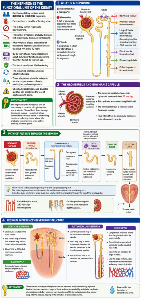
The two kidneys are located on the posterior wall of the abdomen, outside the peritoneal cavity. Each adult kidney weighs about 150 grams and is approximately the size of a clenched fist. Kidney weight is usually greater in men than in women, partly because men generally have a larger body size.
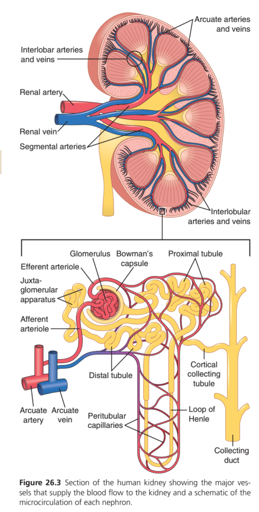
On the medial side of each kidney is a depression called the hilum. Through the hilum pass the renal artery, renal vein, lymphatic vessels, nerves, and the ureter. The renal artery brings blood to the kidney, and the renal vein carries blood away from it. The ureter transports the urine formed by the kidney to the urinary bladder.
Each kidney is surrounded by a tough fibrous capsule that protects the delicate structures inside the kidney.
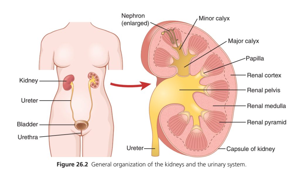
When a kidney is cut from top to bottom, two main regions can be seen: the outer cortex and the inner medulla. The cortex forms the outer part of the kidney, while the medulla forms the inner part.
The medulla is divided into 8 to 10 cone-shaped structures called renal pyramids. The broad base of each pyramid lies near the cortex, while the narrow tip, called the papilla, projects into the renal pelvis.
The renal pelvis is a funnel-shaped expansion at the upper end of the ureter. It collects urine before it enters the ureter.
The outer part of the renal pelvis divides into large chambers called major calyces. Each major calyx further divides into smaller chambers called minor calyces.
The minor calyces collect urine from the papillae of the renal pyramids. From the minor calyces, urine flows into the major calyces, then into the renal pelvis, and finally into the ureter.
The walls of the calyces, renal pelvis, and ureter contain smooth muscle. These muscles contract rhythmically to propel urine toward the urinary bladder.
The urinary bladder stores urine until it is expelled from the body during micturition (urination).
KEY CONCEPT
The kidney consists of an outer cortex and an inner medulla containing renal pyramids. Urine formed in the kidney flows from the papillae → minor calyces → major calyces → renal pelvis → ureter → urinary bladder, where it is stored until urination.
RENAL BLOOD SUPPLY
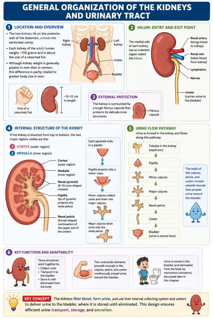
- Blood flow to both kidneys is normally about 22% of the cardiac output.
- In an average adult, renal blood flow is about 1100 mL/min.
- Renal blood flow can vary depending on body size, age, and sex.
- The renal artery enters the kidney through the hilum.
- The renal artery divides into interlobar arteries.
- The interlobar arteries divide into arcuate arteries.
- The arcuate arteries divide into interlobular arteries (radial arteries).
- The interlobular arteries divide into afferent arterioles.
- The afferent arterioles carry blood to the glomerular capillaries.
- Large amounts of fluid and solutes are filtered in the glomerular capillaries.
- Plasma proteins are not filtered.
- Filtration in the glomerular capillaries is the first step in urine formation.
- The capillaries of each glomerulus join together to form the efferent arteriole.
- The efferent arteriole leads to a second capillary network called the peritubular capillaries.
- The peritubular capillaries surround the renal tubules.
- The renal circulation contains two capillary beds arranged in series.
- These are the glomerular capillaries and the peritubular capillaries.
- The two capillary beds are separated by the efferent arteriole.
- The efferent arteriole helps regulate hydrostatic pressure in both capillary beds.
- The hydrostatic pressure in the glomerular capillaries is high (about 60 mm Hg).
- This high pressure causes rapid filtration of fluid.
- The hydrostatic pressure in the peritubular capillaries is low (about 13 mm Hg).
- This low pressure permits rapid fluid reabsorption.
- The kidneys can adjust the resistance of the afferent and efferent arterioles.
- This helps regulate hydrostatic pressure in both capillary networks.
- Changes in hydrostatic pressure can alter the rate of glomerular filtration.
- Changes in hydrostatic pressure can also alter the rate of tubular reabsorption.
- These adjustments help meet the body’s homeostatic needs.
- The peritubular capillaries drain into the venous system.
- The veins run parallel to the arterial vessels.
- The venous vessels form the interlobular veins.
- The interlobular veins join to form the arcuate veins.
- The arcuate veins join to form the interlobar veins.
- The interlobar veins join to form the renal vein.
- The renal vein leaves the kidney beside the renal artery and ureter.
KEY CONCEPT
Blood enters the kidney through the renal artery and passes through two capillary networks in series: the glomerular capillaries for filtration and the peritubular capillaries for reabsorption. Blood then leaves the kidney through the renal vein.
THE NEPHRON IS THE FUNCTIONAL UNIT OF THE KIDNEY
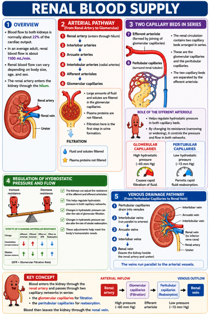
- Each human kidney contains about 800,000 to 1,000,000 nephrons.
- Each nephron is capable of forming urine.
- The kidney cannot regenerate new nephrons.
- The number of nephrons gradually decreases with kidney injury, disease, or normal aging.
- After 40 years of age, the number of functioning nephrons usually decreases by about 10% every 10 years.
- By 80 years of age, many people have about 40% fewer functioning nephrons than they had at 40 years of age.
- This loss is usually not life-threatening.
- The remaining nephrons undergo adaptive changes.
- These adaptations allow the kidneys to excrete proper amounts of water, electrolytes, and waste products.
- Obesity, hypertension, and diabetes mellitus can accelerate the loss of nephrons with aging.
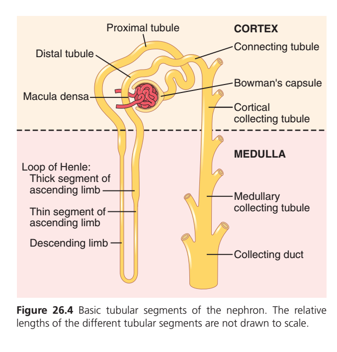
- Each nephron contains a tuft of glomerular capillaries called the glomerulus.
- Large amounts of fluid are filtered from the blood through the glomerulus.
- Each nephron also contains a long tubule.
- The filtered fluid is converted into urine as it passes through the tubule.
- The glomerulus contains a network of branching and interconnected capillaries.
- The glomerular capillaries have a high hydrostatic pressure of about 60 mm Hg.
- The glomerular capillaries are covered by epithelial cells.
- The entire glomerulus is enclosed within Bowman’s capsule.
- Fluid filtered from the glomerular capillaries enters Bowman’s capsule.
- From Bowman’s capsule, the fluid flows into the proximal tubule.
- The proximal tubule is located in the renal cortex.
- From the proximal tubule, the fluid flows into the loop of Henle.
- The loop of Henle extends into the renal medulla.
- Each loop of Henle has a descending limb and an ascending limb.
- The walls of the descending limb and the lower part of the ascending limb are very thin.
- These thin-walled portions are called the thin segment of the loop of Henle.
- As the ascending limb returns toward the cortex, its wall becomes much thicker.
- This portion is called the thick ascending limb.
- At the end of the thick ascending limb is a short segment containing specialized epithelial cells.
- These specialized cells are called the macula densa.
- The macula densa plays an important role in controlling nephron function.
- Beyond the macula densa, fluid enters the distal tubule.
- The distal tubule is located in the renal cortex.
- The distal tubule is followed by the connecting tubule.
- The connecting tubule leads to the cortical collecting tubule.
- The cortical collecting tubule leads to the cortical collecting duct.
- The initial parts of 8 to 10 cortical collecting ducts join together.
- They form a single larger collecting duct.
- The collecting duct extends downward into the medulla.
- In the medulla, it becomes the medullary collecting duct.
- The collecting ducts join to form progressively larger ducts.
- These ducts eventually empty into the renal pelvis through the tips of the renal papillae.
- Each kidney contains about 250 very large collecting ducts.
- Each large collecting duct collects urine from about 4000 nephrons.
KEY CONCEPT
The nephron is the functional unit of the kidney. It consists of a glomerulus and a tubule. Filtered fluid flows through Bowman’s capsule → proximal tubule → loop of Henle → distal tubule → connecting tubule → collecting ducts, where it is gradually converted into urine before entering the renal pelvis.
REGIONAL DIFFERENCES IN NEPHRON STRUCTURE: CORTICAL AND JUXTAMEDULLARY NEPHRONS
- Although all nephrons contain the same basic components, there are structural differences based on their location in the kidney.
- These differences depend on how deep the nephron is located within the kidney.
- Nephrons with glomeruli located in the outer cortex are called cortical nephrons.
- Cortical nephrons have short loops of Henle.
- These loops of Henle extend only a short distance into the medulla.
- About 20% to 30% of all nephrons are juxtamedullary nephrons.
- Juxtamedullary nephrons have glomeruli located deep in the renal cortex near the medulla.
- Juxtamedullary nephrons have long loops of Henle.
- These loops extend deep into the medulla.
- In some cases, the loops of Henle reach the tips of the renal papillae.
- The blood supply of juxtamedullary nephrons differs from that of cortical nephrons.
- In cortical nephrons, the entire tubular system is surrounded by an extensive network of peritubular capillaries.
- In juxtamedullary nephrons, long efferent arterioles extend from the glomeruli into the outer medulla.
- These efferent arterioles divide into specialized peritubular capillaries called vasa recta.
- The vasa recta extend downward into the medulla.
- The vasa recta run alongside the loops of Henle.
- Like the loops of Henle, the vasa recta return toward the cortex.
- The vasa recta eventually empty into the cortical veins.
- This specialized capillary network in the medulla is essential for the formation of concentrated urine.
KEY CONCEPT
There are two main types of nephrons: cortical nephrons and juxtamedullary nephrons. Cortical nephrons have short loops of Henle and are surrounded by peritubular capillaries, whereas juxtamedullary nephrons have long loops of Henle and vasa recta that extend deep into the medulla, helping in the formation of concentrated urine.