


First Step in Urine Formation
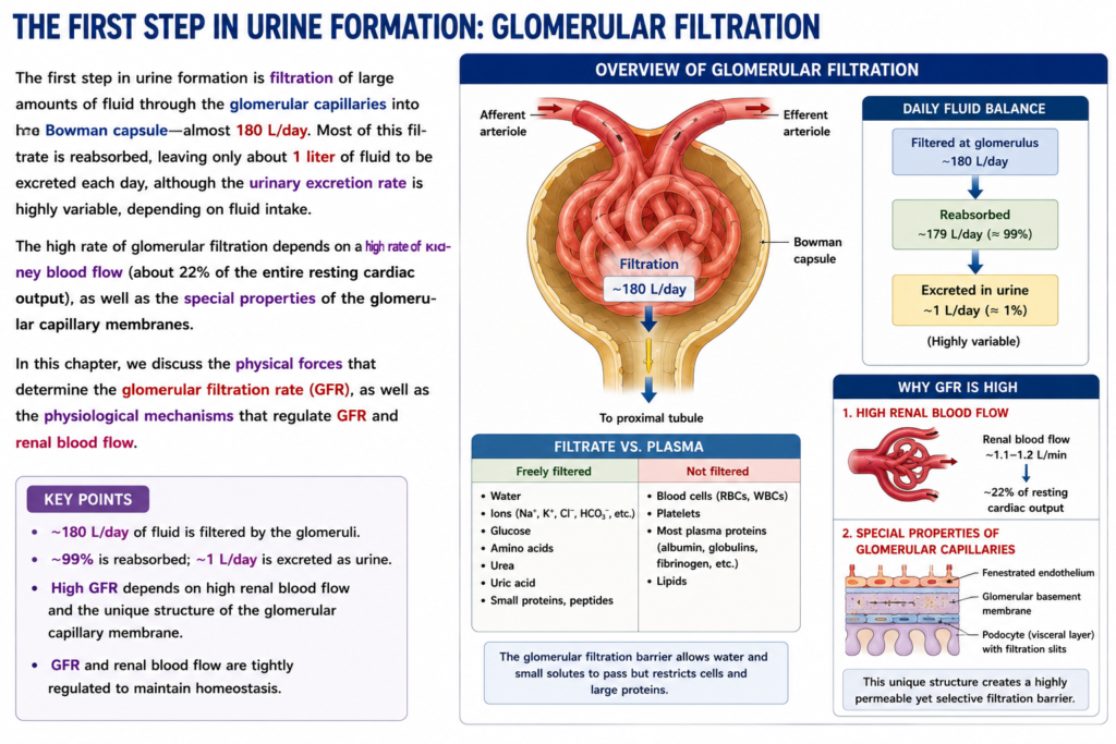
- The first step in making urine is glomerular filtration.
- During this process, a large amount of fluid is filtered from the glomerular capillaries into Bowman capsule.
- About 180 liters of fluid are filtered each day.
Most Filtrate Is Reabsorbed
- Most of the filtered fluid is reabsorbed back into the body.
- Only about 1 liter of fluid remains to be excreted as urine each day.
- The amount of urine produced can vary greatly depending on fluid intake.
Why Glomerular Filtration Is High
- The high filtration rate depends on a high rate of blood flow to the kidneys.
- The kidneys receive about 22% of the total resting cardiac output.
- The high filtration rate also depends on the special properties of the glomerular capillary membranes.
What Determines Filtration Rate
- Physical forces determine the glomerular filtration rate (GFR).
- Physiological mechanisms regulate GFR.
- Physiological mechanisms also regulate renal blood flow.
KEY CONCEPT
Glomerular filtration is the first step in urine formation. About 180 L of fluid are filtered into Bowman capsule each day. Most of this fluid is reabsorbed, and only a small amount is excreted as urine. High kidney blood flow and specialized glomerular capillary membranes help maintain a high filtration rate.
COMPOSITION OF THE GLOMERULAR FILTRATE
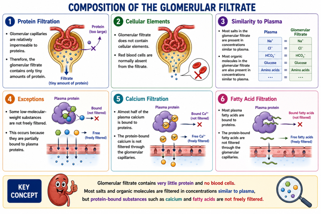
- Like most capillaries, the glomerular capillaries are relatively impermeable to proteins.
- Therefore, the filtered fluid, called the glomerular filtrate, normally contains only very small amounts of protein.
- The glomerular filtrate normally contains no cellular elements, including red blood cells.
- The concentrations of most other substances in the glomerular filtrate are similar to their concentrations in plasma.
- These substances include most salts and organic molecules.
- There are some exceptions to this general rule.
- Some low-molecular-weight substances, such as calcium and fatty acids, are not freely filtered.
- They are not freely filtered because they are partially bound to plasma proteins.
- For example, almost half of the plasma calcium is bound to proteins.
- Most of the plasma fatty acids are also bound to proteins.
- These protein-bound portions cannot pass through the glomerular capillaries and are not filtered.
KEY CONCEPT
Glomerular filtrate contains very little protein and no blood cells. Most salts and organic molecules are filtered in concentrations similar to plasma, but substances bound to plasma proteins, such as much of the calcium and fatty acids, are not freely filtered.
GFR IS ABOUT 20% OF RENAL PLASMA FLOW
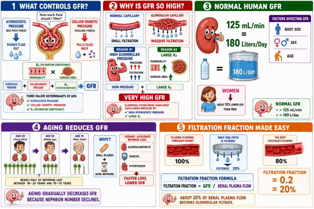
- Similar to other capillaries, the filtration rate of the glomerular capillaries depends on two main factors.
- The first factor is the balance of hydrostatic pressure and colloid osmotic pressure acting across the capillary membrane.
- The second factor is the capillary filtration coefficient (Kf).
- Kf is the product of capillary permeability and filtering surface area.
- Glomerular capillaries filter fluid at a much higher rate than most other capillaries.
- This high filtration rate occurs because of a high glomerular hydrostatic pressure.
- It also occurs because of a large Kf.
- In an average adult human, the glomerular filtration rate (GFR) is about 125 mL/min.
- This is equal to about 180 L/day.
- GFR depends on the body size, sex, and age of the individual.
- The average GFR is about 10% lower in women than in men, even after adjustment for body size.
- This difference becomes smaller at older ages.
- The reason is that GFR declines slightly more with aging in men.
- Studies in healthy men and women show that GFR decreases significantly with normal aging.
- This decrease may occur because of accumulated small injuries and gradual aging of nephrons.
- From young adulthood (18–29 years) to 70–75 years of age, healthy adults lose almost half of their nephrons.
- This loss becomes faster when kidney diseases are present.
- Examples include glomerulonephritis, diabetes, and hypertension.
- The fraction of renal plasma flow that is filtered is called the filtration fraction.
- The average filtration fraction is about 0.2.
- This means that about 20% of the plasma flowing through the kidneys is filtered through the glomerular capillaries.
Filtration Fraction Formula
Filtration Fraction = GFR / Renal Plasma Flow
KEY CONCEPT
GFR depends on hydrostatic pressure, colloid osmotic pressure, and the filtration coefficient (Kf). The average GFR is about 125 mL/min (180 L/day). About 20% of the renal plasma flow is filtered through the glomerular capillaries, giving a filtration fraction of 0.2.
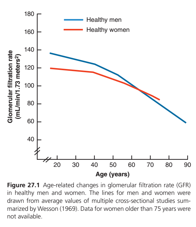
E-RELATED CHANGES IN GLOMERULAR FILTRATION RATE (GFR)
What Does This Graph Show?
- The graph shows how GFR (kidney filtering ability) changes with age.
- Blue line = Healthy men
- Red line = Healthy women
- As age increases, GFR gradually decreases in both men and women.
Blue Line (Healthy Men)
Age 20 Years
- GFR ≈ 135 mL/min/1.73 m²
- Kidneys are filtering very efficiently.
Age 40 Years
- GFR ≈ 124 mL/min/1.73 m²
- Slight decrease from young adulthood.
Age 55 Years
- GFR ≈ 112 mL/min/1.73 m²
- Filtering ability continues to decline gradually.
Age 65 Years
- GFR ≈ 95 mL/min/1.73 m²
- Noticeable reduction in kidney function.
Age 90 Years
- GFR ≈ 60 mL/min/1.73 m²
- Kidney filtering ability is much lower than in young adults.
Summary for Men:
- GFR falls steadily throughout life.
- By old age, men have lost a large portion of their filtering capacity.
Red Line (Healthy Women)
Age 20 Years
- GFR ≈ 120 mL/min/1.73 m²
- Slightly lower than men of the same age.
Age 40 Years
- GFR ≈ 115 mL/min/1.73 m²
- Small age-related decline.
Age 55 Years
- GFR ≈ 104 mL/min/1.73 m²
- Continued gradual decrease.
Age 65 Years
- GFR ≈ 95 mL/min/1.73 m²
- Similar to men at this age.
Age 75 Years
- GFR ≈ 84 mL/min/1.73 m²
- Further decline with aging.
Summary for Women:
- GFR is slightly lower than men during young adulthood.
- The decline is more gradual.
- Data beyond 75 years were not available.
Comparison of Both Lines
- At young ages (20–50 years):
- Men have a higher GFR than women.
- Around 65 years:
- Both lines meet.
- Men and women have nearly the same GFR.
- After 65 years:
- The men’s line drops more steeply.
- Women’s GFR becomes slightly higher than men’s.
Main Message of the Graph
✅ Kidney filtering ability (GFR) decreases naturally with aging.
✅ Young adults have the highest GFR.
✅ Men start with a higher GFR than women.
✅ Both sexes lose kidney function as they grow older.
✅ By 70–90 years of age, GFR is much lower than in young adulthood.
KEY CONCEPT
The graph shows that GFR gradually decreases with age in both healthy men and women. Men begin with a higher GFR, but their decline is steeper. Around 65 years of age, men and women have similar GFR values, and aging causes a significant reduction in kidney filtering capacity in both sexes.
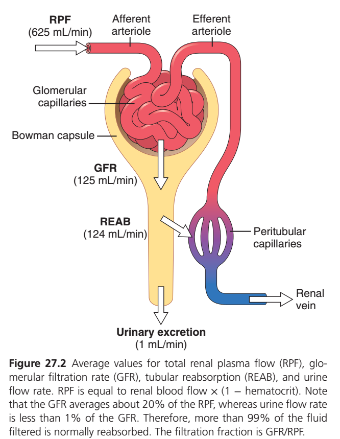
FIGURE 27.2 — THE JOURNEY OF PLASMA THROUGH THE KIDNEY
Step 1: Plasma Enters the Kidney
- Renal Plasma Flow (RPF) = 625 mL/min
- This means 625 mL of plasma enters the kidneys every minute through the afferent arteriole.
Think of RPF as the total plasma delivered to the kidney.
Step 2: Filtration Occurs in the Glomerulus
- Inside the glomerular capillaries, part of the plasma is filtered into Bowman capsule.
- Glomerular Filtration Rate (GFR) = 125 mL/min
This means:
- Out of 625 mL/min entering the kidney,
- 125 mL/min is filtered into the nephron.
Easy Calculation
Filtration Fraction = GFR ÷ RPF
= 125 ÷ 625
= 0.2 (20%)
Meaning: About 20% of the plasma entering the kidney is filtered.
Step 3: Most Filtered Fluid Is Reabsorbed
- The filtered fluid moves through the renal tubules.
- Most of this fluid does not become urine.
- It is returned to the blood through the peritubular capillaries.
Tubular Reabsorption (REAB) = 124 mL/min
This means:
- Out of 125 mL/min filtered
- 124 mL/min is taken back into the blood
Step 4: Very Little Becomes Urine
After reabsorption:
Filtered Fluid = 125 mL/min
Reabsorbed = 124 mL/min
Remaining = 1 mL/min
Therefore:
Urinary Excretion = 1 mL/min
Only a tiny amount leaves the body as urine.
The Big Picture
Imagine 625 Students Enter a Hall
625 students enter
↓
125 students are selected
(GFR)
↓
124 students return back
(Reabsorption)
↓
Only 1 student leaves
(Urine)
Important Relationships
Renal Plasma Flow (RPF)
- Total plasma entering kidneys.
- 625 mL/min
Glomerular Filtration Rate (GFR)
- Plasma filtered into nephron.
- 125 mL/min
Reabsorption (REAB)
- Filtered fluid returned to blood.
- 124 mL/min
Urinary Excretion
- Fluid finally leaving as urine.
- 1 mL/min
Numbers to Remember
| Process | Value |
|---|---|
| Renal Plasma Flow (RPF) | 625 mL/min |
| Glomerular Filtration Rate (GFR) | 125 mL/min |
| Reabsorption (REAB) | 124 mL/min |
| Urinary Excretion | 1 mL/min |
| Filtration Fraction | 20% |
KEY CONCEPT
Every minute, about 625 mL of plasma enters the kidneys. Only 125 mL (20%) is filtered into the nephron. Of this filtered fluid, 124 mL is reabsorbed back into the blood, and only about 1 mL becomes urine. Thus, more than 99% of the filtered fluid is normally reabsorbed.
GLOMERULAR CAPILLARY MEMBRANE
- The glomerular capillary membrane is similar to other capillary membranes.
- However, it has three major layers instead of two.
- The first layer is endothelial cells with a glycocalyx coat on the inner surface of the capillary.
- The glycocalyx is made of glycoproteins.
- The second layer is the basement membrane.
- The third layer is a layer of epithelial cells called podocytes.
- Podocytes surround the outer surface of the capillary basement membrane.
- Together, these three layers form the filtration barrier.
- Despite having three layers, this filtration barrier filters several hundred times more water and solutes than a usual capillary membrane.
- Even with this high filtration rate, only a small amount of plasma proteins is normally filtered.
- The high filtration rate occurs partly because of the special characteristics of the glomerular capillary membrane.
- The capillary endothelium contains thousands of small holes called fenestrae.
- These fenestrae are similar to those found in liver capillaries.
- However, they are smaller than the fenestrae in the liver.
- Although the fenestrae are relatively large, endothelial cell proteins contain many fixed negative charges.
- These negative charges hinder the passage of plasma proteins.
- Surrounding the endothelium is the basement membrane.
- The basement membrane consists of a meshwork of collagen and proteoglycan fibrillae.
- It contains large spaces through which large amounts of water and small solutes can filter.
- The basement membrane greatly hinders the filtration of plasma proteins.
- This occurs partly because of the strong negative electrical charges associated with the proteoglycans.
- The outer layer of the glomerular membrane is formed by podocytes.
- Podocytes line the outer surface of the glomerulus.
- These cells are not continuous.
- They have long foot-like processes called pedicels.
- The pedicels encircle the outer surface of the capillaries.
- The foot processes are separated by gaps called filtration slits.
- These slits are bridged by thin diaphragms.
- The diaphragms contain pores through which the glomerular filtrate moves.
- The filtration slit diaphragm is composed of several proteins.
- These proteins include nephrin and podocin.
- These proteins play a critical role in restricting the filtration of plasma proteins.
- Mutations in the nephrin gene can lead to abnormal or absent filtration slit diaphragms.
- This can cause large increases in the filtration and excretion of plasma proteins.
- This condition is called proteinuria.
- Thus, all layers of the glomerular capillary wall act as a barrier to plasma proteins.
- At the same time, they allow rapid filtration of water and most plasma solutes.
Mesangial Cells
- Another important cell type in the glomerulus is the mesangial cell.
- Mesangial cells lie around and between the glomerular capillaries.
- They produce extracellular matrix and other factors.
- These substances provide structural and functional support to the capillaries.
- Mesangial cells are not part of the glomerular filtration barrier.
- They contain actin- and myosin-based microfilaments.
- These microfilaments contact the glomerular basement membrane.
- Mesangial cells have contractile properties.
- Their contraction may alter blood flow through the glomerular capillaries.
KEY CONCEPT
The glomerular filtration barrier consists of three layers: endothelial cells, basement membrane, and podocytes. These layers allow rapid filtration of water and small solutes while restricting the passage of plasma proteins. Mesangial cells support the glomerular capillaries and may help regulate blood flow through them.
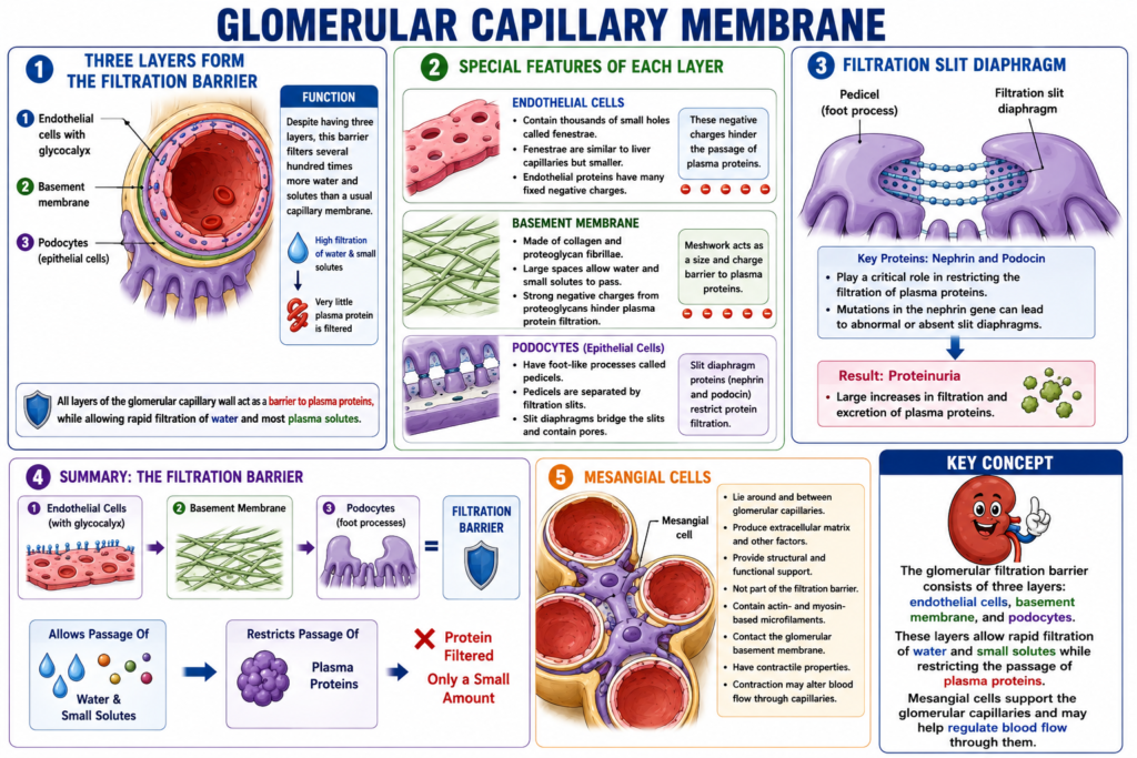
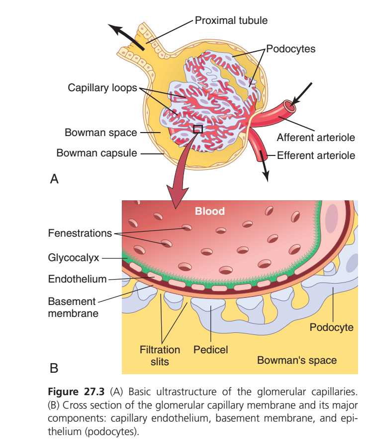
FIGURE 27.3 — STRUCTURE OF THE GLOMERULAR FILTRATION BARRIER
Part A: Overview of the Glomerulus
- Blood enters the glomerulus through the afferent arteriole.
- Inside the glomerulus, blood flows through many capillary loops.
- These capillaries are surrounded by Bowman capsule.
- The space between the capillaries and Bowman capsule is called Bowman’s space.
- The filtered fluid collects in Bowman’s space.
- The filtered fluid then enters the proximal tubule.
- This is the beginning of the renal tubule.
- Blood that is not filtered leaves through the efferent arteriole.
Easy Flow
Afferent arteriole
↓
Glomerular capillaries
↓
Filtration into Bowman’s space
↓
Proximal tubule
Part B: Zoomed View of the Filtration Barrier
This figure shows the three layers that every filtered substance must cross.
Layer 1: Endothelium
- Blood is present inside the capillary.
- The inner lining is called the endothelium.
- The endothelium contains many small holes called fenestrations.
- Water and small solutes can pass through these holes easily.
- The endothelium is covered by a glycocalyx.
- The glycocalyx helps prevent proteins from passing through.
Layer 2: Basement Membrane
- Below the endothelium lies the basement membrane.
- This is the main barrier against plasma proteins.
- Water and small solutes can pass through it.
- Most plasma proteins are restricted.
Layer 3: Podocytes
- The outer layer is formed by podocytes.
- Podocytes have foot-like projections called pedicels.
- Spaces between pedicels are called filtration slits.
- Filtered fluid passes through these slits into Bowman’s space.
Route Followed by Filtered Fluid
What Crosses the Barrier?
Blood
↓
Fenestrations (Endothelium)
↓
Basement Membrane
↓
Filtration Slits (Between Pedicels)
↓
Bowman’s Space
↓
Proximal Tubule
What Is Filtered?
✅ Water
✅ Electrolytes
✅ Glucose
✅ Amino acids
✅ Small solutes
What Is Normally Not Filtered?
❌ Red blood cells
❌ Blood cells
❌ Most plasma proteins
One-Line Memory Trick
“3 Layers, 3 Filters”
- Endothelium with fenestrations
- Basement membrane
- Podocyte filtration slits
Together they allow water and small solutes to pass while preventing most proteins and blood cells from entering the filtrate.
KEY CONCEPT
The glomerular filtration barrier has three layers: fenestrated endothelium, basement membrane, and podocyte filtration slits. Blood enters through the afferent arteriole, filtration occurs across these three layers, the filtrate enters Bowman’s space, and then flows into the proximal tubule. Water and small solutes are filtered, while most proteins and blood cells are retained in the blood.
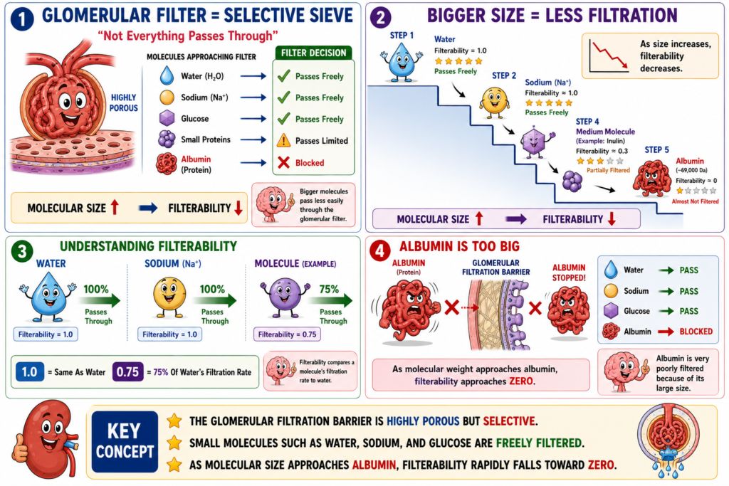
FILTERABILITY OF SOLUTES DECREASES AS THEIR SIZE INCREASES
- The glomerular capillary membrane is thicker than most other capillaries.
- However, it is also much more porous.
- Therefore, it filters fluid at a high rate.
- Despite this high filtration rate, the glomerular filtration barrier is selective.
- It determines which molecules can be filtered.
- This selectivity is based on molecular size and electrical charge.
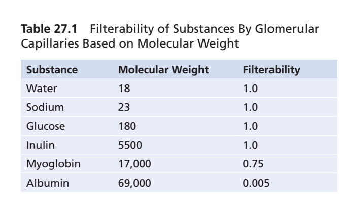
- The effect of molecular size on filtration can be measured by filterability.
- A filterability of 1.0 means that a substance is filtered as freely as water.
- A filterability of 0.75 means that a substance is filtered only 75% as rapidly as water.
- Electrolytes such as sodium are freely filtered.
- Small organic molecules such as glucose are also freely filtered.
- As the molecular weight of a molecule increases, its filterability decreases.
- When the molecular weight approaches that of albumin, filterability decreases rapidly.
- The filterability then approaches zero.
KEY CONCEPT
The glomerular filtration barrier is highly selective. Small molecules such as sodium and glucose are freely filtered, whereas larger molecules are filtered less easily. As molecular size approaches that of albumin, filterability rapidly decreases and becomes almost zero.
NEGATIVELY CHARGED LARGE MOLECULES ARE FILTERED LESS EASILY THAN POSITIVELY CHARGED MOLECULES OF EQUAL SIZE
- The plasma protein albumin has a molecular diameter of about 6 nanometers.
- The pores of the glomerular endothelial cell membrane are about 8 nanometers in diameter.
- Although albumin is smaller than the pore size, it is still restricted from filtration.
- This restriction occurs because albumin carries a negative charge.
- The glomerular capillary wall contains negatively charged glycoproteins.
- These negative charges create electrostatic repulsion against negatively charged albumin.
- Therefore, albumin is prevented from passing easily through the filtration barrier.
- Electrical charge affects the filtration of different molecules by the glomerulus.
- Dextrans are polysaccharides that can be manufactured with neutral, negative, or positive charges.
- For the same molecular radius, positively charged molecules are filtered more easily than negatively charged molecules.
- Neutral dextrans are also filtered more easily than negatively charged dextrans of the same molecular weight.
- These differences occur because the basement membrane and podocytes contain negative charges.
- These negative charges help restrict the filtration of large negatively charged molecules.
- Plasma proteins are important examples of large negatively charged molecules that are restricted from filtration.
KEY CONCEPT
The glomerular filtration barrier is selective not only by size but also by electrical charge. Positively charged molecules are filtered most easily, neutral molecules are filtered moderately, and negatively charged molecules are filtered least easily because the negatively charged basement membrane and podocytes repel them. This helps prevent plasma proteins such as albumin from being filtered.
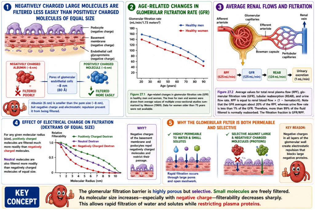
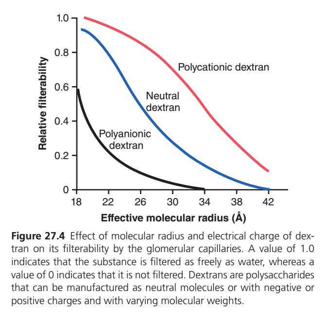
FIGURE 27.4 — EFFECT OF MOLECULAR SIZE AND ELECTRICAL CHARGE ON FILTRATION
What Does the Graph Show?
- The graph shows how molecular size and electrical charge affect filtration through the glomerulus.
- The X-axis shows the effective molecular radius (size).
- The Y-axis shows relative filterability.
Relative Filterability
- 1.0 = filtered as freely as water
- 0 = not filtered at all
Red Line — Polycationic Dextran (Positively Charged)
- This line remains the highest throughout the graph.
- Small positively charged molecules are filtered almost as freely as water.
- As molecular size increases, filtration gradually decreases.
- Even at larger sizes, positively charged molecules are filtered better than the other molecules.
Meaning
✅ Positive charge helps molecules pass through the filtration barrier more easily.
Blue Line — Neutral Dextran
- This line lies between the red and black lines.
- Small neutral molecules are filtered fairly well.
- As size increases, filterability steadily decreases.
- Large neutral molecules are filtered poorly.
Meaning
✅ Neutral molecules are filtered less easily than positively charged molecules but more easily than negatively charged molecules.
Black Line — Polyanionic Dextran (Negatively Charged)
- This line is the lowest.
- Even small negatively charged molecules have reduced filterability.
- As size increases, filterability falls very rapidly.
- Large negatively charged molecules are almost completely blocked.
Meaning
✅ Negative charge strongly reduces filtration.
Effect of Increasing Molecular Size
Small Molecules
- All three types are filtered relatively easily.
- Positively charged molecules are filtered the most.
- Negatively charged molecules are filtered the least.
Large Molecules
- Filterability decreases for all molecules.
- The larger the molecule, the harder it is to filter.
- Eventually filtration approaches zero.
Meaning
✅ Increasing size decreases filtration regardless of charge.
Comparison of the Three Lines
For the Same Size Molecule
Polycationic (Positive)
⬇
Most easily filtered
Neutral
⬇
Moderately filtered
Polyanionic (Negative)
⬇
Least filtered
Why Does This Happen?
- The basement membrane and podocytes contain many negative charges.
- These negative charges attract positive molecules less restrictively.
- They strongly repel negatively charged molecules.
- Therefore, negatively charged molecules are filtered less easily.
Clinical Connection
- Albumin is a large negatively charged plasma protein.
- Although its size is small enough to fit through many pores, its negative charge prevents easy filtration.
- This helps keep albumin in the blood.
KEY CONCEPT
This graph shows that glomerular filtration depends on both molecular size and electrical charge. As molecular size increases, filtration decreases. For molecules of the same size, positively charged molecules are filtered most easily, neutral molecules are filtered moderately, and negatively charged molecules are filtered least easily because the glomerular filtration barrier carries negative charges that repel them.
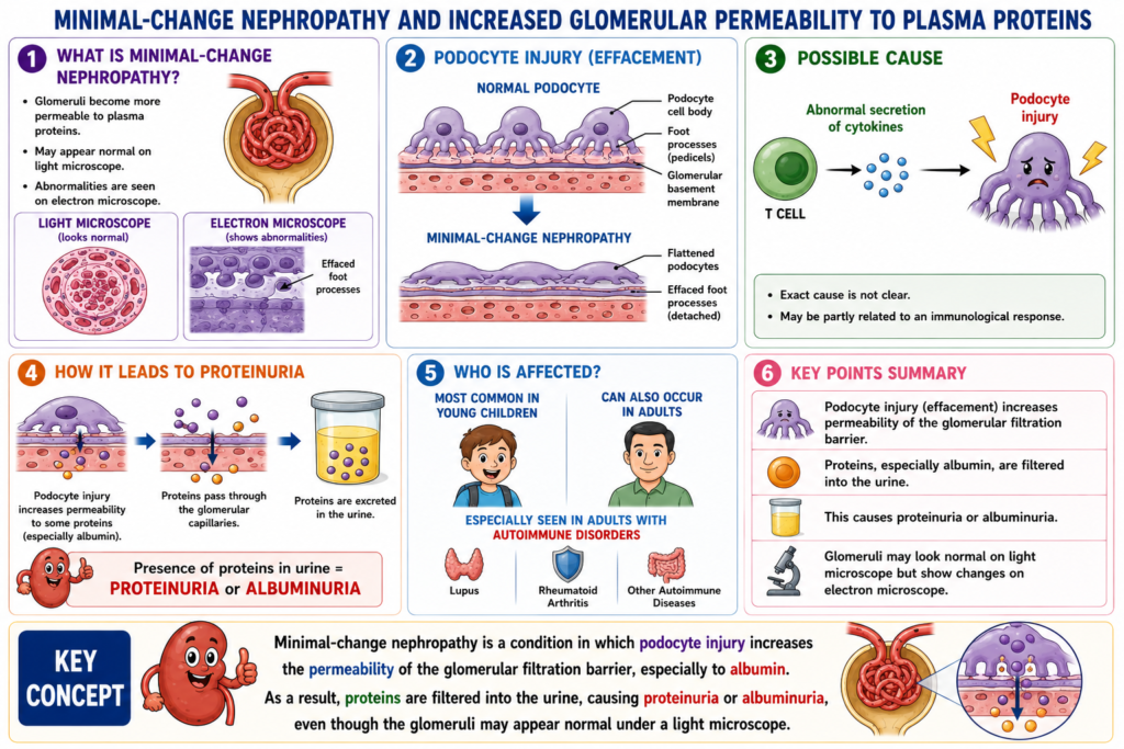
MINIMAL-CHANGE NEPHROPATHY AND INCREASED GLOMERULAR PERMEABILITY TO PLASMA PROTEINS
- In minimal-change nephropathy, the glomeruli become more permeable to plasma proteins.
- The glomeruli may appear normal when examined with a standard light microscope.
- However, when examined with an electron microscope, abnormalities are usually seen.
- The podocytes become flattened.
- The podocyte foot processes may become detached from the glomerular basement membrane.
- This change is called podocyte effacement.
- The exact cause of minimal-change nephropathy is not clear.
- It may be partly related to an immunological response.
- Abnormal T-cell secretion of cytokines may injure the podocytes.
- Podocyte injury increases their permeability to some lower-molecular-weight proteins.
- Albumin is especially affected.
- The increased permeability allows proteins to pass through the glomerular capillaries.
- These proteins are then excreted in the urine.
- The presence of proteins in the urine is called proteinuria or albuminuria.
- Minimal-change nephropathy is most common in young children.
- It can also occur in adults.
- It is especially seen in adults with autoimmune disorders.
KEY CONCEPT
Minimal-change nephropathy is a condition in which podocyte injury increases the permeability of the glomerular filtration barrier, especially to albumin. As a result, proteins are filtered into the urine, causing proteinuria or albuminuria, even though the glomeruli may appear normal under a light microscope.
Glomerular Filtration: The First Step in Urine Formation (Summarized Essay)
Glomerular filtration is the first and one of the most important steps in urine formation. In this process, large amounts of fluid are filtered from the blood through the glomerular capillaries into Bowman’s capsule. The kidneys filter approximately 180 liters of fluid each day, although most of this filtrate is reabsorbed, leaving only about 1–2 liters of urine to be excreted daily. This high filtration rate is possible because the kidneys receive a large blood supply, about 22% of the resting cardiac output, and because of the unique structure of the glomerular filtration membrane.
The fluid that enters Bowman’s capsule is called the glomerular filtrate. It is very similar in composition to plasma, except that it contains almost no proteins and no blood cells. Water, electrolytes, glucose, amino acids, urea, and many other small substances are freely filtered. However, substances that are bound to plasma proteins, such as a portion of calcium and most fatty acids, are filtered less readily. As a result, the glomerular filtrate is essentially a protein-free ultrafiltrate of plasma.
The glomerular filtration rate (GFR) is the amount of filtrate formed each minute and averages about 125 mL/min, or approximately 180 liters per day in a healthy adult. The GFR is influenced by body size, age, and sex. Women generally have a slightly lower GFR than men, and GFR gradually declines with aging because the number of functioning nephrons decreases over time. Diseases such as hypertension, diabetes, and glomerulonephritis can accelerate this decline.
Normally, only about 20% of the plasma flowing through the kidneys is filtered, a value known as the filtration fraction. The remaining plasma continues through the renal circulation without being filtered. Thus, the filtration fraction can be expressed as:
Filtration Fraction = GFR ÷ Renal Plasma Flow
The remarkable filtering ability of the kidneys is due to the specialized structure of the glomerular filtration membrane, which consists of three layers: the capillary endothelium, the basement membrane, and the podocyte layer. These layers together form a highly selective filtration barrier. They allow rapid passage of water and small solutes while preventing significant filtration of plasma proteins and blood cells.
The endothelial layer contains numerous small openings called fenestrations, which allow fluid to pass easily but restrict proteins. Beneath the endothelium lies the basement membrane, a mesh-like structure rich in negatively charged proteoglycans. This membrane acts as a major barrier to protein filtration. The outermost layer consists of specialized epithelial cells called podocytes, whose foot processes create narrow filtration slits covered by slit diaphragms. Proteins such as nephrin and podocin within these diaphragms play a crucial role in preventing excessive protein leakage into the urine.
The filtration barrier is highly selective based on molecular size and electrical charge. Small molecules such as sodium, chloride, glucose, and urea are freely filtered. As molecular size increases, filtration becomes progressively more difficult. Large proteins such as albumin are normally retained in the blood. In addition, negatively charged molecules are filtered less readily than positively charged molecules of the same size because the filtration membrane itself carries strong negative charges that repel negatively charged plasma proteins.
Another important cell type in the glomerulus is the mesangial cell, which provides structural support to the capillaries and helps regulate glomerular blood flow. Although mesangial cells are not part of the filtration barrier, they contribute to normal glomerular function.
A clinical example of altered glomerular filtration is minimal-change nephropathy, a condition in which the glomeruli appear normal under a light microscope but show flattening and fusion of podocyte foot processes under an electron microscope. Damage to the podocytes increases permeability to plasma proteins, especially albumin, leading to proteinuria (albuminuria). This disorder is most common in children and is often related to immune-mediated injury of the podocytes.
In summary, glomerular filtration is the initial step in urine formation and involves the movement of large amounts of protein-free fluid from the blood into Bowman’s capsule. The glomerular filtration membrane provides an efficient yet highly selective barrier that filters water and small solutes while retaining proteins and blood cells. The rate and selectivity of filtration are essential for maintaining fluid balance, removing waste products, and preserving important plasma constituents.
