


Learning Objectives
After completing this lecture, students will be able to:
- Explain parasympathetic and sympathetic innervation of the eye.
- Describe the mechanism and control of accommodation.
- Explain control of pupillary diameter and the pupillary light reflex.
- Describe important pupillary abnormalities in central nervous system disease.
- Explain the major features of Horner syndrome.
Introduction
The eye receives both parasympathetic and sympathetic autonomic nerve fibers.
These autonomic pathways help control two important visual functions:
- Focusing of the lens.
- Size of the pupil.
Parasympathetic activity controls:
- Ciliary muscle contraction for focusing.
- Constriction of the pupil.
Sympathetic activity mainly causes:
- Dilation of the pupil.
Together, these mechanisms help the eye adjust its focus and regulate the amount of light entering the eye.
Main Topic / Core Concept
Autonomic Nerves to the Eyes
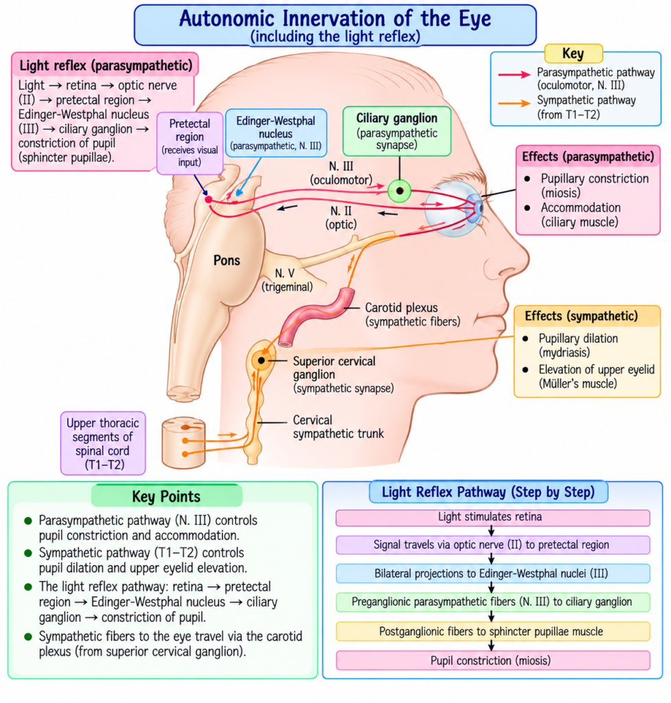
The autonomic innervation of the eye is shown in Fig. 52.11.
The eye receives:
- Parasympathetic nerve fibers.
- Sympathetic nerve fibers.
Parasympathetic Innervation of the Eye
Parasympathetic preganglionic fibers begin in the:
Edinger-Westphal nucleus
Edinger-Westphal nucleus: The visceral nucleus associated with the third cranial nerve that gives rise to parasympathetic fibers controlling important functions of the eye.
Parasympathetic Pathway
Edinger-Westphal nucleus
→ third cranial nerve
→ ciliary ganglion
→ synapse with postganglionic parasympathetic neurons
→ ciliary nerves
→ eyeball
The ciliary ganglion lies immediately behind the eye.
Functions of Parasympathetic Fibers
Parasympathetic nerves excite:
- Ciliary muscle.
- Sphincter muscle of the iris.
Ciliary Muscle
Ciliary muscle stimulation
→ controls focusing of the lens
Sphincter of the Iris
Sphincter muscle stimulation
→ pupil constricts
Key Concept
Parasympathetic stimulation
→ ciliary muscle activation
→ accommodation
and
Parasympathetic stimulation
→ iris sphincter contraction
→ pupil constriction
Sympathetic Innervation of the Eye
Sympathetic innervation begins in the:
- Intermediolateral horn cells of the first thoracic segment of the spinal cord.
Sympathetic Pathway
First thoracic spinal cord
→ sympathetic chain
→ superior cervical ganglion
→ synapse with postganglionic neurons
→ fibers travel along carotid artery and smaller arteries
→ eye
Functions of Sympathetic Fibers
Sympathetic fibers innervate:
- Radial fibers of the iris.
- Several extraocular muscles described in relation to Horner syndrome.
Radial Iris Fibers
Sympathetic stimulation
→ radial fibers contract
→ pupil opens
Comparison of Parasympathetic and Sympathetic Effects
| Feature | Parasympathetic | Sympathetic |
|---|---|---|
| Main origin described | Edinger-Westphal nucleus | First thoracic spinal cord |
| Important ganglion | Ciliary ganglion | Superior cervical ganglion |
| Iris effect | Sphincter contraction | Radial fiber contraction |
| Pupil response | Constriction | Dilation |
| Ciliary muscle | Excited | Not described as the accommodation pathway in this text |
Control of Accommodation — Focusing the Eyes
Accommodation: The mechanism by which the lens system changes its focusing power to maintain clear vision.
Accommodation is essential for:
- A high degree of visual acuity.
It depends on:
- Contraction of the ciliary muscle.
- Relaxation of the ciliary muscle.
Effect of Ciliary Muscle Contraction
Ciliary muscle contracts
→ refractive power of lens increases
Effect of Ciliary Muscle Relaxation
Ciliary muscle relaxes
→ refractive power of lens decreases
Negative Feedback Control of Accommodation
Accommodation is regulated by a negative feedback mechanism.
Negative feedback: Automatic correction of lens power until the image reaches the best possible focus.
Basic Mechanism
Image becomes out of focus
→ focusing error is detected
→ lens strength changes in correct direction
→ image becomes clearer
→ best visual acuity is restored
When the eyes shift suddenly from a far object to a near object:
→ the lens usually achieves best focus within less than 1 second
The precise mechanism is not completely understood, but several important clues help control accommodation.
Clues Used to Control Accommodation
Chromatic Aberration
Chromatic aberration: Difference in the focusing positions of different colors of light.
- Blue light is bent more strongly by the lens than red light.
- Therefore:
- Red rays focus slightly behind blue rays.
The eye can apparently detect which color is in better focus.
Concept
Blue and red rays focus differently
→ eye detects which is clearer
→ accommodation system determines whether lens should become stronger or weaker
Convergence of the Eyes
When viewing a near object:
- The two eyes must converge.
The neural mechanism that causes convergence also produces:
→ simultaneous signal to strengthen the lens
Flow
Near object
→ convergence required
→ accommodation signal also activated
→ lens becomes stronger
Depth of the Fovea
The fovea lies in a slightly deeper depression than the surrounding retina.
Therefore:
- Focus at the depth of the fovea differs from focus at its edges.
This difference may provide information about:
→ whether lens power should increase or decrease
Oscillation of Accommodation
Lens accommodation continually oscillates slightly.
Frequency:
→ up to 2 times per second
When lens strength changes in the correct direction:
→ image becomes clearer
When it changes in the wrong direction:
→ image becomes less clear
This changing clarity may provide a rapid clue about how lens power should be adjusted.
Concept
Small lens-power oscillation
→ image becomes clearer or blurrier
→ brain detects correct direction
→ lens power adjusted appropriately
Cortical Control of Accommodation
The brain areas controlling accommodation closely parallel those controlling eye fixation.
Visual signals are analyzed in:
- Brodmann area 18.
- Brodmann area 19.
Accommodation Pathway
Visual analysis in Brodmann areas 18 and 19
→ pretectal area
→ Edinger-Westphal nucleus
→ parasympathetic fibers
→ ciliary muscle
→ lens focusing
Mechanism / Pathway / Step-by-Step Explanation
Parasympathetic Accommodation Pathway
Need for refocusing
→ visual cortical analysis
→ pretectal area
→ Edinger-Westphal nucleus
→ parasympathetic fibers
→ ciliary muscle
→ change in lens refractive power
→ improved focus
Control of Pupillary Diameter
Pupillary size is controlled by opposing autonomic actions.
Parasympathetic Effect
Parasympathetic stimulation
→ pupillary sphincter muscle contracts
→ pupillary aperture becomes smaller
This is called:
Miosis
Miosis: Constriction of the pupil caused by contraction of the pupillary sphincter.
Sympathetic Effect
Sympathetic stimulation
→ radial iris fibers contract
→ pupil enlarges
This is called:
Mydriasis
Mydriasis: Dilation of the pupil caused by sympathetic stimulation of the radial iris fibers.
Miosis vs Mydriasis
| Feature | Miosis | Mydriasis |
|---|---|---|
| Pupil | Constricted | Dilated |
| Main autonomic pathway | Parasympathetic | Sympathetic |
| Iris muscle involved | Sphincter | Radial fibers |
Pupillary Light Reflex
Pupillary light reflex: Constriction of the pupil when light enters the eye.
Pathway
Light reaches retina
→ visual impulses travel through optic nerve
→ some impulses pass to pretectal nuclei
→ signals pass to Edinger-Westphal nucleus
→ parasympathetic fibers return to eye
→ iris sphincter contracts
→ pupil constricts
In Darkness
Darkness
→ pupillary light reflex inhibited
→ pupil dilates
Function of the Pupillary Light Reflex
The pupillary light reflex helps the eye adapt rapidly to changes in illumination.
The pupil can vary approximately between:
- 1.5 mm at the small end.
- 8 mm at the large end.
The brightness of light reaching the retina changes approximately with the square of pupillary diameter.
Easy Mathematical Concept
Maximum pupil diameter:
8 mm
Minimum pupil diameter:
1.5 mm
Diameter ratio:
8 ÷ 1.5 ≈ 5.3
Because retinal brightness varies with the square of diameter:
5.3 × 5.3 ≈ 28
This is approximately:
30 : 1
Therefore, the pupillary reflex can change the amount of light entering the eye by approximately 30-fold.
Key Concept
Pupil diameter changes
→ amount of entering light changes greatly
→ contributes to light and dark adaptation
Pupillary Reflexes in Central Nervous System Diseases
Some central nervous system diseases can interfere with transmission of visual signals from the retina to the Edinger-Westphal nucleus.
This may block the pupillary reflex.
Conditions mentioned in the provided text include:
- Central nervous system syphilis.
- Chronic alcoholism.
- Encephalitis.
- Multiple sclerosis.
- Lyme disease.
Site of the Block
The block usually occurs in:
- Pretectal region of the brain stem.
It may also result from:
- Destruction of small fibers in the optic nerves.
Effect of Loss of Inhibitory Signals
The final fibers passing through the pretectal pathway to the Edinger-Westphal nucleus are mainly inhibitory.
If these inhibitory signals are lost:
Loss of inhibition
→ Edinger-Westphal nucleus remains chronically active
→ pupils remain mostly constricted
At the same time:
→ pupils fail to respond normally to light
Pupillary Reaction to Accommodation
Even when the pupillary light reflex is impaired, the pupil may still constrict through another pathway.
When the eyes look at a near object:
- Lens accommodation occurs.
- Eyes converge.
- Mild pupillary constriction occurs at the same time.
This is called:
Pupillary reaction to accommodation
Flow
Near object
→ accommodation
- convergence
→ mild pupillary constriction
Argyll Robertson Pupil
Argyll Robertson pupil: A small pupil that fails to respond to light but can still constrict during accommodation.
Important features:
- Very small pupil.
- Fails to respond to light.
- Still responds to accommodation.
It is an important diagnostic sign of central nervous system disease such as:
- Syphilis.
Key Concept
Light
→ little or no pupillary response
Near-object accommodation
→ pupil still constricts
Horner Syndrome — Oculosympathetic Paresis
Horner syndrome: Clinical condition produced by interruption of sympathetic nerve supply to the eye.
It is also called:
Oculosympathetic paresis
The interruption frequently occurs in the:
- Cervical sympathetic chain.
Persistent Pupillary Constriction
Normally:
Sympathetic fibers
→ pupillary dilator muscle
→ pupil dilation
In Horner syndrome:
Sympathetic pathway interrupted
→ dilator muscle loses stimulation
→ affected pupil remains smaller than the opposite pupil
Drooping of the Superior Eyelid
The superior eyelid is partly maintained in an open position by:
- Smooth muscle fibers.
- These fibers receive sympathetic innervation.
If sympathetic nerves are damaged:
→ superior eyelid cannot open as widely
→ eyelid droops
Dilated Blood Vessels
Loss of sympathetic innervation causes:
→ blood vessels on the affected side of the face and head remain persistently dilated
Loss of Sweating
Sweating requires sympathetic nerve signals.
Therefore:
Sympathetic interruption
→ sweating cannot occur normally
→ affected side of face and head does not sweat
Main Features of Horner Syndrome
| Sympathetic Loss | Result |
|---|---|
| Loss of pupillary dilator stimulation | Persistently small pupil |
| Loss of sympathetic eyelid support | Drooping superior eyelid |
| Loss of vascular sympathetic effect | Persistent vasodilation |
| Loss of sympathetic sweating signals | Absence of sweating on affected side |
Original Educational Figure / Diagram
Autonomic Control of the Eye
Parasympathetic pathway
Edinger-Westphal nucleus
→ CN III
→ ciliary ganglion
→ ciliary nerves
→ ciliary muscle + iris sphincter
Results:
Ciliary muscle
→ accommodation
Iris sphincter
→ miosis
Sympathetic pathway
T1 spinal cord
→ sympathetic chain
→ superior cervical ganglion
→ carotid arterial pathway
→ eye
→ radial iris fibers
Result:
Radial iris contraction
→ mydriasis
Pupillary Light Reflex
Light
→ retina
→ optic nerve
→ pretectal nuclei
→ Edinger-Westphal nucleus
→ parasympathetic fibers
→ iris sphincter
→ pupil constriction
Key Concept
Autonomic control of the eye can be remembered as:
Parasympathetic
Edinger-Westphal nucleus
→ ciliary ganglion
→ ciliary muscle + iris sphincter
→ accommodation + pupil constriction
Sympathetic
T1 spinal cord
→ superior cervical ganglion
→ radial iris fibers
→ pupil dilation
For light:
Light enters eye
→ pretectal pathway
→ Edinger-Westphal nucleus
→ parasympathetic stimulation
→ miosis
Clinical Importance / Clinical Correlation
- Accommodation is necessary for clear focusing and high visual acuity.
- Abnormal central nervous system transmission can interfere with the pupillary light reflex.
- A pupil that does not respond to light but responds to accommodation may indicate disease affecting the pupillary reflex pathway.
- The Argyll Robertson pupil is described as an important diagnostic sign in central nervous system disease such as syphilis.
- Interruption of sympathetic innervation produces Horner syndrome.
- Horner syndrome affects:
- Pupil size.
- Eyelid position.
- Facial blood vessels.
- Sweating.
High-Yield / Exam Points
- Parasympathetic preganglionic fibers arise from the Edinger-Westphal nucleus.
- They travel through:
- Third cranial nerve.
- Ciliary ganglion.
- Ciliary nerves.
- Parasympathetic stimulation:
- Activates ciliary muscle.
- Constricts pupil.
- Sympathetic fibers begin in the first thoracic spinal cord segment.
- They synapse in the superior cervical ganglion.
- Sympathetic stimulation:
- Activates radial iris fibers.
- Dilates pupil.
- Ciliary muscle contraction:
- Increases lens refractive power.
- Ciliary muscle relaxation:
- Decreases lens refractive power.
- Accommodation for a new fixation distance can occur in less than 1 second.
- Important accommodation clues:
- Chromatic aberration.
- Convergence.
- Foveal depth.
- Lens-power oscillations.
- Accommodation oscillation:
- Up to 2 times/sec.
- Miosis = pupil constriction.
- Mydriasis = pupil dilation.
- Pupillary light reflex pathway:
- Retina → optic nerve → pretectal nuclei → Edinger-Westphal nucleus → parasympathetic nerves → iris sphincter.
- Pupil diameter:
- About 1.5–8 mm.
- Pupillary reflex changes retinal illumination by approximately:
- 30-fold.
- Argyll Robertson pupil:
- Small.
- No normal light response.
- Accommodation response preserved.
- Horner syndrome:
- Caused by interruption of sympathetic fibers to the eye.
Common Student Mistakes
- Do not confuse the autonomic control of the iris muscles.
- Sphincter → parasympathetic.
- Radial fibers → sympathetic.
- Do not confuse miosis with mydriasis.
- Miosis → small pupil.
- Mydriasis → large pupil.
- Do not think accommodation is simply a pupillary response.
- Accommodation primarily involves adjustment of lens refractive power through the ciliary muscle.
- Do not confuse the light reflex with the accommodation reaction.
- Light reflex begins with retinal illumination.
- Accommodation reaction occurs during near fixation with accommodation and convergence.
- Do not confuse the Edinger-Westphal nucleus with the superior cervical ganglion.
- Edinger-Westphal → parasympathetic pathway.
- Superior cervical ganglion → sympathetic pathway.
Quick Revision
Parasympathetic
Edinger-Westphal nucleus
→ CN III
→ ciliary ganglion
→ ciliary nerves
Effects:
→ ciliary muscle activation
→ accommodation
→ iris sphincter activation
→ miosis
Sympathetic
T1 spinal cord
→ sympathetic chain
→ superior cervical ganglion
→ eye
Effect:
→ radial iris contraction
→ mydriasis
Accommodation
Ciliary muscle contracts
→ lens refractive power increases
Ciliary muscle relaxes
→ lens refractive power decreases
Clues:
→ chromatic aberration
→ convergence
→ foveal depth
→ accommodation oscillation
Pupillary Light Reflex
Light
→ retina
→ optic nerve
→ pretectal nuclei
→ Edinger-Westphal nucleus
→ parasympathetic fibers
→ sphincter contraction
→ pupil constricts
Argyll Robertson Pupil
Light response absent
- accommodation response preserved
- small pupil
Horner Syndrome
Sympathetic interruption
→ small pupil
→ drooping upper eyelid
→ dilated facial/head vessels
→ loss of sweating on affected side
Final Take-Home Concept / Summary
Autonomic control of the eye depends on coordinated parasympathetic and sympathetic pathways.
The parasympathetic pathway begins in the:
Edinger-Westphal nucleus
and travels through:
CN III
→ ciliary ganglion
→ ciliary nerves
It controls:
- Ciliary muscle.
- Iris sphincter.
Therefore:
Parasympathetic activity
→ accommodation + miosis
The sympathetic pathway begins in:
T1 spinal cord
and travels through:
Sympathetic chain
→ superior cervical ganglion
→ eye
It stimulates the radial iris fibers and produces:
→ mydriasis
Accommodation uses negative feedback to maintain clear focus.
Blur or change in fixation distance
→ focusing clues detected
→ lens power adjusted
→ visual acuity improved
The pupillary light reflex follows:
Light
→ retina
→ pretectal nuclei
→ Edinger-Westphal nucleus
→ parasympathetic fibers
→ iris sphincter
→ pupil constriction
Neurological disease can interrupt this pathway.
A small pupil that fails to react to light but still reacts to accommodation is described as an:
→ Argyll Robertson pupil
Loss of sympathetic innervation produces:
→ Horner syndrome
with:
- Persistent pupillary constriction.
- Drooping upper eyelid.
- Vasodilation.
- Loss of sweating on the affected side.
References / Sources
- Guyton and Hall Textbook of Medical Physiology, 15th Edition, Chapter 52.
