


Learning Objectives
After studying this topic, students will be able to:
- Explain the major functions of amacrine cells.
- Describe retinal convergence and differences between central and peripheral retina.
- Compare important types of retinal ganglion cells.
- Explain ganglion-cell responses to light, contrast, and color.
- Understand how retinal signals begin to be analyzed before reaching the brain.
Introduction
- Amacrine cells are the most structurally and functionally diverse neurons in the retina.
- Many amacrine cells act as interneurons and help analyze visual information before it leaves the retina.
- Ganglion cells receive the processed retinal information and send it toward the brain through their optic nerve fibers.
- Although the retina contains about 100 million rods and 3 million cones, it contains only about 1.6 million ganglion cells.
- The central and peripheral retina differ greatly in:
- Visual acuity
- Light sensitivity
- Degree of photoreceptor convergence
- Different ganglion-cell types are specialized for different visual functions, including:
- Movement
- Fine details
- Color
- Contrast
- Rapid changes in visual images
- Ganglion cells transmit their output by repetitive action potentials.
- Retinal processing of contrast and color therefore begins before visual signals reach the brain.
Interneuron: A neuron that helps process and analyze signals between other neurons.
Convergence: The arrangement in which signals from several photoreceptors come together onto a smaller number of ganglion-cell pathways.
AMACRINE CELLS AND THEIR FUNCTIONS
Amacrine cells: Structurally and functionally diverse retinal neurons that help analyze visual signals before those signals leave the retina.
- More than 60 types of amacrine cells have been identified.
- They have been identified by:
- Morphological methods
- Histochemical methods
- Molecular methods
- Several types have been functionally characterized.
- Their functions are different from one another.
Amacrine Cells in the Rod Pathway
- One type of amacrine cell forms part of the direct pathway for rod vision.
Pathway
Rod
→ bipolar cell
→ amacrine cell
→ ganglion cell
Key Concept
Amacrine cells are therefore not only lateral-processing cells.
In the pure rod pathway, an amacrine cell forms an actual part of the direct route carrying the visual signal toward the ganglion cell.
Amacrine Cells Responding to the Onset of Light
- One type responds strongly when a continuing visual signal begins.
- Its response is initially strong.
- The response then rapidly disappears.
Concept
Visual signal starts
→ strong amacrine-cell response
→ response rapidly fades
Amacrine Cells Responding to the Offset of Light
- Other amacrine cells respond strongly when a visual signal ends.
- Their response also fades rapidly.
Concept
Visual signal stops
→ strong response
→ response rapidly fades
Amacrine Cells Responding to Both On and Off
- Some amacrine cells respond when light is:
- Turned on
- Turned off
- These cells therefore signal a change in illumination.
- The direction of the change does not matter.
Concept
Light ON
or
Light OFF
→ amacrine-cell response
Therefore:
Change in illumination itself is detected.
Directionally Sensitive Amacrine Cells
Directionally sensitive amacrine cell: An amacrine cell that responds when a spot moves across the retina in a particular direction.
- Some amacrine cells respond to movement of a spot across the retina.
- Their response depends on the direction of movement.
- Therefore, they are called directionally sensitive.
Key Concept
Moving spot
→ movement in a specific direction
→ selected amacrine cells respond strongly
Overall Function of Amacrine Cells
- Many or most amacrine cells act as interneurons.
- They help analyze visual signals within the retina.
- This processing occurs before the signals leave the retina.
Key Concept
Visual signal enters retinal circuitry
→ amacrine cells analyze particular features
→ processed information reaches ganglion cells
→ signal leaves retina
GANGLION CELLS AND OPTIC NERVE FIBERS
Ganglion cells: Retinal output neurons whose fibers form pathways carrying visual signals from the retina toward the brain.
The retina contains approximately:
- 100 million rods
- 3 million cones
- Only about 1.6 million ganglion cells
Therefore, many photoreceptors must converge onto ganglion-cell pathways.
Average Photoreceptor Convergence
On average:
- About 60 rods
- About 2 cones
converge on each ganglion cell and its optic nerve fiber.
Easy Numerical Concept
For rods:
100 million rods ÷ 1.6 million ganglion cells
≈ 62.5 rods per ganglion cell
This corresponds approximately to the stated average of:
→ 60 rods
For cones:
3 million cones ÷ 1.6 million ganglion cells
≈ 1.9 cones per ganglion cell
This corresponds approximately to:
→ 2 cones
Key Concept
Many photoreceptors
→ fewer ganglion cells
→ photoreceptor signals converge before leaving the retina
CENTRAL AND PERIPHERAL RETINA
Major differences exist between the central and peripheral retina.
| Feature | Central Retina / Fovea | Peripheral Retina |
|---|---|---|
| Photoreceptor convergence | Much less convergence | Greater convergence |
| Rods and cones | Become progressively slender toward fovea | Greater rod contribution |
| Central fovea | About 35,000 slender cones, no rods | Rods and cones are present |
| Optic fibers | Nearly equal to number of cones in central fovea | Many photoreceptors may converge onto one fiber |
| Visual function emphasized | High visual acuity | Greater sensitivity to weak light |
High Visual Acuity in the Central Retina
As the fovea is approached:
- Fewer rods and cones converge on each optic nerve fiber.
- Photoreceptors become more slender.
- These changes progressively increase visual acuity.
Visual acuity: The ability to represent fine visual detail accurately.
In the central fovea:
- There are about 35,000 slender cones.
- There are no rods.
- The number of optic nerve fibers from this region is almost equal to the number of cones.
This relationship is shown in Fig. 51.12.
Key Concept
Less convergence
→ more separate cone pathways
→ greater visual acuity
Therefore:
Central retina
→ very high visual acuity
Peripheral retina
→ much poorer visual acuity
GREATER LIGHT SENSITIVITY OF THE PERIPHERAL RETINA
- The peripheral retina is much more sensitive to weak light than the central retina.
This occurs partly because:
- Rods are 30 to 300 times more sensitive to light than cones.
The sensitivity is further increased because:
- As many as 200 rods may converge onto a single optic nerve fiber in the peripheral retina.
- Their signals can summate.
- This produces stronger stimulation of peripheral ganglion cells and their optic nerve fibers.
Summation: Addition of signals from multiple rods so that their combined effect produces stronger stimulation.
Key Concept
Many rods
→ converge onto one optic nerve pathway
→ rod signals add together
→ stronger ganglion-cell stimulation
→ greater sensitivity to weak lightRETINAL GANGLION CELLS AND THEIR RESPECTIVE FIELDS
Different retinal ganglion cells respond to different features of visual information.
W, X, AND Y GANGLION CELLS
Studies in cats identified three distinct types of ganglion cells:
- W cells
- X cells
- Y cells
They differ in both structure and function.
| Feature | W Cells | X Cells | Y Cells |
|---|---|---|---|
| Signal conduction | Slow | Not emphasized as fastest | 50 m/sec or faster |
| Main retinal input | Mostly rods | At least one cone | Widespread retinal areas |
| Field size | Broad peripheral fields | Small fields | Broad dendritic fields |
| Main function | Directional movement and crude rod vision | Fine detail and color vision | Rapid changes and new visual events |
| Spatial accuracy | Broad field | High, discrete retinal location | Poorer localization |
| Special feature | Important in dark conditions | Represents fine retinal detail | Alerts CNS rapidly to new visual events |
W Cells
- W cells transmit signals slowly through their optic nerve fibers.
- They receive most of their excitation from rods.
- Rod signals reach them through:
- Small bipolar cells
- Amacrine cells
- W cells have broad fields in the peripheral retina.
- They are sensitive to directional movement.
- They are probably important for crude rod vision under dark conditions.
Key Concept
Rod input
→ W cell
→ slow signaling
→ directional movement detection
→ crude vision in darkness
X Cells
- X cells have small receptive fields.
- Their dendrites do not spread widely through the retina.
- Their signals therefore represent discrete retinal locations.
- They transmit fine details of visual images.
- Every X cell receives input from at least one cone.
- Therefore, X-cell transmission is probably responsible for color vision.
Key Concept
Small field
→ precise retinal location
→ fine visual detail
Cone input
→ contribution to color vision
Y Cells
- Y cells are the largest of these ganglion cells.
- They transmit signals to the brain at 50 m/sec or faster.
- Their dendritic fields are broad.
- They receive signals from widespread retinal areas.
- They respond strongly to rapid changes in visual images.
- They inform the central nervous system almost immediately when a new visual event occurs anywhere in the visual field.
- However, they do not specify its location with great accuracy.
- Instead, they provide clues that cause the eyes to move toward the exciting visual event.
Key Concept
New rapid visual event
→ Y cells respond quickly
→ CNS is alerted almost immediately
→ eyes can move toward the event
P AND M GANGLION CELLS
In primates, including humans, a different classification is commonly used.
- As many as 20 types of retinal ganglion cells have been described.
- Different types respond to different features of the visual scene.
These features include:
- Specific directions of movement
- Specific orientations
- Fine details
- Increase in light
- Decrease in light
- Particular colors
The two most extensively studied general classes are:
- Parvocellular (P) cells
- Magnocellular (M) cells
P Cells
P cells: Ganglion cells specialized especially for fine visual detail and color.
They are also called:
- Beta cells
- Midget ganglion cells in the central retina
They project to the parvocellular, or small-cell, layer of the lateral geniculate nucleus of the thalamus.
M Cells
M cells: Ganglion cells particularly sensitive to low-contrast stimuli and rapid movement.
They are also called:
- Alpha cells
- Parasol cells
They project to the magnocellular, or large-cell, layer of the lateral geniculate nucleus.
The lateral geniculate nucleus then relays information from the optic tract toward the visual cortex.
Comparison of P and M Cells
| Feature | P Cells | M Cells |
|---|---|---|
| Receptive field | Much smaller | Larger |
| Axonal conduction | Much slower | Faster |
| Response duration | Can be sustained | Much more transient |
| Color sensitivity | Generally sensitive to color | Not sensitive to color stimuli |
| Low-contrast black-and-white stimuli | Relatively insensitive | Highly sensitive |
| Main visual role | Fine detail and different colors | Low contrast and rapid movement |
Key Concept
P cells
Fine detail
- color
- sustained response
→ detailed visual information
M cells
Low contrast
- rapid movement
- transient response
→ rapid changing visual information
PHOTOSENSITIVE RETINAL GANGLION CELLS
A third type of photosensitive retinal ganglion cell has also been described.
- These ganglion cells contain their own photopigment called melanopsin.
Melanopsin: The photopigment contained within this type of photosensitive retinal ganglion cell.
- Less is known about these cells.
- They appear to send signals mainly to nonvisual areas of the brain.
- An important destination is the suprachiasmatic nucleus of the hypothalamus.
Suprachiasmatic nucleus: The master circadian pacemaker described in the provided text.
- These signals presumably help control circadian rhythms.
Circadian rhythms: Physiological changes synchronized with night and day.
Key Concept
Photosensitive retinal ganglion cells
→ melanopsin
→ suprachiasmatic nucleus
→ help synchronize physiological changes with night and day
EXCITATION OF THE GANGLION CELLS
Spontaneous, Continuous Action Potentials
- Long optic nerve fibers extend from ganglion cells into the brain.
- Because of this long distance, electrotonic conduction used by rods, cones, and bipolar cells is no longer suitable.
- Ganglion cells therefore transmit their signals using repetitive action potentials.
Action potential: The electrical impulse used by ganglion cells to transmit signals over the long optic nerve pathway.
- Ganglion cells continue firing even when they are not being stimulated.
- Their spontaneous firing rate varies from approximately:
5 to 40 impulses per second
- Visual signals are then superimposed on this continuous background activity.
Key Concept
Ganglion cell at rest
→ continuous background firing
Visual stimulus occurs
→ firing pattern changes on top of background activity
TRANSMISSION OF CHANGES IN LIGHT INTENSITY — THE ON-OFF RESPONSE
Many ganglion cells are especially responsive to changes in light intensity.
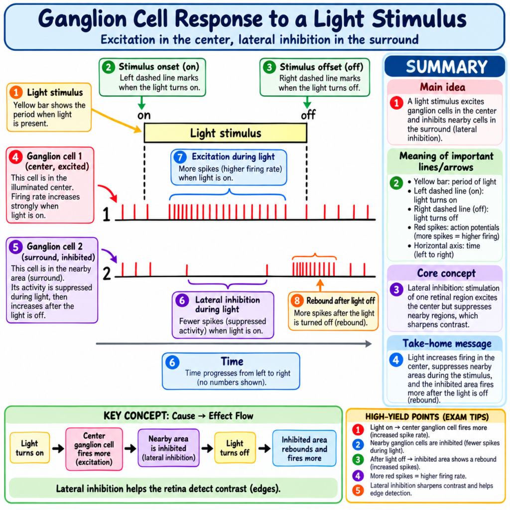
This response is demonstrated in Fig. 51.14.
Light Turned On
- When light is first turned on:
- Rapid impulses occur for a fraction of a second.
- These impulses then rapidly decrease.
Concept
Light ON
→ rapid burst of ganglion-cell impulses
→ response quickly declines
Lateral Ganglion Cell Response
- A ganglion cell located lateral to the light spot is markedly inhibited when the light is turned on.
- This occurs because of lateral inhibition.
When the light is turned off:
- The opposite responses occur.
Basis of Opposite Responses
The opposite responses to light are produced by:
- Depolarizing bipolar cells
- Hyperpolarizing bipolar cells
The transient nature of the response is probably produced at least partly by:
- Amacrine cells
because many amacrine cells themselves have transient responses.
Importance of Detecting Change
- The ability to detect changes in light intensity is strongly developed in:
- Peripheral retina
- Central retina
Example
A tiny gnat flying across the visual field
→ detected immediately
The same gnat sitting still
→ may remain below the threshold of visual detection
Key Concept
The retina responds strongly to change.
Movement/change in visual stimulus
→ strong detection
Unchanging small stimulus
→ may be much less noticeable
TRANSMISSION OF CONTRASTS IN THE VISUAL SCENE
Role of Lateral Inhibition
Lateral inhibition: Inhibitory signaling through lateral retinal pathways that helps detect and enhance contrast.
- Many ganglion cells respond mainly to contrast borders.
- Contrast detection appears to be a major way in which the pattern of a visual scene is transmitted to the brain.
Uniform Illumination
When flat light is applied equally across the retina:
- All photoreceptors are stimulated approximately equally.
- Contrast-type ganglion cells are neither strongly stimulated nor inhibited.
Why?
Direct Pathway
Photoreceptor
→ depolarizing bipolar cell
→ excitatory effect
Lateral Pathways
Photoreceptor
→ hyperpolarizing bipolar pathway / horizontal cells
→ inhibitory effect
Therefore:
Direct excitation
- lateral inhibition
→ approximately neutralize one another
Retinal Circuit During Uniform Light
A circuit illustrating this principle is shown in Fig. 51.15.
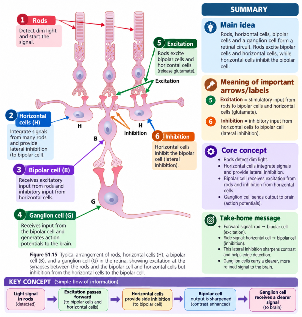
- A central photoreceptor excites a depolarizing bipolar cell.
- Photoreceptors on either side connect to the same bipolar cell through inhibitory horizontal cells.
- If all three photoreceptors are stimulated simultaneously:
- Direct excitation occurs.
- Lateral inhibition also occurs.
- Lateral inhibitory signals neutralize the direct excitatory signal.
Key Concept
Equal illumination everywhere
→ excitation + lateral inhibition balance
→ little contrast signal
CONTRAST BORDER
Now consider a bright-dark border.
- The central photoreceptor is stimulated by a bright spot.
- One lateral photoreceptor remains in darkness.
The bright central receptor:
→ excites the direct bipolar pathway.
The dark lateral receptor:
→ does not stimulate its horizontal cell.
Therefore:
Horizontal cell remains unstimulated
→ it does not inhibit the bipolar cell
→ bipolar cell receives extra excitation
Concept
Bright center + dark side
→ strong direct excitation
- reduced lateral inhibition
→ stronger contrast signal
Thus, where visual contrast exists:
- Direct and lateral pathways accentuate the difference.
Key Concept
Uniform light
→ excitation and inhibition tend to cancel
Contrast border
→ cancellation is reduced
→ difference becomes stronger
Therefore:
Lateral inhibition provides contrast detection and contrast enhancement.
TRANSMISSION OF COLOR SIGNALS BY GANGLION CELLS
Ganglion cells also participate in retinal color processing.
White Signal
- A ganglion cell may receive signals from several cones or only a few cones.
- When all three cone types stimulate the same ganglion cell:
- Red cones
- Green cones
- Blue cones
the signal transmitted by that ganglion cell is similar for any spectral color.
Therefore:
- This ganglion-cell signal does not distinguish between different colors.
- It acts as a white signal.
Key Concept
Red + green + blue cone input to the same ganglion cell
→ no specific color discrimination
→ white signal
OPPONENT COLOR SIGNALING
Some ganglion cells show an opposite pattern.
- One cone color excites the ganglion cell.
- Another cone color inhibits it.
Red-Green Opponent Response
A common example is:
Red cone input
→ excitation
Green cone input
→ inhibition
or the reverse:
Green cone input
→ excitation
Red cone input
→ inhibition
Key Concept
Red and green can act as opponent colors in selected ganglion-cell pathways.
Blue-Yellow Opponent Response
A similar reciprocal relationship occurs between:
- Blue cones
and
- The combination of red and green cones, which are both excited by yellow.
Therefore:
Blue
↔ Yellow
form another reciprocal excitation-inhibition relationship.
MECHANISM OF COLOR OPPOSITION
The opposing effect is produced through two retinal pathways.
Excitatory Route
One cone type
→ depolarizing bipolar cell
→ direct excitatory pathway
→ ganglion-cell excitation
Inhibitory Route
Opponent cone type
→ hyperpolarizing bipolar cell
→ indirect inhibitory pathway
→ ganglion-cell inhibition
Key Concept
One color
→ excites ganglion cell
Opponent color
→ inhibits same ganglion cell
This allows the retina to begin distinguishing one color from another.
IMPORTANCE OF COLOR CONTRAST SIGNALING
- Opponent-color mechanisms provide a way for the retina to begin differentiating colors.
- Each color-contrast ganglion cell:
- Is excited by one color.
- Is inhibited by the opponent color.
- Therefore, color analysis begins within the retina.
- It is not entirely performed by the brain.
Key Concept
Cone signals
→ opponent retinal pathways
→ color differences begin to be analyzed
→ ganglion-cell output carries processed color information onward
CLINICAL / FUNCTIONAL IMPORTANCE
- Amacrine cells help analyze visual signals before they leave the retina.
- Central retinal organization supports high visual acuity.
- Peripheral retinal organization provides high sensitivity to weak light.
- W, X, and Y cells process different aspects of visual information.
- P cells are important for fine detail and color information.
- M cells are important for low-contrast stimuli and rapid movement.
- Photosensitive ganglion cells contribute to physiological synchronization with night and day.
- Ganglion cells detect changes in light intensity.
- Lateral inhibition enhances visual contrast.
- Opponent ganglion-cell pathways allow color differentiation to begin in the retina.
HIGH-YIELD POINTS
- More than 60 amacrine-cell types have been identified.
- Amacrine cells may respond to:
- Light onset
- Light offset
- Both onset and offset
- Directional movement
- Many amacrine cells function as retinal interneurons.
- Retina contains about:
- 100 million rods
- 3 million cones
- 1.6 million ganglion cells
- Average convergence is approximately:
- 60 rods
- 2 cones
per ganglion-cell pathway.
- Central fovea contains about 35,000 slender cones and no rods.
- Reduced convergence in the fovea produces high visual acuity.
- Rods are 30–300 times more sensitive to light than cones.
- As many as 200 rods may converge onto one peripheral optic nerve fiber.
- W cells:
- Slow
- Rod-related
- Directional movement
- Crude dark vision
- X cells:
- Small fields
- Fine detail
- Cone input
- Color vision
- Y cells:
- Largest
- 50 m/sec or faster
- Rapid changes
- Rapid alerting of CNS
- P cells:
- Small fields
- Fine detail
- Color-sensitive
- Sustained responses
- M cells:
- Faster
- Low-contrast sensitive
- Movement-sensitive
- Transient responses
- Photosensitive ganglion cells contain melanopsin.
- Ganglion cells fire spontaneously at approximately 5–40 impulses/sec.
- Visual signals are superimposed on this background activity.
- Lateral inhibition enhances contrast.
- Uniform illumination tends to produce balanced excitation and inhibition.
- Contrast borders produce stronger ganglion-cell signals.
- Red-green and blue-yellow opponent pathways participate in retinal color analysis.
- Color analysis begins in the retina.
COMMON STUDENT MISTAKES
- Do not assume all amacrine cells have the same function.
- More than 60 types exist, with different responses.
- Do not think the fovea is more sensitive to weak light because it has more rods.
- The central fovea has no rods.
- Do not confuse visual acuity with weak-light sensitivity.
- Central retina → greater acuity.
- Peripheral retina → greater weak-light sensitivity.
- Do not think greater photoreceptor convergence produces better fine detail.
- Greater convergence supports sensitivity but peripheral acuity is poorer.
- Do not confuse W, X, and Y cells:
- W → crude rod vision and directional movement
- X → fine detail and color
- Y → rapid changes
- Do not confuse P and M cells:
- P → fine detail and color
- M → low contrast and rapid movement
- Do not think ganglion cells remain electrically silent without visual stimulation.
- They continuously fire at approximately 5–40 impulses/sec.
- Do not think a uniform bright field necessarily strongly excites contrast ganglion cells.
- Direct excitation can be neutralized by lateral inhibition.
- Do not think color analysis begins only in the brain.
- Opponent-color analysis already begins in the retina.
QUICK REVISION
Amacrine Cells
More than 60 types
→ different functions
Possible responses:
Light ON
→ transient response
Light OFF
→ transient response
Light ON or OFF
→ signals illumination change
Movement in specific direction
→ directional response
Overall
→ analyze visual signals before they leave retina
Central Retina
Low convergence
- slender cones
- nearly one optic fiber for each central foveal cone
→ high visual acuity
Peripheral Retina
Rods highly sensitive
- many rods converge
- signals summate
→ high sensitivity to weak light
W, X, Y Cells
W
→ slow
→ rods
→ directional movement
→ crude dark vision
X
→ small field
→ fine detail
→ cone input
→ color
Y
→ very fast
→ broad field
→ rapid visual changes
→ alerts CNS
P and M Cells
P cells
→ small field
→ slower
→ sustained
→ color
→ fine detail
M cells
→ larger field
→ faster
→ transient
→ low contrast
→ rapid movement
Ganglion-Cell Firing
No visual stimulus
→ spontaneous firing at 5–40/sec
Visual stimulus
→ change superimposed on background firing
On-Off Response
Light changes
→ strong transient ganglion-cell response
Moving tiny object
→ easily detected
Stationary tiny object
→ may remain undetected
Contrast
Uniform illumination
→ excitation + inhibition tend to cancel
Contrast border
→ direct excitation becomes stronger relative to inhibition
→ contrast enhanced
Color
All three cone types stimulate same ganglion cell
→ white signal
One cone excites + opponent cone inhibits
→ color contrast signal
Important opponent pairs:
Red ↔ Green
Blue ↔ Yellow
→ color analysis begins in retina
CONCEPTUAL SUMMARY
Amacrine cells and ganglion cells perform important processing of visual information within the retina.
Amacrine cells are highly diverse.
Different amacrine cells may detect:
Light onset
→ ON response
Light offset
→ OFF response
Either change
→ illumination-change response
Movement in a particular direction
→ direction-sensitive response
Therefore, many amacrine cells act as interneurons that analyze visual signals before those signals leave the retina.
The retina contains enormous numbers of photoreceptors but far fewer ganglion cells.
About:
100 million rods
+ 3 million cones
→ only about 1.6 million ganglion cells
Therefore, retinal signals show convergence.
In the central retina:
Less convergence
→ more precise retinal representation
→ high visual acuity
In the peripheral retina:
Highly sensitive rods
- greater convergence
- signal summation
→ high sensitivity to weak light
Ganglion cells are also functionally specialized.
W cells
→ crude rod vision and directional movement
X cells
→ fine detail and color
Y cells
→ very rapid detection of new visual events
In primates:
P cells
→ fine detail + color
M cells
→ low contrast + rapid movement
A separate photosensitive ganglion-cell system containing melanopsin sends signals toward nonvisual areas such as the suprachiasmatic nucleus and helps synchronize physiological changes with night and day.
Ganglion cells continuously produce action potentials even without stimulation:
5–40 impulses/sec
Visual information is represented by changes imposed on this background firing.
The retina is especially effective at detecting change.
Light ON or OFF
→ transient changes in ganglion-cell firing
Movement
→ rapidly detected
Contrast is enhanced by lateral inhibition.
Uniform illumination
→ direct excitation and lateral inhibition tend to cancel
Contrast border
→ imbalance between direct and lateral pathways
→ stronger contrast signal
Color analysis also begins in the retina.
All three cone types acting similarly on one ganglion cell
→ white signal
Opponent cone signals
→ one color excites
→ another inhibits
Examples:
Red ↔ Green
Blue ↔ Yellow
Therefore:
Visual information is not simply passed unchanged through the retina.
Amacrine and ganglion cells already analyze movement, change, detail, contrast, light intensity, and color before retinal output reaches the brain.
Final Take-Home Concept
Central retina → low convergence → high acuity
Peripheral retina → rod sensitivity + convergence → better weak-light sensitivity
P cells → fine detail and color
M cells → low contrast and rapid movement
Ganglion cells → continuous action potentials carrying processed retinal output
Lateral inhibition → contrast enhancement
Opponent colors → retinal color differentiation begins before signals reach the brain
Reference Guyton and Hall Textbook of Medical Physiology 15th Edition, page. 659 Chapter51
