


Learning Objectives
After studying this topic, students will be able to:
- Identify the major retinal neurons and their functions.
- Compare the cone and rod visual pathways.
- Explain electrotonic conduction in the retina.
- Describe the role of lateral inhibition in visual contrast.
- Differentiate depolarizing and hyperpolarizing bipolar cells.
Introduction
- The retina contains several types of neurons that receive, modify, and transmit visual signals.
- Visual information begins in the rods and cones and finally leaves the retina through the ganglion cells.
- Signals can travel:
- Vertically through the retinal layers.
- Horizontally or laterally within the retinal layers.
- Rod and cone pathways are not exactly the same.
- Cone signals travel through a newer and faster visual pathway.
- Rod signals use a different pathway with additional neuronal connections.
- Most retinal neurons transmit signals by electrotonic conduction, whereas ganglion cells transmit visual signals by action potentials.
- Lateral connections in the retina help maintain proper visual contrast.
Retinal neural pathway: The sequence of neuronal connections through which visual signals pass from photoreceptors toward ganglion cells and then into the brain.
RETINAL NEURONAL CONNECTIONS
Definition: Retinal neuronal connections are the pathways through which visual signals pass between the different neuronal cells of the retina.
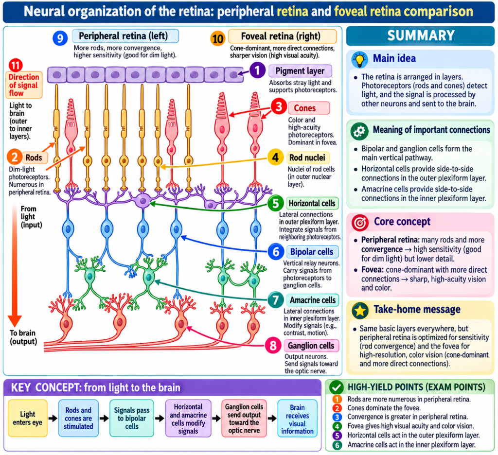
Fig. 51.12 shows the essential neural connections of the retina, with the peripheral retinal circuit on the left and the foveal retinal circuit on the right.
The important neuronal cells are described below.
Photoreceptors
Photoreceptors: The rods and cones that begin the transmission of visual signals in the retina.
- Photoreceptors consist of:
- Rods
- Cones
- They transmit visual signals to the outer plexiform layer.
- In this layer, they form synapses with:
- Bipolar cells
- Horizontal cells
Synapse: The site where one neuronal cell communicates with another neuronal cell.
Outer plexiform layer: The retinal layer in which rods and cones communicate with bipolar and horizontal cells.
Signal Flow
Rods and cones
→ outer plexiform layer
→ bipolar cells + horizontal cells
Horizontal Cells
Horizontal cells: Retinal neurons that transmit signals horizontally in the outer plexiform layer.
- They receive signals from:
- Rods
- Cones
- They transmit these signals horizontally toward bipolar cells.
- Their outputs are inhibitory.
- They play an important role in controlling the lateral spread of visual signals.
- They also help maintain proper visual contrast.
Signal Flow
Rods/cones
→ horizontal cells
→ lateral influence on bipolar cells
Bipolar Cells
Bipolar cells: Retinal neurons that transmit signals vertically from photoreceptors and horizontal cells toward the inner plexiform layer.
- They receive signals from:
- Rods
- Cones
- Horizontal cells
- They carry these signals vertically toward the inner plexiform layer.
- In the inner plexiform layer, bipolar cells form synapses with:
- Ganglion cells
- Amacrine cells
Inner plexiform layer: The retinal layer where bipolar cells communicate with ganglion cells and amacrine cells.
Signal Flow
Rods/cones/horizontal cells
→ bipolar cells
→ inner plexiform layer
→ ganglion cells + amacrine cells
Amacrine Cells
Amacrine cells: Retinal neurons that transmit signals between bipolar and ganglion cells and also provide horizontal connections in the inner plexiform layer.
- Amacrine cells transmit signals in two ways.
Direct Direction
Bipolar cells
→ amacrine cells
→ ganglion cells
Horizontal Direction
- Amacrine cells can also transmit signals horizontally in the inner plexiform layer.
- Signals may pass from bipolar cell axons toward:
- Ganglion cell dendrites
- Other amacrine cells
Dendrites: Parts of neuronal cells that receive signals from other neurons.
Ganglion Cells
Ganglion cells: Retinal output neurons that transmit visual signals from the retina through the optic nerve into the brain.
- Ganglion cells receive processed retinal signals.
- They form the output pathway of the retina.
- Their signals travel:
Retina
→ optic nerve
→ brain
Optic nerve: The pathway through which ganglion cell signals leave the retina and travel into the brain.
Interplexiform Cells
Interplexiform cell: A less prominent retinal neuron that carries signals backward from the inner plexiform layer to the outer plexiform layer.
- This is a sixth type of retinal neuronal cell.
- It is not prominent.
- It is not shown in the figure.
- It transmits signals in a retrograde direction.
Retrograde direction: Signal transmission backward from the inner plexiform layer toward the outer plexiform layer.
Direction
Inner plexiform layer
→ interplexiform cell
→ outer plexiform layer
- These signals are inhibitory.
- They are believed to control the lateral spread of visual signals produced by horizontal cells in the outer plexiform layer.
- Their possible role is to help control the degree of contrast in the visual image.
Inhibitory signal: A signal that reduces or limits neuronal activity or signal spread.
Key Concept
The retina contains neurons that transmit signals in different directions:
Photoreceptors
→ begin visual signaling
Bipolar cells
→ mainly carry signals vertically
Horizontal cells
→ carry signals laterally in the outer plexiform layer
Amacrine cells
→ carry signals between bipolar and ganglion cells and laterally in the inner plexiform layer
Ganglion cells
→ carry retinal output to the brain
Interplexiform cells
→ carry inhibitory signals backward toward the outer plexiform layer
CONE AND ROD VISUAL PATHWAYS
The Cone Pathway Functions Differently From the Rod Pathway
- The retina contains:
- An older type of vision based on rod vision.
- A newer type of vision based on cone vision.
- The neurons and nerve fibers carrying cone visual signals are considerably larger than those carrying rod signals.
- Cone signals are conducted to the brain two to five times more rapidly than rod signals.
- The circuitry of the cone and rod systems is also slightly different.
Key Concept
Cone pathway
→ larger neurons and nerve fibers
→ faster conduction to the brain
Rod pathway
→ smaller conducting elements
→ slower conduction
FOVEAL CONE PATHWAY
Foveal pathway: The direct visual pathway in the foveal retina representing the newer and faster cone system.
- The foveal pathway contains three neurons in its direct pathway:
Cones
→ bipolar cells
→ ganglion cells
- Horizontal cells transmit inhibitory signals laterally in the outer plexiform layer.
- Amacrine cells transmit signals laterally in the inner plexiform layer.
Direct Cone Pathway
Cone
→ bipolar cell
→ ganglion cell
Key Concept
The direct foveal cone pathway uses only three neurons:
Cone → Bipolar cell → Ganglion cell
This represents the newer and faster visual system described in the provided text.
PERIPHERAL RETINAL PATHWAY
Peripheral retinal pathway: The neural circuitry in the peripheral retina, where both rods and cones are present.
- The peripheral retina contains both:
- Rods
- Cones
- Three bipolar cells are demonstrated in the described circuitry.
- The middle bipolar cell connects only with rods.
- This represents the type of visual system present in many lower animals.
Pure Rod Visual Pathway
- In the pure rod pathway:
- The bipolar cell does not pass its output directly to the ganglion cell.
- Its output passes first to amacrine cells.
- Amacrine cells then relay the signal to ganglion cells.
Therefore, pure rod vision contains four neurons in the direct visual pathway:
Rods
→ bipolar cells
→ amacrine cells
→ ganglion cells
- Horizontal cells and amacrine cells also provide lateral connections.
Key Concept
Pure cone pathway
Cone
→ bipolar cell
→ ganglion cell
Pure rod pathway
Rod
→ bipolar cell
→ amacrine cell
→ ganglion cell
Therefore:
- Cone direct pathway = 3 neuronal levels
- Pure rod direct pathway = 4 neuronal levels
Mixed Rod and Cone Connections
- The other two bipolar cells in the peripheral retinal circuitry connect with:
- Rods
- Cones
- Their outputs travel by two pathways:
- Directly to ganglion cells.
- Through amacrine cells before reaching ganglion cells.
Signal Flow
Rods/cones
→ bipolar cells
→ ganglion cells
or
Rods/cones
→ bipolar cells
→ amacrine cells
→ ganglion cells
NEUROTRANSMITTERS RELEASED BY RETINAL NEURONS
Neurotransmitter: A chemical substance used for synaptic transmission between neuronal cells.
- All neurotransmitter substances used in the retina have not been completely identified.
- However, both:
- Rods
- Cones
Glutamate: The transmitter released by rods and cones at their synapses with bipolar cells.
Amacrine Cell Transmitters
- Histological and pharmacological studies have demonstrated many types of amacrine cells.
- These cells secrete at least eight types of transmitter substances.
- Examples mentioned in the text include:
- Gamma-aminobutyric acid (GABA)
- Glycine
- Dopamine
- Acetylcholine
- Indolamine
- These normally function as inhibitory transmitters.
Inhibitory transmitter: A transmitter that normally produces an inhibitory effect on neuronal signaling.
Other Retinal Transmitters
- The transmitters used by:
- Bipolar cells
- Horizontal cells
- Interplexiform cells
- At least some horizontal cells release inhibitory transmitters.
Key Concept
Rods and cones
→ release glutamate
Many amacrine cells
→ release inhibitory transmitter substances
Some horizontal cells
→ also release inhibitory transmitters
ELECTROTONIC CONDUCTION IN THE RETINA
Electrotonic conduction: Direct flow of electric current through neuronal cytoplasm and nerve processes from the point of excitation to the output synapse, without the use of action potentials.
- Most retinal neurons transmit their visual signals by electrotonic conduction.
- They do not usually use action potentials.
Ganglion Cells and Action Potentials
Action potential: The type of signal used consistently by ganglion cells to transmit visual information through the optic nerve into the brain.
- Ganglion cells are the only retinal neurons that always transmit visual signals by action potentials.
- Their action potentials travel:
Ganglion cells
→ optic nerve
→ brain
- Action potentials have occasionally also been recorded in amacrine cells.
- However, the importance of these amacrine cell action potentials is questionable.
- Other retinal neurons normally conduct their visual signals by electrotonic conduction.
How Electrotonic Conduction Works
- Electrotonic conduction involves direct electric current flow.
- It does not depend on action potentials.
- Current flows through:
- Neuronal cytoplasm
- Nerve processes
- The current travels from the site of excitation to the output synapses.
Electrotonic Conduction in Rods and Cones
- Even photoreceptors use electrotonic conduction.
- Signal conduction from their outer segments to their synaptic bodies occurs by direct current flow.
When light acts on a rod or cone:
Light
→ hyperpolarization in the outer segment
→ direct electric current flows through cytoplasm
→ almost the same degree of hyperpolarization reaches the synaptic body
- No action potential is required.
Hyperpolarization: The electrical response produced in the outer segment of a rod or cone in response to light.
Electrotonic Conduction in Bipolar and Horizontal Cells
- A transmitter released from a rod or cone can stimulate:
- A bipolar cell
- A horizontal cell
- The resulting signal again travels from input to output through direct electric current flow.
- It does not require an action potential.
Key Concept
In most retinal neurons:
Stimulus
→ change in electrical potential
→ direct current spreads through the cell
→ signal reaches output synapse
No action potential is required.
IMPORTANCE OF ELECTROTONIC CONDUCTION
- Electrotonic conduction allows graded conduction of signal strength.
Graded conduction: Signal strength can vary according to the strength of the stimulus rather than behaving in an all-or-none manner.
- In rods and cones:
- Greater illumination is associated with a stronger hyperpolarizing output signal.
- The strength of the output signal is directly related to the intensity of illumination.
- The response is therefore graded.
- It is not all-or-none as an action potential would be.
Conceptual Example
Lower illumination
→ smaller hyperpolarizing signal
Greater illumination
→ stronger hyperpolarizing signal
Therefore:
Intensity of illumination
→ determines the strength of the photoreceptor output signal
Key Concept
Electrotonic conduction
→ permits different signal strengths
→ signal strength can match illumination intensity
Action potential
→ would behave in an all-or-none manner
LATERAL INHIBITION TO ENHANCE VISUAL CONTRAST
Lateral inhibition: Inhibition of surrounding retinal pathways that prevents excitation from spreading widely and helps preserve contrast borders in the visual image.
Function of Horizontal Cells
- Horizontal cells connect laterally between:
- Synaptic bodies of rods and cones.
- Dendrites of bipolar cells.
- Their outputs are always inhibitory.
- Their lateral connections produce lateral inhibition.
- This mechanism is also important in other sensory systems.
- In the retina, it helps transmit visual patterns with proper visual contrast.
Visual contrast: The distinction between different areas or borders within the visual image.
Effect of a Small Spot of Light
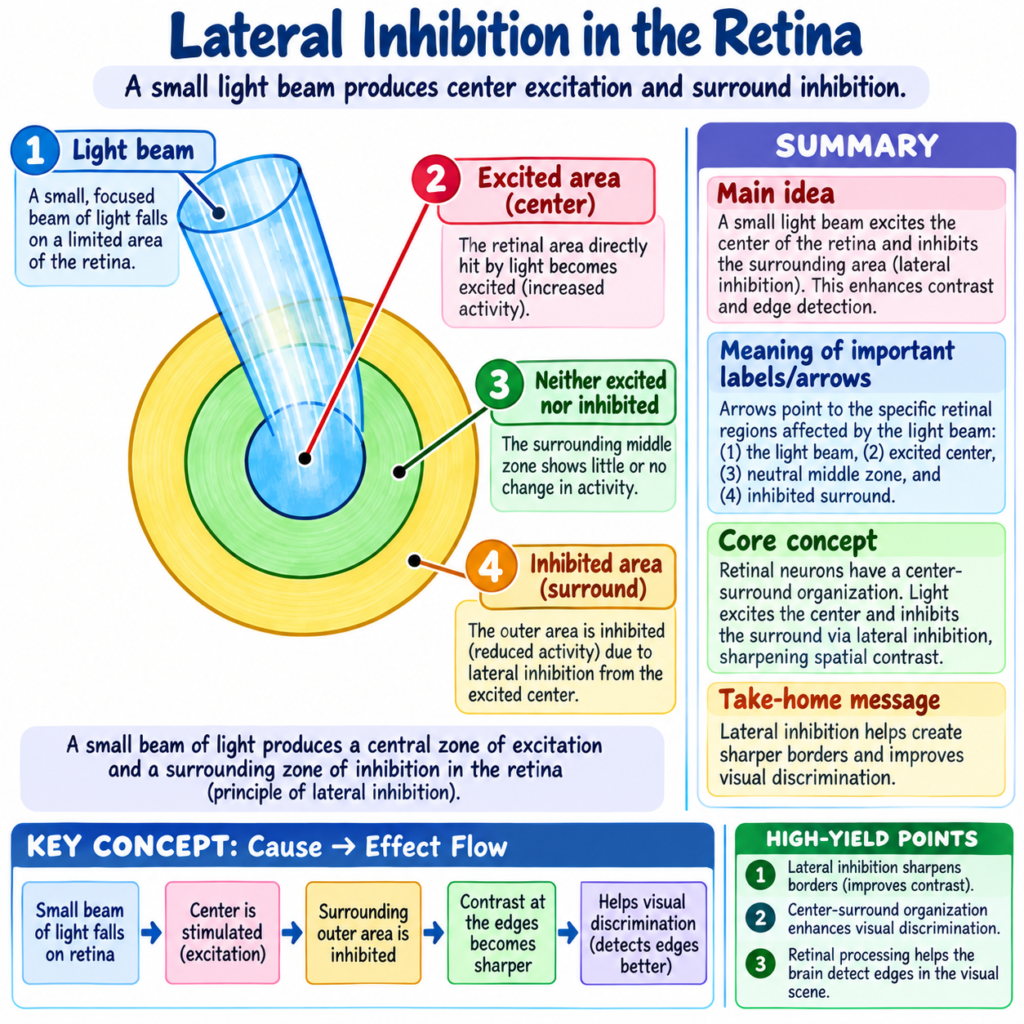
Fig. 51.13 demonstrates a minute spot of light focused on the retina.
- The visual pathway at the central area where the light falls becomes excited.
- An area to the side becomes inhibited.
Therefore:
Central illuminated area
→ excitation
Surrounding area
→ inhibition
Why Lateral Inhibition Is Important
- Dendritic and axonal trees in the plexiform layers could allow excitation to spread widely through the retina.
- Horizontal cells oppose this spread.
- They inhibit the surrounding areas.
- This prevents excessive lateral spread of the excitatory signal.
- This process is essential for high visual accuracy when transmitting contrast borders in the visual image.
Key Concept
Light stimulates one retinal area
→ central pathway becomes excited
→ horizontal cells inhibit surrounding pathways
→ excessive signal spread is reduced
→ contrast border remains clearer
Role of Amacrine Cells in Lateral Inhibition
- Some amacrine cells probably provide additional lateral inhibition.
- This occurs in the inner plexiform layer.
- Their effect may further enhance visual contrast.
Key Concept
Horizontal cells
→ lateral inhibition in outer plexiform layer
Some amacrine cells
→ additional lateral inhibition in inner plexiform layer
Together
→ help enhance visual contrast
DEPOLARIZING AND HYPERPOLARIZING BIPOLAR CELLS
Two types of bipolar cells produce opposing responses in the visual pathway:
- Depolarizing bipolar cells
- Hyperpolarizing bipolar cells
Depolarizing Bipolar Cells
Depolarizing bipolar cell: A bipolar cell that depolarizes when rods and cones are excited.
Hyperpolarizing Bipolar Cells
Hyperpolarizing bipolar cell: A bipolar cell that hyperpolarizes when rods and cones are excited.
Therefore, when photoreceptors are stimulated:
Some bipolar cells
→ depolarize
Other bipolar cells
→ hyperpolarize
POSSIBLE EXPLANATIONS FOR OPPOSITE BIPOLAR RESPONSES
The text provides two possible explanations.
Different Types of Bipolar Cells
- The bipolar cells may be entirely different cell types.
- One type may respond to glutamate by depolarizing.
- The other type may respond to glutamate by hyperpolarizing.
Concept
Same transmitter from rods/cones
→ different bipolar cell types
→ opposite electrical responses
Direct and Indirect Pathways
Another possibility is:
- One bipolar cell receives direct excitation from rods and cones.
- Another receives its signal indirectly through a horizontal cell.
- Horizontal cells are inhibitory.
- Therefore, passage through a horizontal cell can reverse the polarity of the electrical response.
Polarity: The direction of the electrical response—whether the cell depolarizes or hyperpolarizes.
Concept
Rods/cones
→ direct bipolar pathway
→ one electrical response
Rods/cones
→ horizontal inhibitory cell
→ bipolar cell
→ reversed electrical response
IMPORTANCE OF OPPOSING BIPOLAR RESPONSES
- Regardless of the exact mechanism, the two types of bipolar responses are important.
- They allow:
- About half of the bipolar cells to transmit positive signals.
- The other half to transmit negative signals.
- Both positive and negative signals are used in transmitting visual information to the brain.
Key Concept
Bipolar cells produce opposite responses
→ positive and negative visual signals can be transmitted
→ both contribute to visual information reaching the brain
BIPOLAR CELLS AND CONTRAST BORDERS
- The reciprocal relationship between depolarizing and hyperpolarizing bipolar cells provides another mechanism for lateral inhibition.
- This mechanism is additional to the horizontal cell mechanism.
- Depolarizing and hyperpolarizing bipolar cells lie immediately next to each other.
- Their opposing responses help separate contrast borders in the visual image.
- This can occur even when a contrast border lies exactly between two adjacent photoreceptors.
- In comparison, horizontal-cell lateral inhibition operates over a much greater distance.
Key Concept
Adjacent depolarizing and hyperpolarizing bipolar cells
→ opposite signals
→ better separation of nearby contrast borders
Horizontal cells
→ lateral inhibition over a greater distance
CLINICAL IMPORTANCE
- Proper retinal neural connections are important for accurate transmission of visual information.
- Cone signals are transmitted more rapidly than rod signals.
- Electrotonic conduction allows retinal signal strength to vary according to illumination intensity.
- Horizontal-cell inhibition limits lateral spread of excitation.
- This helps preserve visual contrast and improves accuracy in transmitting contrast borders.
- Opposing bipolar-cell responses provide an additional mechanism for separating contrast borders.
HIGH-YIELD POINTS
- Rods and cones are the photoreceptors of the retina.
- Photoreceptors communicate with bipolar and horizontal cells in the outer plexiform layer.
- Bipolar cells carry signals vertically toward the inner plexiform layer.
- Amacrine cells transmit signals between bipolar and ganglion cells and laterally within the inner plexiform layer.
- Ganglion cells carry the final retinal output through the optic nerve to the brain.
- Interplexiform cells transmit inhibitory signals backward from the inner to the outer plexiform layer.
- Cone visual signals travel to the brain two to five times faster than rod signals.
- Direct foveal cone pathway:
Cone → Bipolar cell → Ganglion cell
- Pure rod pathway:
Rod → Bipolar cell → Amacrine cell → Ganglion cell
- Rods and cones release glutamate.
- Many amacrine-cell transmitters are inhibitory.
- Ganglion cells always use action potentials.
- Most other retinal neurons normally use electrotonic conduction.
- Electrotonic conduction allows graded signal strength.
- Photoreceptor hyperpolarization varies with the intensity of illumination.
- Horizontal cells provide lateral inhibition.
- Lateral inhibition improves visual contrast.
- Some amacrine cells may provide additional lateral inhibition.
- Bipolar cells may either depolarize or hyperpolarize.
- Opposite bipolar responses provide positive and negative visual signals.
- Opposing bipolar cells also help separate contrast borders.
COMMON STUDENT MISTAKES
- Do not think all retinal neurons transmit signals by action potentials.
- Ganglion cells always use action potentials.
- Most other retinal neurons use electrotonic conduction.
- Do not confuse horizontal and bipolar signal directions.
- Horizontal cells → lateral transmission
- Bipolar cells → mainly vertical transmission
- Do not forget the difference between the direct cone and pure rod pathways.
- Cone pathway:
- Pure rod pathway:
- Do not assume horizontal cells are excitatory.
- Their outputs are described as inhibitory.
- Do not think electrotonic signals are all-or-none.
- They allow graded signal strength.
- Do not confuse depolarizing and hyperpolarizing bipolar cells.
- One group depolarizes.
- The other group hyperpolarizes.
- Do not think only horizontal cells contribute to contrast.
- Some amacrine cells may also provide lateral inhibition.
- Opposing bipolar cells provide another contrast-separating mechanism.
QUICK REVISION
Retinal Signal Flow
Photoreceptor
→ bipolar cell
→ ganglion cell
→ optic nerve
→ brain
With lateral modification by:
- Horizontal cells
- Amacrine cells
Main Retinal Neurons
Photoreceptors
→ rods and cones
→ begin visual signaling
Horizontal cells
→ lateral inhibitory signaling in outer plexiform layer
Bipolar cells
→ vertical signal transmission
Amacrine cells
→ bipolar-to-ganglion and lateral signaling in inner plexiform layer
Ganglion cells
→ retinal output to brain
Interplexiform cells
→ inhibitory backward signaling from inner to outer plexiform layer
Cone Pathway
Cone
→ bipolar
→ ganglion
- Three neurons
- Newer system
- Faster system
Pure Rod Pathway
Rod
→ bipolar
→ amacrine
→ ganglion
- Four neurons in the direct pathway
Neurotransmitters
Rods + cones
→ glutamate
Many amacrine cells
→ inhibitory transmitters
Retinal Electrical Signaling
Most retinal neurons
→ electrotonic conduction
Ganglion cells
→ action potentials
Electrotonic Conduction
More illumination
→ stronger photoreceptor hyperpolarization
→ stronger graded output
Lateral Inhibition
Central light stimulation
→ central excitation
Horizontal cells
→ surrounding inhibition
Result
→ improved transmission of contrast borders
Bipolar Cells
Depolarizing bipolar cells
→ depolarize
Hyperpolarizing bipolar cells
→ hyperpolarize
Opposing signals
→ positive + negative visual signaling
→ improved separation of contrast borders
CONCEPTUAL SUMMARY
The neural retina contains several interconnected neuronal cells that process visual signals before these signals leave the eye.
Visual information begins in the rods and cones.
Rods and cones
→ transmit signals to bipolar and horizontal cells in the outer plexiform layer.
Bipolar cells
→ carry signals vertically toward the inner plexiform layer.
Amacrine cells
→ modify and relay signals between bipolar cells and ganglion cells and also provide lateral connections.
Ganglion cells
→ form the final output pathway of the retina
→ transmit signals through the optic nerve
→ brain.
Interplexiform cells
→ carry inhibitory signals backward from the inner plexiform layer to the outer plexiform layer
→ may help control lateral signal spread and visual contrast.
The cone and rod pathways differ.
Foveal cone pathway:
Cone
→ Bipolar cell
→ Ganglion cell
It represents the newer and faster visual system.
Pure rod pathway:
Rod
→ Bipolar cell
→ Amacrine cell
→ Ganglion cell
Cone signals reach the brain two to five times faster than rod signals.
Rods and cones release glutamate at their synapses with bipolar cells.
Most retinal neurons do not rely on action potentials.
Instead:
Electrical change
→ direct current flow through the cell
→ output synapse
This is electrotonic conduction.
Its major importance is that:
Illumination intensity changes
→ degree of photoreceptor hyperpolarization changes
→ output signal strength changes
Therefore, retinal signaling can be graded.
Horizontal cells provide lateral inhibition.
Central retinal stimulation
→ excitation
Surrounding retinal pathways
→ inhibition
This prevents excessive spread of excitation and helps preserve visual contrast.
Some amacrine cells may add further lateral inhibition.
Finally, bipolar cells can show two opposite responses:
Photoreceptor stimulation
→ some bipolar cells depolarize
→ other bipolar cells hyperpolarize
These opposing responses allow positive and negative visual signals to be transmitted and provide an additional mechanism for separating contrast borders.
Final Take-Home Concept
Photoreceptors receive light → retinal neurons process the signal → ganglion cells send the final signal to the brain.
Cone pathway = Cone → Bipolar → Ganglion
Pure rod pathway = Rod → Bipolar → Amacrine → Ganglion
Most retinal neurons = electrotonic graded conduction
Ganglion cells = action potentials
Horizontal-cell inhibition + opposing bipolar responses = better visual contrast
Reference Guyton and Hall Textbook of Medical Physiology 15th Edition, page. 656, Chapter 51
