


Learning Objectives
After studying this topic, students will be able to:
- Describe the aqueous humor and vitreous humor.
- Explain the formation and outflow of aqueous humor.
- Understand the normal intraocular pressure and its regulation.
- Explain how glaucoma develops and causes loss of vision.
Introduction
The eye is filled with intraocular fluid.
This fluid produces enough pressure inside the eyeball to keep the eye properly distended.
The intraocular fluid is divided into two main portions:
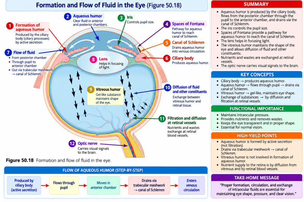
- Aqueous humor — located in front of the lens.
- Vitreous humor — located between the posterior surface of the lens and the retina.
This arrangement is shown in Fig. 50.18.
The balance between the formation and reabsorption of aqueous humor helps regulate the volume and pressure of the intraocular fluid.
Intraocular Fluid
The eye contains intraocular fluid that maintains enough pressure to keep the eyeball distended.
The intraocular fluid has two portions.
Aqueous Humor
Aqueous humor lies in front of the lens.
It is a freely flowing fluid.
Vitreous Humor
Vitreous humor lies between the posterior surface of the lens and the retina.
It is also called the vitreous body.
Unlike aqueous humor, vitreous humor is a gelatinous mass.
It is held together by a fine fibrillar network made mainly of proteoglycan molecules.
Water and dissolved substances can slowly diffuse through the vitreous humor.
However, there is very little actual fluid flow through it.
Key Concept
Aqueous humor = freely flowing fluid
Vitreous humor = gelatinous mass with little fluid flow
Aqueous humor is continuously:
Formed → circulated → reabsorbed
The balance between its formation and reabsorption determines the total volume and pressure of intraocular fluid.
Formation of Aqueous Humor by the Ciliary Body
Aqueous humor is formed at an average rate of approximately:
2–3 μL/min
Almost all aqueous humor is secreted by the ciliary processes.
The ciliary processes are linear folds that project from the ciliary body into the space behind the iris.
This is the region where the lens ligaments and ciliary muscle attach to the eyeball.
A cross-section of the ciliary processes is shown in Fig. 50.19.
Because the ciliary processes are highly folded, they have a large total surface area.
Their
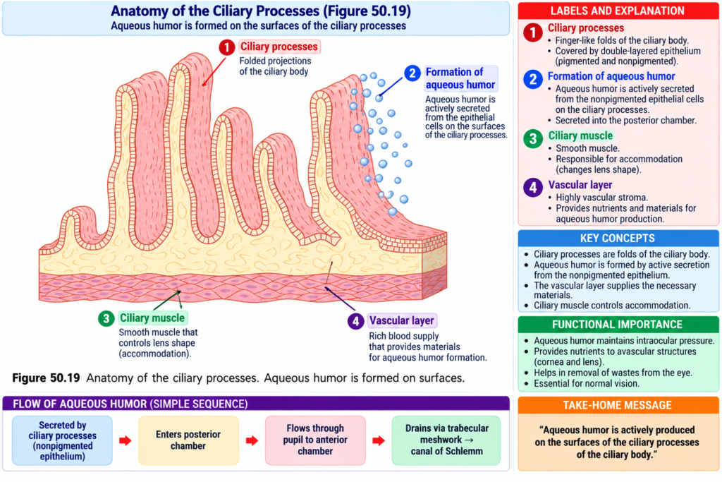
total surface area is approximately:
6 cm² in each eye
This is a large surface area compared with the small size of the ciliary body.
The surfaces of the ciliary processes are covered by highly secretory epithelial cells.
Immediately beneath these cells is a highly vascular area.
How Aqueous Humor Is Secreted
Aqueous humor is formed almost completely by active secretion from the epithelium of the ciliary processes.
The process begins with the active transport of sodium ions into the spaces between epithelial cells.
Simple Sequence
Sodium ions are actively transported
↓
Chloride and bicarbonate ions follow sodium
↓
This maintains electrical neutrality
↓
These ions increase the movement of water by osmosis
↓
Water moves from the blood capillaries into the spaces between epithelial cells
↓
The resulting fluid moves from the ciliary processes into the eye chambers
Several nutrients are also transported across the epithelium.
These include:
- Amino acids
- Ascorbic acid
- Glucose
They are transported by either:
- Active transport
- Facilitated diffusion
Key Concept
The main sequence of aqueous humor formation is:
Active Na⁺ transport → Cl⁻ and bicarbonate follow → water follows by osmosis → aqueous humor is formed
Example
When sodium is actively transported into the spaces between epithelial cells, chloride and bicarbonate move with it.
The presence of these ions causes water to move from nearby blood capillaries by osmosis.
This produces the aqueous fluid.
Outflow of Aqueous Humor From the Eye
After aqueous humor is produced by the ciliary processes, it follows a definite pathway.
Flow of Aqueous Humor
Ciliary processes
↓
Through the pupil
↓
Anterior chamber
↓
Angle between the cornea and iris
↓
Trabecular meshwork
↓
Canal of Schlemm
↓
Extraocular veins
This flow pathway is shown in Fig. 50.18.
The anatomical structures present at the angle between the iris and cornea are shown in Fig. 50.20.
The spaces between the trabeculae extend from the anterior chamber to the canal of Schlemm.
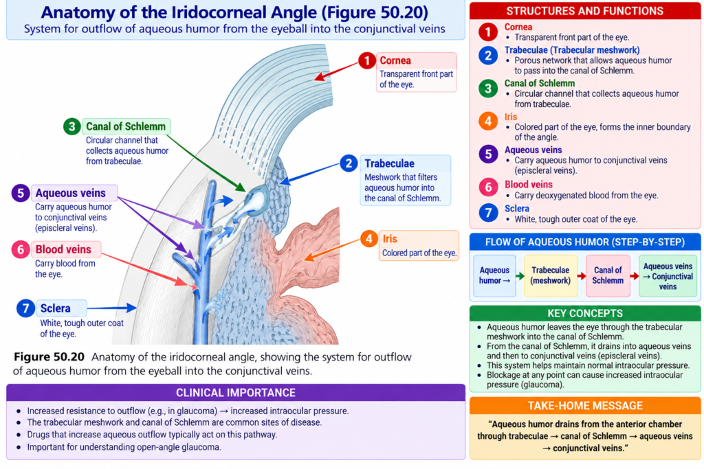
Canal of Schlemm
The canal of Schlemm is a thin-walled vein.
It runs around the circumference of the eye.
Its endothelial membrane is very porous.
Because of this high porosity, the following can pass from the anterior chamber into the canal:
- Large protein molecules
- Small particulate material
- Material as large as red blood cells
Although the canal of Schlemm is actually a venous blood vessel, so much aqueous humor normally enters it that it usually contains aqueous humor rather than blood.
Small veins leading from the canal of Schlemm to the larger veins of the eye also usually contain aqueous humor.
These veins are called:
Aqueous veins
Key Concept
Normal aqueous humor drainage:
Anterior chamber → trabecular spaces → canal of Schlemm → aqueous veins → larger veins of the eye
Intraocular Pressure
The average normal intraocular pressure is approximately:
15 mm Hg
The normal range is approximately:
12–20 mm Hg
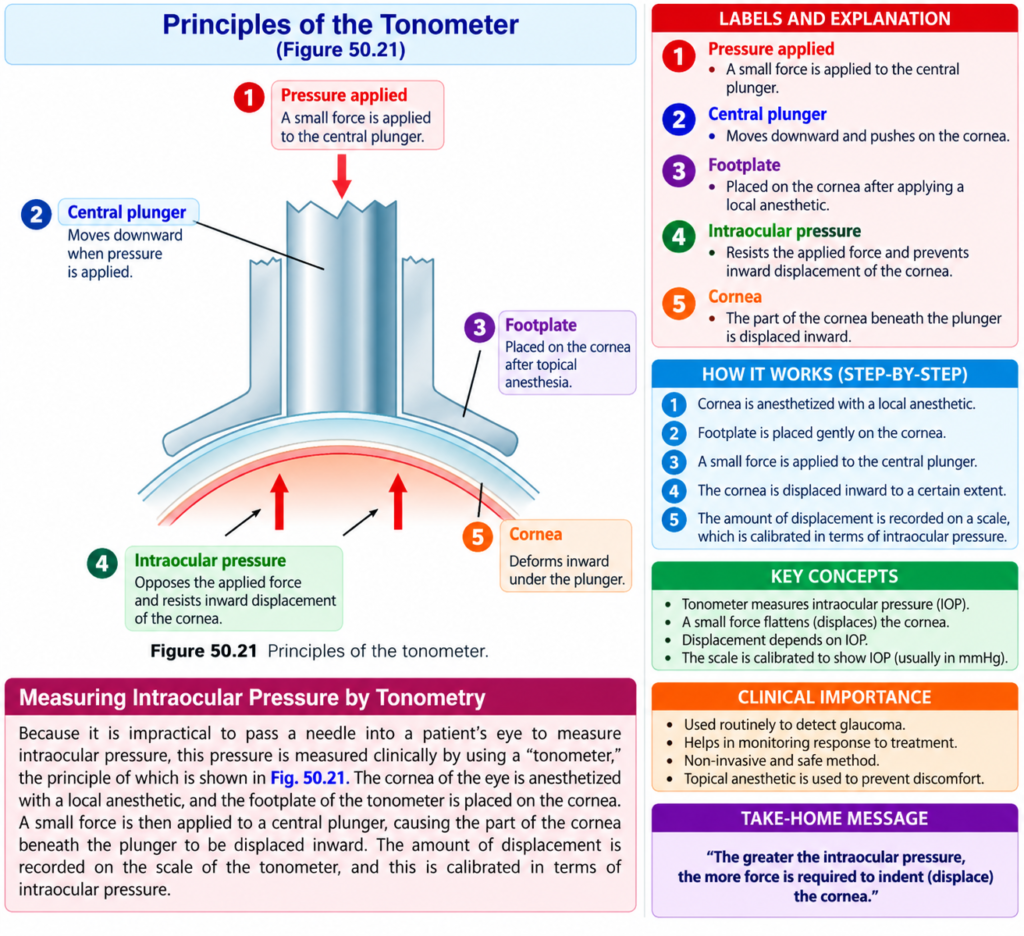
Measuring Intraocular Pressure by Tonometry
It is impractical to insert a needle into the eye simply to measure intraocular pressure.
Therefore, intraocular pressure is measured clinically using a:
Tonometer
The basic principle is shown in Fig. 50.21.
First, the cornea is anesthetized using a local anesthetic.
The footplate of the tonometer is then placed on the cornea.
A small force is applied to a central plunger.
This pushes part of the cornea slightly inward.
The amount of inward displacement is measured on the scale of the tonometer.
The scale is calibrated to indicate the intraocular pressure.
Key Concept
Tonometer measures intraocular pressure by measuring how much the cornea is displaced inward by an applied force.
Regulation of Intraocular Pressure
In the normal eye, intraocular pressure remains fairly constant.
It usually stays within approximately:
±2 mm Hg of its normal level
The average normal level is about:
15 mm Hg
The main factor controlling intraocular pressure is the resistance to the outflow of aqueous humor.
Aqueous humor must move from the anterior chamber through the trabecular meshwork before entering the canal of Schlemm.
The trabecular openings are very small.
Their diameter is approximately:
2–3 micrometers
These small openings provide resistance to fluid outflow.
As intraocular pressure increases, the flow of fluid into the canal of Schlemm also increases markedly.
At an intraocular pressure of approximately:
15 mm Hg
the amount of fluid leaving through the canal of Schlemm averages approximately:
2.5 μL/min
This is approximately equal to the amount of fluid entering from the ciliary body.
Key Concept
Normal intraocular pressure is maintained when:
Aqueous humor entering the eye ≈ aqueous humor leaving the eye
At approximately 15 mm Hg:
Outflow ≈ 2.5 μL/min
and this approximately equals the inflow from the ciliary body.
Example
If aqueous humor leaves the eye at approximately the same rate at which it is produced, intraocular pressure remains near its normal level.
Mechanism for Cleansing the Trabecular Spaces and Intraocular Fluid
Debris can sometimes enter the aqueous humor.
This may happen after:
- Hemorrhage into the eye
- Intraocular infection
The debris may accumulate in the trabecular spaces.
If these spaces become blocked, adequate reabsorption of aqueous humor may be prevented.
This can sometimes cause glaucoma.
The eye has mechanisms that help keep these drainage spaces clean.
Large numbers of phagocytic cells are present on the surfaces of the trabecular plates.
Outside the canal of Schlemm is a layer of interstitial gel.
This layer contains many reticuloendothelial cells.
These cells can engulf debris and digest it into small molecular substances.
These smaller substances can then be absorbed.
Therefore, this phagocytic system helps keep the trabecular spaces clean.
The surfaces of the iris and other surfaces behind the iris are also covered by an epithelium that can phagocytize:
- Proteins
- Small particles
This helps maintain a clear aqueous fluid.
Key Concept
Phagocytic cells remove debris → trabecular spaces remain clear → aqueous humor can continue to drain
Glaucoma Causes High Intraocular Pressure and Is a Principal Cause of Blindness
Glaucoma is one of the most common causes of blindness.
It is an eye disorder in which the intraocular pressure becomes abnormally high.
The pressure may sometimes suddenly increase to:
60–70 mm Hg
Pressures above approximately:
25–30 mm Hg
can cause loss of vision if maintained for a long period.
Extremely high pressures can cause blindness within:
Days or even hours
How High Pressure Damages Vision
As intraocular pressure increases, the axons of the optic nerve are compressed where they leave the eyeball at the optic disc.
This compression is believed to block the normal axonal flow of cytoplasm from retinal neuronal cell bodies into optic nerve fibers.
This reduces the appropriate nutrition of the nerve fibers.
Eventually, the affected nerve fibers can die.
Compression of the retinal artery may also contribute to neuronal damage.
The retinal artery enters the eyeball at the optic disc.
Its compression may reduce nutrition to the retina.
Simple Sequence
Increased intraocular pressure
↓
Compression of optic nerve axons at the optic disc
↓
Axonal flow is blocked
↓
Nutrition of nerve fibers decreases
↓
Nerve fibers die
↓
Loss of vision
Another possible mechanism is:
High pressure
↓
Compression of retinal artery
↓
Reduced nutrition to retina
↓
Additional neuronal damage
Cause of Increased Pressure in Glaucoma
In most cases of glaucoma, abnormally high pressure results from increased resistance to the outflow of aqueous humor.
The main problem occurs in the pathway:
Trabecular spaces → canal of Schlemm
This occurs at the iridocorneal junction.
Acute Condition
During acute inflammation of the eye:
- White blood cells
- Tissue debris
can block the trabecular spaces.
This can cause an acute rise in intraocular pressure.
Chronic Condition
In chronic conditions, especially in older people, the trabecular spaces may become blocked by fibrous occlusion.
This can also increase resistance to aqueous humor drainage.
Key Concept
The basic problem in most glaucoma described here is:
Increased resistance to aqueous humor outflow
↓
Reduced drainage
↓
Increased intraocular pressure
↓
Optic nerve damage
↓
Loss of vision
Treatment of Glaucoma
Glaucoma can sometimes be treated using eye drops.
The drug in the drops diffuses into the eyeball.
The drug can help by:
- Reducing aqueous humor secretion
or
- Increasing aqueous humor absorption
If drug treatment fails, surgery may be used.
Surgical procedures can:
- Open the trabecular spaces
or
- Create channels that allow fluid to flow from inside the eyeball into the subconjunctival space outside the eyeball
These procedures can effectively reduce intraocular pressure.
Clinical Note or Importance
Tonometry
Intraocular pressure is measured clinically with a tonometer rather than by inserting a needle into the eye.
Normal intraocular pressure averages approximately:
15 mm Hg
with a normal range of:
12–20 mm Hg
Glaucoma
Glaucoma is clinically important because abnormally high intraocular pressure can damage the optic nerve and cause blindness.
Pressure may rise as high as:
60–70 mm Hg
Pressure above:
25–30 mm Hg
can cause loss of vision if it remains elevated for a long period.
Very high pressure can produce blindness within days or hours.
High-Yield Points
- Intraocular fluid helps keep the eyeball distended.
- Aqueous humor is a freely flowing fluid in front of the lens.
- Vitreous humor is a gelatinous mass behind the lens with very little fluid flow.
- Aqueous humor is mainly secreted by the ciliary processes.
- Normal aqueous humor formation is approximately 2–3 μL/min.
- Sodium transport is followed by chloride and bicarbonate, and then water follows by osmosis.
- Aqueous humor flows through the pupil into the anterior chamber.
- It then passes through the trabecular meshwork into the canal of Schlemm.
- Normal intraocular pressure averages approximately 15 mm Hg.
- The usual normal range is 12–20 mm Hg.
- Resistance in the trabecular meshwork is the main factor controlling aqueous humor outflow.
- At about 15 mm Hg, outflow through the canal of Schlemm averages approximately 2.5 μL/min.
- Blockage of trabecular spaces can increase intraocular pressure.
- Glaucoma can cause optic nerve damage and blindness.
- Pressures above 25–30 mm Hg, when sustained, can cause loss of vision.
- Glaucoma may be treated by reducing aqueous humor secretion, increasing absorption, or surgically improving drainage.
Common Student Mistakes
- Confusing aqueous humor with vitreous humor.
- Forgetting that aqueous humor is freely flowing, whereas vitreous humor is gelatinous.
- Confusing the site of aqueous humor formation with its drainage site.
- Remember:
Formation = ciliary processes
Drainage = trabecular meshwork → canal of Schlemm
- Forgetting that the main control of intraocular pressure described here is resistance to aqueous humor outflow.
- Confusing normal intraocular pressure with the much higher pressures that may occur in glaucoma.
- Forgetting that blockage of trabecular spaces reduces aqueous humor drainage and therefore increases intraocular pressure.
Quick Revision
Two Intraocular Fluids
Aqueous humor
- In front of lens
- Freely flowing
Vitreous humor
- Behind lens
- Gelatinous
- Little fluid flow
Formation of Aqueous Humor
Ciliary processes
↓
Na⁺ actively transported
↓
Cl⁻ and bicarbonate follow
↓
Water follows by osmosis
↓
Aqueous humor forms
Rate:
2–3 μL/min
Drainage Pathway
Ciliary processes
↓
Pupil
↓
Anterior chamber
↓
Iridocorneal angle
↓
Trabecular meshwork
↓
Canal of Schlemm
↓
Aqueous veins
↓
Larger veins
Normal Intraocular Pressure
Average = 15 mm Hg
Normal range = 12–20 mm Hg
Glaucoma
Reduced aqueous humor outflow
↓
Increased intraocular pressure
↓
Optic nerve compression
↓
Nerve fiber damage
↓
Loss of vision
Conceptual Easiest Summary
The eye contains intraocular fluid, which helps maintain enough pressure to keep the eyeball distended.
This fluid has two main parts.
The aqueous humor is a freely flowing fluid located in front of the lens.
The vitreous humor is a gelatinous material located between the posterior surface of the lens and the retina.
Aqueous humor is continuously produced and reabsorbed.
It is produced mainly by the ciliary processes at approximately 2–3 μL/min.
Its secretion begins when sodium ions are actively transported between epithelial cells.
Chloride and bicarbonate ions follow the sodium, and water then follows by osmosis.
The aqueous humor then passes through the pupil into the anterior chamber.
From there, it moves toward the angle between the cornea and iris.
It passes through the trabecular meshwork, enters the canal of Schlemm, and then drains into veins.
The normal intraocular pressure averages approximately 15 mm Hg, with a normal range of 12–20 mm Hg.
The pressure depends mainly on the resistance to aqueous humor outflow through the trabecular meshwork.
If the trabecular spaces become blocked, aqueous humor cannot drain properly.
This can increase intraocular pressure.
The eye contains phagocytic cells that normally help remove debris from these drainage spaces.
In glaucoma, resistance to aqueous humor outflow becomes abnormally high.
This increases intraocular pressure.
The increased pressure can compress the optic nerve fibers at the optic disc, interfere with their nutrition, and eventually cause nerve fiber death and loss of vision.
Glaucoma may be treated with drugs that reduce aqueous humor secretion or increase its absorption.
If drug treatment is unsuccessful, surgical procedures can improve aqueous humor drainage and reduce intraocular pressure.
Final Concept
Normal eye:
Aqueous humor formation ≈ aqueous humor drainage → normal intraocular pressure
Glaucoma:
Reduced drainage → increased intraocular pressure → optic nerve damage → loss of vision
Reference Guyton Textbook Physiology 15th Edition
