


2. LEARNING OBJECTIVES
- Explain visual acuity.
- Describe clinical measurement of visual acuity.
- Explain the main mechanisms of depth perception.
3. INTRODUCTION
Visual acuity describes how well the eye can distinguish separate points.
The ability to determine how far an object is from the eye is called depth perception.
Depth perception mainly depends on:
- retinal image size,
- moving parallax,
- stereopsis.
4. CONCEPT IN SIMPLE AND EASIEST WORDS
A. VISUAL ACUITY
Theoretically, light coming from a distant point source should form:
An infinitely small point on the retina.
However, the lens system of the eye is never completely perfect.
Therefore, even with maximum resolution of the normal eye:
Retinal light spot → about 11 micrometers in total diameter
The light spot is:
Brightest in the center
and:
Gradually becomes less bright toward the edges.
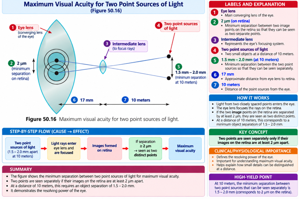
This appearance of two-point images is shown in Fig. 50.16.
Foveal Cones and Visual Acuity
The fovea is the central part of the retina where vision is most highly developed.
The average diameter of cones in the fovea is about:
1.5 micrometers
This is approximately:
One-seventh of the diameter of the light spot.
Even though the entire light spot is larger than one cone, its:
Center is bright
while its:
Edges gradually fade.
Because of this, a person can normally distinguish two separate points when their centers are approximately:
2 micrometers apart on the retina.
This distance is only slightly greater than the width of one foveal cone.
NORMAL VISUAL ACUITY
The normal visual acuity of the human eye for distinguishing two point sources of light is approximately:
25 seconds of arc
This means:
If light rays from two separate points enter the eye with an angle of at least:
25 seconds between them
the person can usually recognize them as:
Two separate points
rather than one point.
Example
A person with normal visual acuity looks at two bright pinpoint light spots from:
10 meters away.
The person can just distinguish them as two separate spots when the spots are approximately:
1.5–2 mm apart.VEA AND MAXIMUM VISUAL ACUITY
The diameter of the fovea is less than:
0.5 mm
or
<500 micrometers
Therefore, maximum visual acuity occurs within less than:
2 degrees of the visual field.
Outside the foveal region:
Visual acuity becomes progressively poorer.
As the retinal periphery is approached:
Visual acuity decreases by more than 10-fold.
The reason given in the text is:
More and more rods and cones connect with each optic nerve fiber in the nonfoveal peripheral retina.
This is discussed further in Chapter 52.
CLINICAL METHOD FOR STATING VISUAL ACUITY
Visual acuity is usually tested using a chart containing:
Letters of different sizes.
The chart is placed:
20 feet away
from the person being tested.
20/20 Vision
If a person can see letters at 20 feet that normally should be visible at 20 feet:
Visual acuity = 20/20
This means:
Normal vision
20/200 Vision
If a person can see only letters at 20 feet that normally should be visible at:
200 feet
the person’s visual acuity is:
20/200
Meaning of the Visual Acuity Fraction
Clinical visual acuity is expressed as a mathematical fraction.
This fraction represents:
Ratio of two distances
and also represents:
Person’s visual acuity compared with normal visual acuity. DETERMINATION OF DISTANCE FROM THE EYE — DEPTH PERCEPTION
The ability to determine the distance of objects is called:
Depth Perception
A person normally determines distance by three main mechanisms:
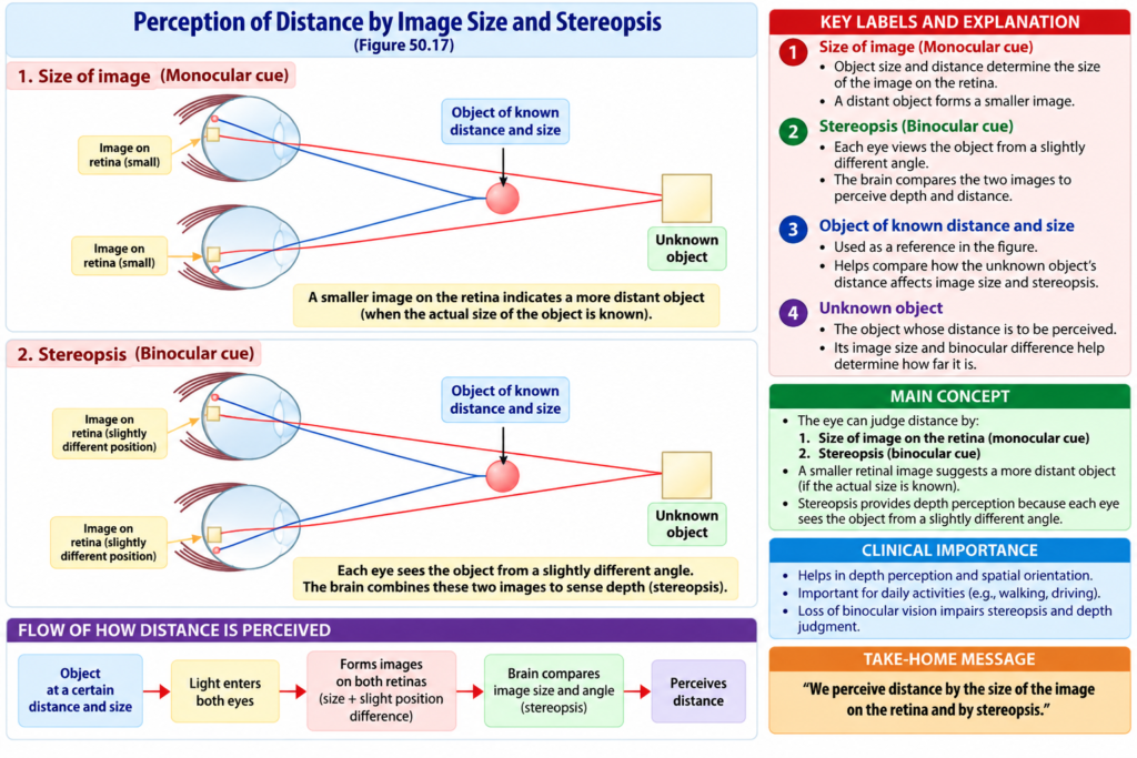
1. DETERMINATION OF DISTANCE BY SIZE OF RETINAL IMAGES
If the actual size of an object is already known, its distance can be estimated from:
Size of its image on the retina.
Example
Suppose a person being viewed is known to be:
6 feet tall.
The distance of that person can be estimated from:
Size of the person’s retinal image.
A person does not consciously calculate this retinal image size.
Instead:
Brain has learned to calculate object distance automatically from image size
when the actual dimensions of the object are known.
2. DETERMINATION OF DISTANCE BY MOVING PARALLAX
Moving parallax means:
Displacement in the apparent position of an object when it is viewed along two different lines of sight.
If a person looks into the distance while keeping the eyes and head completely still:
No moving parallax is perceived.
However, when the person moves the head from one side to the other:
Nearby objects
Their images:
Move rapidly across the retinas.
Distant objects
Their images:
Remain almost completely stationary.
Example of Moving Parallax
If the head moves:
1 inch sideways
and an object is only:
1 inch in front of the eye,
its image moves:
Almost all the way across the retinas.
In contrast, if an object is:
200 feet away,
its image:
Does not move perceptibly.
Therefore:
Nearby object → greater retinal image movement
Distant object → little or almost no retinal image movement
Using moving parallax:
Relative distances of different objects can be judged even when only one eye is used.
3. DETERMINATION OF DISTANCE BY STEREOPSIS — BINOCULAR VISION
Another way of perceiving parallax is:
Binocular vision
The two eyes are separated from each other by:
A little more than 2 inches.
Therefore, each eye views an object from a slightly different position.
As a result:
Images formed on the two retinas are different from each other.
Near Object Example
An object located:
1 inch in front of the nose
forms an image:
On the left side of the retina of the left eye
but:
On the right side of the retina of the right eye.
Distant Object Example
A small object located:
20 feet in front of the nose
forms images at:
Closely corresponding points near the centers of the two retinas.
This type of binocular parallax is demonstrated in Fig. 50.17.
The red spot and yellow square form reversed retinal positions because they are located at different distances in front of the eyes.
This binocular parallax is present whenever:
Both eyes are being used.
IMPORTANCE OF STEREOPSIS
Binocular parallax, or stereopsis, gives a person with two eyes:
Much greater ability to judge relative distances of nearby objects
than a person using only one eye.
However:
Stereopsis becomes virtually useless for depth perception at distances beyond 50–200 feet.
5. FIGURE / TABLE NUMBERS
The figure numbers have been placed only once at the exact relevant concepts in the explanation above.
No table number appears in the provided text.
6. MATHEMATICAL EQUATIONS / FORMULAS
The provided text describes visual acuity as a mathematical fraction:
Visual Acuity = Test Distance / Distance at which the letters should normally be seen
Example 1: 20/20 Vision
[
\frac{20}{20}=1
]
Meaning:
The person sees at 20 feet what a person with normal vision should see at 20 feet.
Therefore:
20/20 = Normal vision
Example 2: 20/200 Vision
[
\frac{20}{200}=\frac{1}{10}
]
The person sees at:
20 feet
what a person with normal vision should be able to see at:
200 feet.
Therefore, this fraction represents the person’s visual acuity relative to normal visual acuity.
7. KEY CONCEPTS WITH EXAMPLES
Key Concept 1 — Resolution of Two Points
Two separate light points can normally be distinguished when their centers are approximately:
2 micrometers apart on the retina.
Example
At a distance of 10 meters:
Two bright points about 1.5–2 mm apart can just be distinguished separately.
Key Concept 2 — Fovea
Fovea → area of highest visual acuity
Maximum visual acuity occurs within less than:
2 degrees of the visual field.
Key Concept 3 — Peripheral Retina
Moving toward the retinal periphery:
Visual acuity progressively decreases
and may decrease by:
More than 10-fold.
Key Concept 4 — Clinical Visual Acuity
20/20 → normal vision
20/200 → person sees at 20 feet what should normally be visible at 200 feet
Key Concept 5 — Depth Perception
Three main mechanisms are:
Known retinal image size + moving parallax + stereopsisey Concept 6 — Moving Parallax
Example
Nearby object → image moves rapidly across retina when head moves
Distant object → image remains almost stationary
Moving parallax can work with:
One eye
Key Concept 7 — Stereopsis
The two eyes receive slightly different retinal images because they are separated by more than:
2 inches.
This binocular difference helps judge:
Relative distances of nearby objects.
8. CLINICAL NOTE / IMPORTANCE
Visual Acuity Testing
Visual acuity is clinically tested using:
Letters of different sizes placed 20 feet away.
20/20 → normal vision
20/200 → only letters normally visible at 200 feet can be seen at 20 feet
Importance of the Fovea
The fovea provides:
Maximum visual acuity.
Outside the fovea:
Visual acuity becomes progressively poorer.
Importance of Moving Parallax
Moving parallax allows a person to estimate:
Relative distances of objects even with one eye.
Importance of Stereopsis
Stereopsis provides:
Greater ability to judge nearby relative distances when both eyes are used.
Its usefulness becomes very small beyond:
50–200 feet.9. HIGH-YIELD POINTS
- Normal retinal light spot diameter ≈ 11 µm.
- Foveal cone diameter ≈ 1.5 µm.
- Two retinal points can be distinguished at about 2 µm separation.
- Normal visual acuity ≈ 25 seconds of arc.
- At 10 m, two light points can just be distinguished when about 1.5–2 mm apart.
- Fovea diameter is <0.5 mm.
- Maximum visual acuity occurs within <2° of visual field.
- Peripheral visual acuity decreases by more than 10-fold.
- 20/20 = normal vision.
- 20/200 = sees at 20 feet what normally should be seen at 200 feet.
- Depth perception depends mainly on retinal image size, moving parallax, and stereopsis.
- Moving parallax can work with one eye.
- Stereopsis requires both eyes.
- Stereopsis is virtually useless beyond 50–200 feet.
10. COMMON STUDENT MISTAKES
Mistake 1
Thinking the focused retinal point is infinitely small in the real eye.
Correct: The normal retinal light spot has a diameter of about 11 micrometers because the optical system is not perfect.
Mistake 2
Thinking maximum visual acuity occurs throughout the retina.
Correct: Maximum visual acuity occurs in the small foveal area.
Mistake 3
Thinking 20/200 is better vision than 20/20.
Correct: In 20/200 vision, the person must be at 20 feet to see letters normally visible at 200 feet.
Mistake 4
Thinking all depth perception requires two eyes.
Correct: Moving parallax can determine relative distance even with one eye.
Mistake 5
Thinking stereopsis is equally useful at all distances.
Correct: Stereopsis is virtually useless for depth perception beyond 50–200 feet.
Mistake 6
Confusing moving parallax with stereopsis.
Moving parallax → depends on apparent movement when viewing position changes
Stereopsis → depends on different retinal images from the two eyes
11. QUICK REVISION
Visual Acuity
Normal retinal spot → ~11 µm
Foveal cone → ~1.5 µm
Two points distinguished → ~2 µm retinal separation
Normal angular resolution → ~25 seconds of arc
Fovea
Fovea → highest visual acuity
Peripheral retina → progressively poorer visual acuity
Clinical Measurement
20/20 → normal
20/200 → sees at 20 ft what normal person sees at 200 ft
Depth Perception
Three mechanisms:
1. Known retinal image size
2. Moving parallax
3. Stereopsis
Moving Parallax
Near object → large apparent movement
Far object → little movement
Can work with one eye
Stereopsis
Two eyes → different retinal images → better judgment of nearby relative distance
Beyond 50–200 ft → virtually useless12. SUMMARY
Visual acuity is the ability of the eye to distinguish separate points.
Although a distant point theoretically should form an infinitely small retinal image, the normal optical system produces a light spot about 11 micrometers in diameter.
The foveal cones are approximately 1.5 micrometers in diameter, and two separate points can normally be distinguished when their retinal centers are about 2 micrometers apart.
Normal visual acuity corresponds to an angular separation of approximately 25 seconds of arc. At 10 meters, two bright points can just be distinguished when they are about 1.5–2 millimeters apart.
The fovea provides maximum visual acuity within less than 2 degrees of the visual field. Visual acuity becomes progressively poorer toward the retinal periphery.
Clinically, visual acuity is expressed as a fraction. 20/20 represents normal vision, while 20/200 means that a person sees at 20 feet what should normally be visible at 200 feet.
Depth perception is the ability to judge the distance of objects. It depends mainly on retinal image size of known objects, moving parallax, and stereopsis.
Moving parallax can judge relative distance using only one eye. Stereopsis uses the different images formed on the two retinas and gives much greater ability to judge nearby relative distances, but it becomes virtually useless beyond 50–200 feet.
13. CLINICALLY CONCEPTUAL MCQs
MCQ 1
During visual acuity testing, a person standing 20 feet from the eye chart can read only the letters that a person with normal visual acuity should be able to read at 200 feet. What is this person’s visual acuity?
A. 20/10
B. 20/20
C. 20/40
D. 20/100
E. 20/200
Correct Answer: E. 20/200
Explanation
The clinical method expresses visual acuity as a fraction representing the ratio of two distances.
If the person sees at:
20 feet
only the letters that normally should be visible at:
200 feet
the visual acuity is:
20/200
By contrast:
20/20 represents normal vision.
MCQ 2
A person closes one eye and moves the head from side to side while looking at several objects. Nearby objects appear to move rapidly across the retina, while distant objects remain almost stationary. Which mechanism is being used to judge the relative distances of the objects?
A. Stereopsis
B. Moving parallax
C. Visual acuity fraction
D. Foveal cone diameter
E. Pupillary accommodation
Correct Answer: B. Moving parallax
Explanation
With moving parallax:
Head moves from side to side → nearby retinal images move rapidly → distant retinal images remain almost stationary.
This allows a person to determine the relative distances of objects:
Even when only one eye is used.
Stereopsis, in contrast, depends on the different images formed on the two retinas when both eyes are used.
14. FINAL EXAM CONCEPT
Complete Conceptual Flow
Visual acuity → ability to distinguish separate points
Fovea → maximum visual acuity
20/20 → normal vision
20/200 → sees at 20 ft what should normally be seen at 200 ft
Depth Perception
Known object size → retinal image size gives distance
Head movement → moving parallax → relative distance even with one eye
Two eyes → different retinal images → stereopsis → better nearby depth judgment
Distance beyond 50–200 ft → stereopsis becomes virtually useless
Reference Guyton Physiology 15th Edition. Page # 644| Chapter # 50
