


1. Learning Objective
By the end of this topic, students should be able to:
- Understand the main refractive surfaces and refractive power of the eye.
- Explain how an image is formed on the retina.
- Understand the mechanism and nervous control “in accommodation”
2. Introduction
The eye works like an optical system.
Different surfaces of the eye bend light so that an image can be focused on the retina.
The cornea provides most of the refractive power of the eye, while the internal lens has the special ability to change its curvature for accommodation.
3. Concept in Simple and Easy Words
Refractive Interfaces of the Eye
The lens system of the eye has four refractive interfaces (Fig. 50.9):
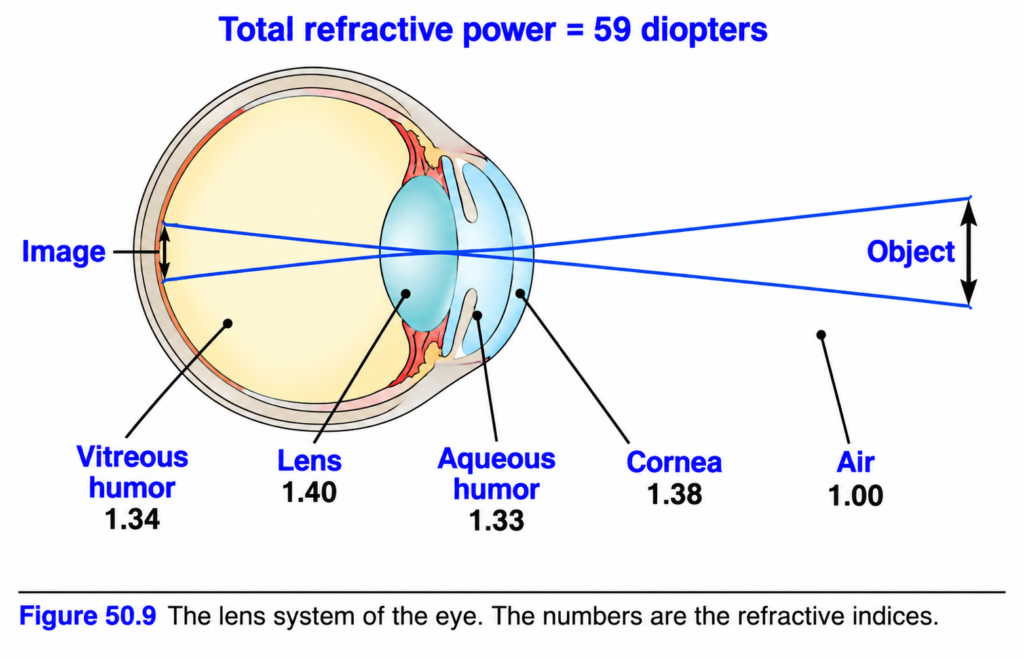
- Air → anterior surface of cornea
- Posterior surface of cornea → aqueous humor
- Aqueous humor → anterior surface of lens
- Posterior surface of lens → vitreous humor
Each of these interfaces is involved in the refraction of light.
Refractive Indices
The refractive indices are:
- Air = 1
- Cornea = 1.38
- Aqueous humor = 1.33
- Crystalline lens = about 1.40
- Vitreous humor = 1.34
Reduced Eye
Instead of considering all the refractive surfaces separately, they can be added together mathematically and considered as a single lens system.
This simplified optical model is called the reduced eye.
It makes simple optical calculations easier.
In the reduced eye:
- One refractive surface is considered.
- Its central point is 17 mm in front of the retina.
- Total refractive power for distant vision is 59 diopters.
Refractive Power of the Cornea
About two-thirds of the total 59 diopters of refractive power is produced by the anterior surface of the cornea.
Therefore, most of the refractive power of the eye comes from the cornea rather than from the internal lens.
Why?
The refractive index of the cornea is markedly different from the refractive index of air.
In comparison, the refractive index of the eye lens is not greatly different from the refractive indices of the aqueous humor and vitreous humor.
Therefore, the corneal surface produces much more refraction.
Refractive Power of the Internal Lens
The internal lens normally has a refractive power of about 20 diopters when it is surrounded by fluid on both sides.
This is about one-third of the total refractive power of the eye.
Although its normal refractive power is less than that of the cornea, the internal lens is important because its curvature can change.
Nervous signals from the brain can increase the curvature of the lens.
This change produces accommodation.
Formation of an Image on the Retina
A glass lens can focus an image on paper.
In the same way, the lens system of the eye focuses an image on the retina.
The image formed on the retina is:
- Inverted
- Reversed
However, we normally perceive the object in an upright position.
This occurs because the brain is trained to consider the inverted retinal image as normal.
MECHANISM OF ACCOMMODATION
Accommodation means increasing the refractive power of the lens by changing its shape.
In children, the refractive power of the lens can increase from:
20 diopters → about 34 diopters
Therefore:
Accommodation = 34 − 20 = 14 diopters
So, the lens can add about 14 diopters of refractive power.
To produce this accommodation, the lens changes:
Moderately convex lens → very convex lens
A more convex lens has greater refractive power.
Structure of the Young Lens
In a young person, the lens has:
- A strong elastic capsule
- Viscous, protein-containing, transparent fluid inside it
When there is no tension on the lens capsule, the lens naturally becomes almost spherical.
This spherical shape occurs mainly because of the elastic retraction of the lens capsule.
Role of Suspensory Ligaments
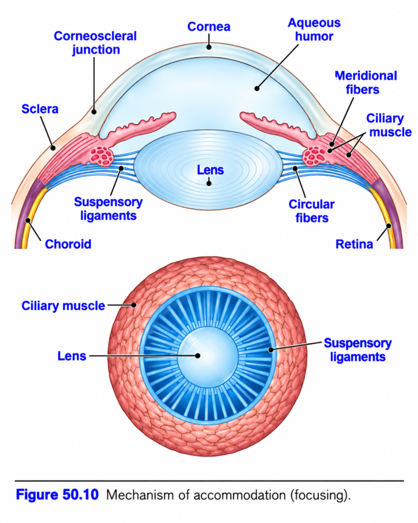
About 70 suspensory ligaments are attached radially around the lens (Fig. 50.10).
These ligaments pull the edges of the lens toward the outer part of the eyeball.
The ligaments remain under tension because of their attachments at the anterior border of the choroid and retina.
This tension pulls on the lens.
As a result:
Ligament tension → lens becomes relatively flat
Thus, under normal conditions, tension in the suspensory ligaments keeps the lens relatively flat.
Role of the Ciliary Muscle
The ciliary muscle is located near the lateral attachment of the lens ligaments.
It contains two groups of smooth muscle fibers:
- Meridional fibers
- Circular fibers
Meridional Fibers
The meridional fibers extend from the peripheral ends of the suspensory ligaments toward the corneoscleral junction.
When these fibers contract:
Ciliary muscle contracts
→ ligament attachment moves medially toward the cornea
→ tension on suspensory ligaments decreases
→ tension on lens decreases
Circular Fibers
The circular fibers are arranged around the attachments of the ligaments.
When they contract, they act like a sphincter.
Their contraction decreases the diameter of the circle formed by the ligament attachments.
Therefore:
Circular muscle contracts
→ diameter of ligament attachment circle decreases
→ ligaments pull less strongly on lens capsule
Final Effect of Ciliary Muscle Contraction
Contraction of either group of ciliary muscle fibers relaxes the pull of the suspensory ligaments on the lens.
Therefore:
Ciliary muscle contracts
→ suspensory ligaments relax
→ lens capsule is released from tension
→ natural elasticity of lens makes it more spherical
The lens becomes more rounded, similar to a balloon.
Accommodation Is Controlled by Parasympathetic Nerves
The ciliary muscle is controlled almost completely by parasympathetic nerve signals.
These signals reach the eye through the third cranial nerve from the third nerve nucleus in the brain stem.
Parasympathetic stimulation causes both groups of ciliary muscle fibers to contract.
Therefore:
Parasympathetic stimulation
→ ciliary muscle contracts
→ lens ligaments relax
→ lens becomes thicker
→ refractive power increases
Focusing on Near Objects
When the refractive power of the lens increases, the eye can focus on objects that are nearer.
Therefore:
Greater refractive power → focus on nearer objects
As a distant object moves progressively closer to the eye, progressively more parasympathetic impulses must reach the ciliary muscle.
This allows the object to remain continuously in focus.
Sympathetic Effect
Sympathetic stimulation can relax the ciliary muscle.
However, this effect is very weak.
Therefore, sympathetic stimulation plays almost no role in normal accommodation.
4. Mathematical Concept
The passage contains one simple accommodation calculation:
Initial lens power
20 D
Maximum stated lens power in children
34 D
Increase in refractive power
34 D − 20 D = 14 D
Therefore:
Accommodation = 14 diopters
Simple Concept
Accommodation represents the amount by which the refractive power of the lens can increase.
5. Key Concepts with Examples
Key Concept 1: Most refractive power comes from the cornea
Total refractive power of the eye:
59 D
About two-thirds comes from the anterior surface of the cornea.
Concept: The cornea contributes most of the refractive power of the eye.
Key Concept 2: The internal lens provides less power but can change its power
Normal internal lens power:
About 20 D
Its curvature can increase in response to nervous signals.
Example: During accommodation, its power can increase from 20 D to about 34 D in children.
Key Concept 3: Lens shape determines accommodation
Lens flatter → lower refractive power
Lens more spherical/thicker → higher refractive power
Key Concept 4: Ciliary muscle contraction does not tighten the lens ligaments
Instead:
Ciliary muscle contraction
→ ligament tension decreases
→ lens becomes more spherical
Key Concept 5: Parasympathetic activity produces accommodation
Parasympathetic stimulation
→ ciliary muscle contraction
→ relaxed suspensory ligaments
→ thicker lens
→ increased refractive power
→ focus on nearer object
6. Clinical Importance
The ability of the internal lens to change its curvature allows the eye to focus on objects at different distances.
The lens becomes thicker and increases its refractive power when focusing on nearer objects.
The retinal image itself is inverted and reversed, but the brain normally perceives the object as upright.
7. High-Yield Points
- The eye has four refractive interfaces.
- Total refractive power of the reduced eye for distant vision = 59 D.
- About two-thirds of the refractive power is produced by the anterior surface of the cornea.
- Internal lens refractive power is normally about 20 D.
- In children, lens power can increase to about 34 D.
- Maximum increase described here = 14 D of accommodation.
- Retinal image is inverted and reversed.
- Suspensory ligament tension keeps the lens relatively flat.
- Ciliary muscle contraction reduces ligament tension.
- Reduced ligament tension allows the lens to become more spherical.
- Accommodation is controlled mainly by parasympathetic nerves through the third cranial nerve.
- Increased lens refractive power allows focusing on nearer objects.
- Sympathetic influence on normal accommodation is very weak.
8. Common Student Mistakes
Mistake 1
Thinking that most refractive power comes from the lens.
Correct concept:
Most refractive power comes from the anterior surface of the cornea.
Mistake 2
Thinking that ciliary muscle contraction increases tension in the suspensory ligaments.
Correct sequence:
Ciliary muscle contracts
→ ligament tension decreases
→ lens becomes more spherical
Mistake 3
Thinking that a flat lens is used for greater accommodation.
Correct concept:
More spherical/thicker lens → greater refractive power
Mistake 4
Thinking that sympathetic stimulation is the main control of accommodation.
Correct concept:
Accommodation is controlled almost entirely by parasympathetic stimulation.
Mistake 5
Thinking that the image formed on the retina is upright.
Correct concept:
The retinal image is inverted and reversed.
9. Quick Revision
Four refractive interfaces
→ bend light through the eye.
Reduced eye
→ simplified single optical system.
Total power
→ 59 D
Main refractive surface
→ anterior cornea.
Internal lens
→ about 20 D
Accommodation in children
→ 20 D to 34 D
→ increase = 14 D
Suspensory ligaments tense
→ lens flat.
Ciliary muscle contracts
→ ligaments relax.
Ligaments relax
→ lens becomes spherical/thicker.
Lens thicker
→ refractive power increases.
Greater refractive power
→ focus on nearer objects.
Main nerve control
→ parasympathetic through third cranial nerve.
10. Summary
The eye contains several refractive interfaces that bend light and focus an image on the retina.
The simplified reduced eye has a total refractive power of about 59 diopters, with about two-thirds of this power provided by the anterior surface of the cornea.
The internal lens provides about 20 diopters, but its major importance is its ability to change curvature.
The image formed on the retina is inverted and reversed, although the brain perceives it normally as upright.
During accommodation, the ciliary muscle contracts, reducing tension in the suspensory ligaments.
The natural elasticity of the lens then makes it more spherical and thicker.
This increases the refractive power of the lens and allows the eye to focus on nearer objects.
Accommodation is controlled mainly by parasympathetic nerves through the third cranial nerve.
Reference Guyton physiology 15th Edition. Page 640 | Chapter 50
