


- Mitochondrial DNA (mtDNA) contains genes that encode enzymes needed for oxidative phosphorylation → energy production.
- Mitochondrial DNA shows maternal inheritance.
- Reason:
- ovum contains many mitochondria in its cytoplasm
- sperm contains few or no mitochondria
- therefore, the zygote receives its mitochondrial DNA essentially from the mother
- So: Affected mother → can transmit mitochondrial mutation to both sons and daughters
- But: Affected father → does not transmit mitochondrial genes to his children
- Mitochondrial genetic diseases are rare.
- Because mitochondria are important for energy production, mutations mainly damage tissues with high energy requirements, especially:
- CNS
- skeletal muscle
- cardiac muscle
- liver
- kidney
- Leber hereditary optic neuropathy is the classic example.
- It is a neurodegenerative disorder causing: progressive bilateral loss of central vision → eventual blindness
KEY CONCEPT
- Mitochondrial disorders = maternal inheritance.
- Mother transmits mtDNA → sons + daughters.
- Father does not transmit mtDNA.
- mtDNA mutation → impaired oxidative phosphorylation → high-energy tissues affected most.
- Prototype → Leber hereditary optic neuropathy.
CONCEPTUAL EXAMPLES
- Mother has an mtDNA mutation → both her son and daughter may inherit it.
- Father has an mtDNA mutation → children do not inherit it from him.
- Mitochondrial energy production fails → highly energy-dependent nervous tissue is affected → progressive visual loss in Leber hereditary optic neuropathy.
Diseases Caused by Alterations of Imprinted Regions: Prader-Willi and Angelman Syndromes
- Normally, each autosomal gene has two copies:
- one from the mother
- one from the father
- For most genes, the maternal and paternal copies function similarly.
- However, some genes behave differently depending on which parent they came from.
- This parent-specific gene control is called genomic imprinting.
- Genomic imprinting = one parental copy of a gene is selectively silenced/inactivated during formation of sperm or ova.
- Maternal imprinting:
- maternal allele is silenced
- paternal allele remains active
- Paternal imprinting:
- paternal allele is silenced
- maternal allele remains active
- At the molecular level, gene silencing occurs mainly through:
- DNA promoter methylation
- modification of histone proteins
- These changes make the gene transcriptionally inactive.
- Imprinting occurs during gametogenesis:
- in sperm
- or in ova
- After fertilization, the imprint is maintained in the somatic cells of the offspring.
- Two classic disorders caused by abnormal imprinting are:
- Prader-Willi syndrome
- Angelman syndrome
KEY CONCEPT
- Genomic imprinting = parent-of-origin–specific gene silencing.
- Maternal imprinting → maternal allele OFF.
- Paternal imprinting → paternal allele OFF.
- Mechanism → DNA methylation + histone modification → gene silencing.
- Important examples → Prader-Willi and Angelman syndromes.
CONCEPTUAL EXAMPLE
- Same gene is inherited from both parents, but one copy is normally switched off.
- If the active parental copy is lost or abnormal, the remaining imprinted copy cannot compensate → disease develops.
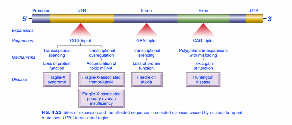
FIG. 4.23 — TRIPLET REPEAT EXPANSION DISEASES
🧠 Simplest idea
Repeated DNA triplets become excessively expanded. The disease depends mainly on WHERE the repeat expands: UTR, intron, or exon.
Whole figure in one flow
Repeat expansion → abnormal gene expression/protein → disease
🎨 COLOR / STRUCTURE GUIDE
- 🔵 Dark blue = promoter / intron regions
- 🟡 Yellow = UTR (untranslated region)
- 🟢 Green = exon
- 🔺 Red triangles = site where repeat expansion occurs
- 5′ → 3′ = direction of the gene
1️⃣ CGG repeat in 5′ UTR → Fragile X
The red triangle points to a CGG triplet expansion in the 5′ UTR.
Full mutation
Large CGG expansion
→ DNA methylation
→ transcriptional silencing of FMR1
→ ↓ FMRP protein
→ loss of protein function
⬇️
Fragile X syndrome
🧠 Memory:
Fragile X = CGG → gene OFF
2️⃣ Smaller CGG expansion / premutation
The same FMR1 region can produce a different problem when the repeat is in the premutation range.
CGG premutation
→ abnormal ↑ transcription
→ excessive/toxic FMR1 mRNA
→ cellular toxicity
⬇️
Can cause:
- Fragile X–associated tremor/ataxia syndrome (FXTAS)
- Fragile X–associated primary ovarian insufficiency (FXPOI)
Key difference:
Full mutation → too little protein
Premutation → too much toxic mRNA
3️⃣ GAA repeat in INTRON → Friedreich ataxia
🔺 Red triangle in the intron
GAA repeat expansion
→ interferes with transcription
→ gene silencing
→ ↓ frataxin protein
⬇️
Friedreich ataxia
🧠 Memory:
Friedreich = GAA in intron → frataxin ↓
4️⃣ CAG repeat in EXON → Huntington disease
🔺 Red triangle lies inside an exon, so the repeat becomes part of the protein-coding sequence.
Why important?
CAG codes for glutamine (Q).
Therefore:
CAG CAG CAG…
→ many glutamines
→ polyglutamine tract
→ protein misfolding/aggregation
→ toxic gain of function
⬇️
Huntington disease
🧠 Memory:
Huntington = CAG → poly-Q → toxic protein
⭐ MOST IMPORTANT COMPARISON
| Repeat + Location | Main mechanism | Disease |
|---|---|---|
| CGG — 5′ UTR | Gene silencing → protein loss | Fragile X syndrome |
| CGG premutation — 5′ UTR | Toxic mRNA | FXTAS / FXPOI |
| GAA — intron | Transcriptional silencing → frataxin loss | Friedreich ataxia |
| CAG — exon | Polyglutamine misfolding → toxic gain | Huntington disease |
🧠 Fastest exam recall
CGG → Fragile X
GAA → Friedreich ataxia
CAG → Huntington
🔥 One-line memory
UTR CGG = gene OFF/toxic RNA; intron GAA = gene OFF; exon CAG = toxic protein.
Prader-Willi and Angelman Syndromes
- Prader-Willi syndrome is characterized by:
- intellectual disability
- short stature
- hypotonia → reduced muscle tone
- obesity
- small hands and feet
- hypogonadism
- In about 60%–75% of cases, there is deletion of chromosome 15: del(15)(q11;q13)
- In Prader-Willi syndrome, this deletion occurs on the paternally derived chromosome 15.
- Therefore: Paternal 15q11–q13 deletion → Prader-Willi syndrome
- Angelman syndrome involves deletion of the same chromosome 15 region, but the abnormal chromosome comes from the mother.
- Therefore: Maternal 15q11–q13 deletion → Angelman syndrome
- Angelman syndrome is characterized by:
- intellectual disability
- ataxic gait
- seizures
- inappropriate laughter
- Thus, the same chromosomal deletion causes different diseases depending on whether it comes from the father or mother.
- This demonstrates the parent-of-origin effect produced by genomic imprinting.
KEY CONCEPT
- Same deletion, different parent → different disease.
- Paternal deletion → Prader-Willi syndrome.
- Maternal deletion → Angelman syndrome.
Easy memory:
P = Paternal → Prader-Willi
M = Maternal → Angelman
CONCEPTUAL EXAMPLES
- Child loses the paternal 15q11–q13 region → Prader-Willi → hypotonia + obesity + hypogonadism.
- Child loses the maternal 15q11–q13 region → Angelman → intellectual disability + ataxia + seizures + inappropriate laughter.
- This difference proves that some genes on chromosome 15 are expressed differently depending on their parental origin.
Pathogenesis
- Prader-Willi and Angelman syndromes involve the same region of chromosome 15, but different genes are normally active depending on the parent of origin (Fig. 4.24).
- In the 15q12 region, a group of genes on the maternal chromosome is normally imprinted (silenced).
- Therefore, for these genes: Maternal copy OFF → paternal copy is the functional copy
- If the paternal 15q region is deleted: paternal active genes lost + maternal genes already silenced → no functional gene expression → Prader-Willi syndrome
- The most likely important genes in Prader-Willi syndrome are a cluster encoding small nucleolar RNAs (snoRNAs).
- snoRNAs help in RNA processing.
- A different gene in the same region, UBE3A, shows the opposite imprinting pattern.
- For UBE3A: Paternal UBE3A OFF → maternal UBE3A is active
- UBE3A encodes a ubiquitin ligase.
- Ubiquitin ligase: adds ubiquitin to cellular proteins → marks them for proteasomal degradation
- If the maternal UBE3A gene is deleted: maternal active UBE3A lost + paternal UBE3A already silenced → ↓ UBE3A → Angelman syndrome
- The neurologic manifestations of Angelman syndrome mainly result from lack of UBE3A expression in certain brain regions.
- Some patients have no visible chromosome deletion but still develop disease because of uniparental disomy.
- Uniparental disomy = both copies of a chromosome pair are inherited from one parent only.
- In Prader-Willi syndrome: two maternal chromosome 15s → no paternal active snoRNA genes → Prader-Willi syndrome
- In Angelman syndrome: two paternal chromosome 15s → no active maternal UBE3A → Angelman syndrome
KEY CONCEPT
- Prader-Willi: paternal gene expression is needed.
Paternal deletion OR two maternal chromosome 15s → Prader-Willi. - Angelman: maternal UBE3A expression is needed.
Maternal deletion OR two paternal chromosome 15s → Angelman. - Same chromosome region, but different parental origin → different disease because of genomic imprinting.
CONCEPTUAL EXAMPLES
- Maternal copy already OFF + paternal copy deleted → no functional Prader-Willi-region genes → Prader-Willi syndrome.
- Paternal UBE3A already OFF + maternal UBE3A deleted → no functional UBE3A → Angelman syndrome.
- Both chromosome 15 copies come from mother → maternal uniparental disomy → Prader-Willi.
- Both chromosome 15 copies come from father → paternal uniparental disomy → Angelman.
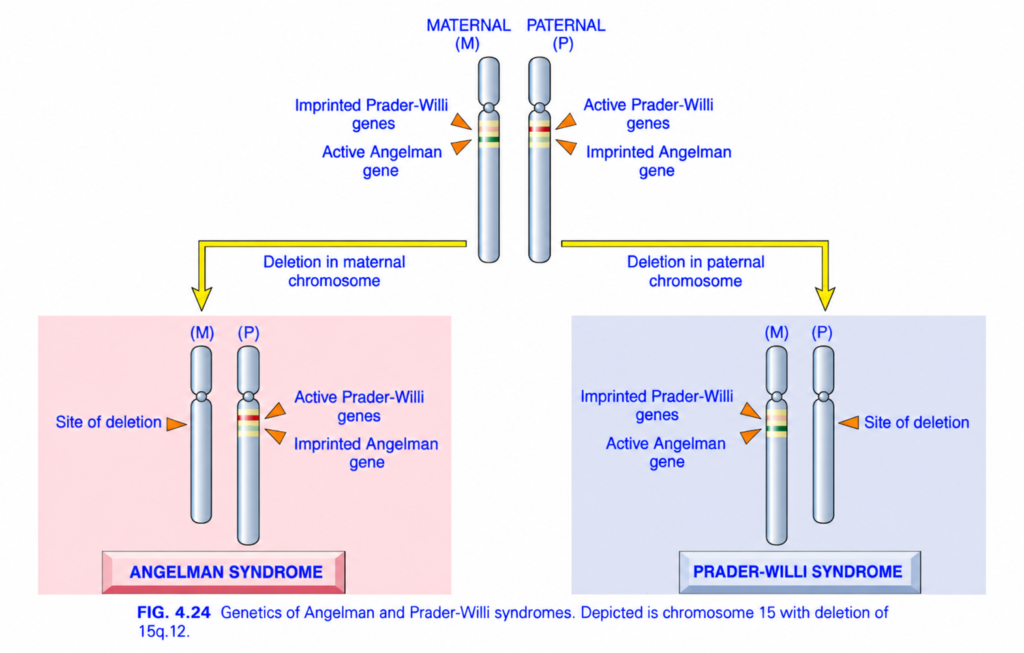
FIG. 4.24 — ANGELMAN vs PRADER-WILLI SYNDROME
🧠 Simplest idea
Same chromosome region (15q11–q13), but disease depends on whether the deletion comes from the MOTHER or FATHER.
Imprinted = gene is switched OFF / silenced
1️⃣ TOP — NORMAL CHROMOSOME 15
There are two copies:
- M = maternal chromosome 15
- P = paternal chromosome 15
But both copies are not equally active because of genomic imprinting.
🟦 Maternal chromosome
- Prader-Willi genes = imprinted → OFF
- Angelman gene (UBE3A) = active → ON
🟦 Paternal chromosome
- Prader-Willi genes = active → ON
- Angelman gene = imprinted → OFF
🧠 Key concept
For these genes, the body depends on only one parent’s active copy.
2️⃣ LEFT YELLOW ARROW — Deletion in MATERNAL chromosome
The maternal chromosome loses the important 15q region.
But remember:
Angelman gene
- Maternal copy = normally ACTIVE
- Paternal copy = normally IMPRINTED/OFF
So:
Maternal deletion
→ active UBE3A lost
→ paternal UBE3A is already silent
→ essentially no functional UBE3A
⬇️
ANGELMAN SYNDROME
🧠 Memory:
Angelman = Mother missing
A = Angelman → Active gene normally from mom
3️⃣ Why doesn’t maternal deletion cause Prader-Willi?
Because the maternal Prader-Willi genes were already imprinted/OFF.
The paternal chromosome still has:
Active Prader-Willi genes
So their function remains.
4️⃣ RIGHT YELLOW ARROW — Deletion in PATERNAL chromosome
Now the paternal 15q region is deleted.
Remember:
Prader-Willi genes
- Paternal copies = normally ACTIVE
- Maternal copies = normally IMPRINTED/OFF
So:
Paternal deletion
→ active Prader-Willi genes lost
→ maternal copies are already silent
→ no functional expression
⬇️
PRADER-WILLI SYNDROME
🧠 Memory:
Prader-Willi = Papa missing
P = Prader-Willi → Paternal genes normally active
5️⃣ Why doesn’t paternal deletion cause Angelman?
Because the paternal Angelman/UBE3A copy is normally imprinted/OFF.
The maternal chromosome still provides:
Active UBE3A
So Angelman syndrome does not result from this paternal deletion.
🎨 ARROW / COLOR GUIDE
- 🟦 Long chromosomes = chromosome 15
- M = maternal copy
- P = paternal copy
- 🟧 Orange triangles = important gene region / deletion site
- 🟨 Large yellow arrows = consequence of maternal vs paternal deletion
- 🟥 Left lower box = Angelman syndrome
- 🟦 Right lower box = Prader-Willi syndrome
⭐ MOST IMPORTANT COMPARISON
| Deleted chromosome | Active gene lost | Disease |
|---|---|---|
| Maternal 15q | UBE3A / Angelman gene | Angelman syndrome |
| Paternal 15q | Prader-Willi genes | Prader-Willi syndrome |
🧠 Fastest exam recall
Maternal deletion → Angelman
Mom’s active UBE3A gone
Paternal deletion → Prader-Willi
Papa’s active PWS genes gone
🔥 One-line memory
“Angelman = Mother missing; Prader-Willi = Papa missing.”
