


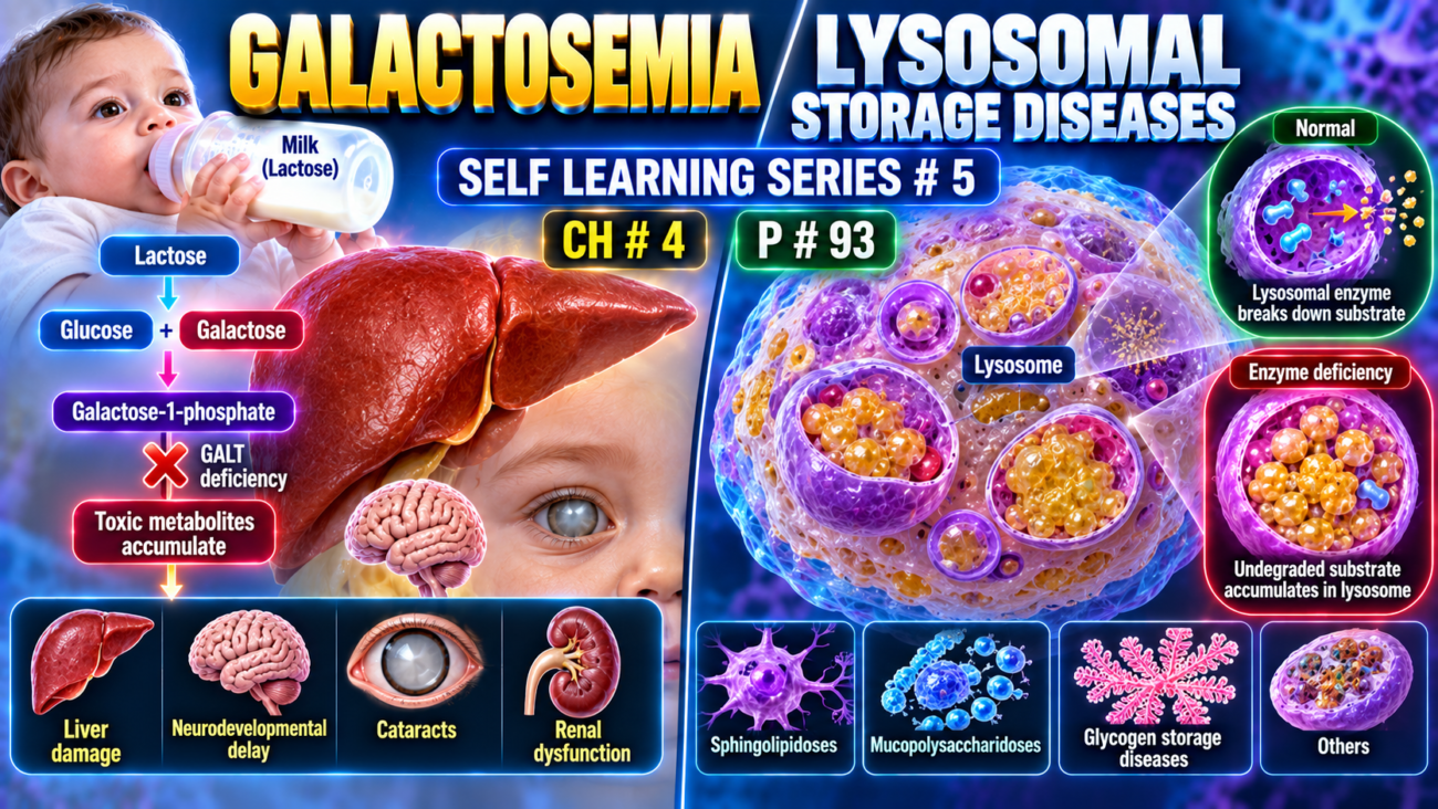
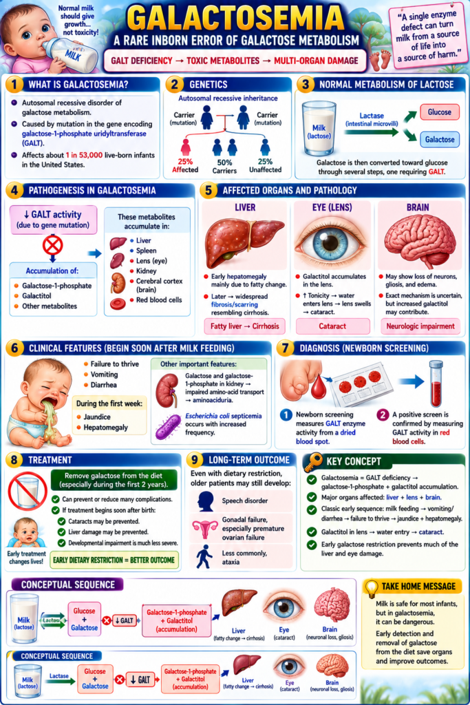
- Galactosemia is an autosomal recessive disorder of galactose metabolism.
- It is caused by mutation of the gene encoding galactose-1-phosphate uridyltransferase (GALT).
- It affects about 1 in 53,000 live-born infants in the United States.
- Normally:
- Milk contains lactose.
- Lactase splits lactose in intestinal microvilli into:
- glucose
- galactose
- Galactose is then converted toward glucose through several steps, one requiring GALT.
- In galactosemia:
↓ GALT → accumulation of galactose-1-phosphate + galactitol and other metabolites. - These metabolites accumulate in:
- liver
- spleen
- lens
- kidney
- cerebral cortex
- red blood cells
- The liver, eyes, and brain are most severely affected.
- Liver:
- early hepatomegaly is mainly due to fatty change.
- later → widespread fibrosis/scarring resembling cirrhosis.
- Eye:
- galactitol accumulates in the lens.
- ↑ tonicity → water enters lens → lens swells → cataract.
- Brain:
- may show loss of neurons, gliosis, and edema.
- The exact mechanism is uncertain, but increased galactitol may contribute.
- Clinical features begin soon after milk feeding:
- failure to thrive
- vomiting
- diarrhea
- During the first week:
- jaundice
- hepatomegaly
- Galactose and galactose-1-phosphate accumulation in the kidney → impaired amino-acid transport → aminoaciduria.
- Escherichia coli septicemia occurs with increased frequency.
- Newborn screening measures GALT enzyme activity from a dried blood spot.
- A positive screen is confirmed by measuring GALT activity in red blood cells.
- Removing galactose from the diet, especially during the first 2 years, can prevent or reduce many complications.
- If treatment begins soon after birth:
- cataracts may be prevented
- liver damage may be prevented
- developmental impairment is much less severe
- Even with dietary restriction, older patients may still develop:
- speech disorder
- gonadal failure, especially premature ovarian failure
- less commonly, ataxia
KEY CONCEPT
- Galactosemia = GALT deficiency → galactose-1-phosphate + galactitol accumulation.
- Major organs affected:
liver + lens + brain. - Classic early sequence:
milk feeding → vomiting/diarrhea → failure to thrive → jaundice + hepatomegaly. - Galactitol in lens → water entry → cataract.
- Early galactose restriction prevents much of the liver and eye damage.
CONCEPTUAL EXAMPLES
- Milk lactose → glucose + galactose → GALT cannot process galactose properly → toxic metabolites accumulate.
- Galactitol builds up in lens → pulls in water → cataract.
- Metabolites accumulate in liver → fatty change → later fibrosis/cirrhosis.
- Galactose removed early from diet → less toxic buildup → major complications reduced.
Lysosomal Storage Diseases
- Lysosomes are the cell’s digestive system and contain hydrolytic enzymes that break down complex substances such as:
- sphingolipids
- mucopolysaccharides
- These materials reach lysosomes from:
- damaged intracellular organelles through autophagy
- outside the cell through endocytosis or phagocytosis
- If a lysosomal enzyme is inheritedly deficient:
substrate cannot be completely degraded → partially digested insoluble material accumulates inside lysosomes (Fig. 4.12). - Lysosomes become enlarged and numerous → interfere with normal cell function.
- Lysosomal dysfunction also impairs autophagy → accumulation of:
- polyubiquitinated proteins
- dysfunctional mitochondria
- Defective mitochondria → ↑ free radicals → may trigger apoptosis.
- About 70 lysosomal storage diseases have been identified.
- They may result from defects in:
- lysosomal enzymes
- proteins involved in substrate degradation
- endosomal sorting
- lysosomal membrane integrity
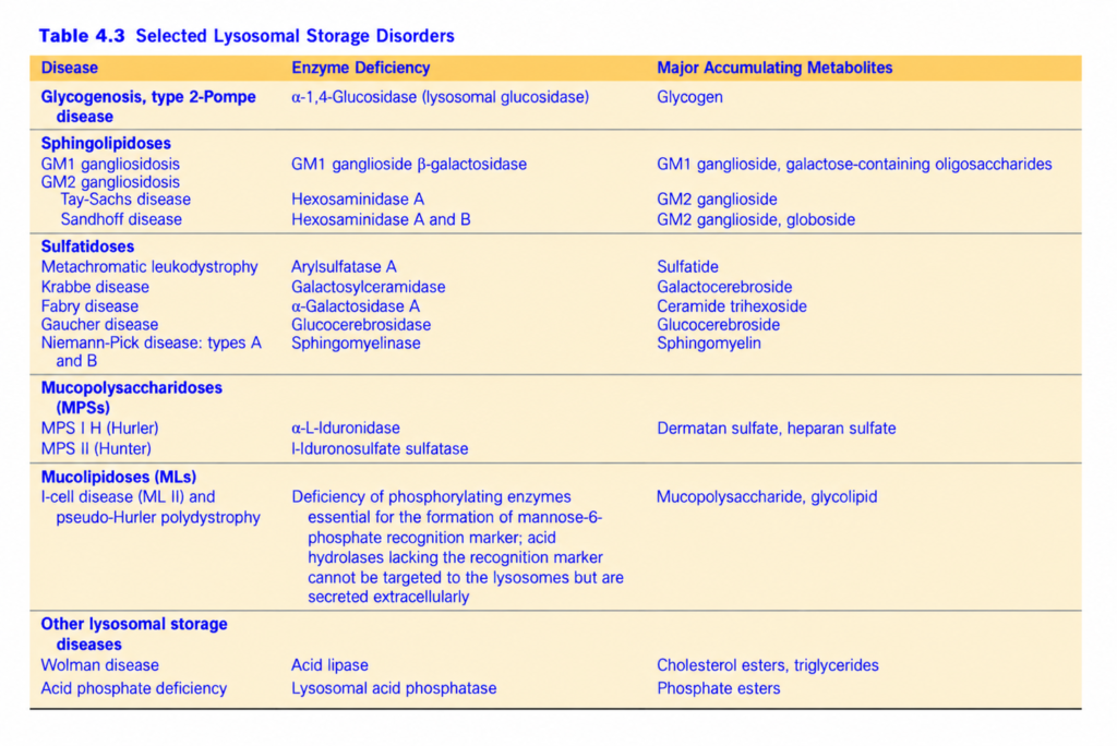
- Lysosomal storage diseases are classified according to the stored substrate and accumulated metabolite (Table 4.3).
- Each disorder usually results from deficiency of a specific enzyme.
- Combined frequency is about 1 in 2500 live births.
- Lysosomal dysfunction may also be linked with more common neurologic diseases:
- Gaucher disease carrier state → increased risk of Parkinson disease
- Niemann-Pick type C → associated with increased risk of Alzheimer disease
- These associations reflect the many functions of lysosomes, including:
- autophagy
- immunity through fusion with phagosomes
- cell membrane repair
- Common features of most lysosomal storage diseases include:
- autosomal recessive inheritance
- usually affect infants and young children
- storage in mononuclear phagocytes → hepatosplenomegaly
- frequent CNS involvement → neuronal damage
- cell injury caused by both stored material and secondary effects such as macrophage activation and cytokine release
KEY CONCEPT
- Lysosomal enzyme defect → substrate cannot be degraded → material accumulates in lysosomes → enlarged lysosomes + impaired cell function.
- Impaired autophagy → defective mitochondria and proteins also accumulate.
- Common pattern:
autosomal recessive + childhood onset + hepatosplenomegaly + CNS involvement.
CONCEPTUAL EXAMPLES
- Missing lysosomal enzyme → undegraded lipid remains inside lysosome → progressive intracellular storage.
- Storage in macrophages of liver and spleen → hepatosplenomegaly.
- Storage in neurons → neuronal dysfunction → CNS symptoms.
- Failed autophagy → abnormal mitochondria accumulate → free-radical injury + apoptosis.
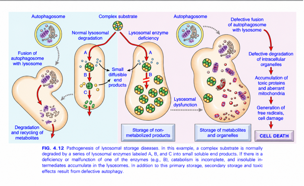
FIG. 4.12 — LYSOSOMAL STORAGE DISEASES
🧠 Simplest idea
Lysosome = cell’s recycling/digestive bag.
If a lysosomal enzyme is missing → material cannot be completely broken down → it accumulates → lysosome swells → autophagy also fails → toxic proteins + damaged mitochondria accumulate → cell injury/death.
Whole figure in one flow
Enzyme deficiency → incomplete substrate breakdown → lysosomal storage → lysosomal dysfunction → defective autophagy → toxic material + abnormal mitochondria → free radicals → CELL DEATH1️⃣ LEFT — NORMAL AUTOPHAGY
🔵/yellow round sac = autophagosome
It surrounds old/damaged cell parts such as:
- mitochondria
- membranes
- other organelles
⬇️ Gray dotted arrow
Autophagosome + lysosome fuse
The lysosome then digests these damaged structures.
⬇️ Red arrow
Result:
Degradation + recycling of metabolites
🧠 Think:
Autophagy = cell cleans and recycles its own old parts.
2️⃣ MIDDLE LEFT — NORMAL LYSOSOMAL DEGRADATION
🟢🟡 Ball = complex substrate
= a large molecule that must be broken down step-by-step.
The lysosome contains different enzymes:
A → B → C
Each enzyme performs one step.
Complex substrate
⬇️ enzyme A
smaller intermediate
⬇️ enzyme B
smaller intermediate
⬇️ enzyme C
🟢 small diffusible end products
These small products can leave the lysosome and be reused.
Easy idea:
A, B, C = three scissors cutting one big molecule into tiny usable pieces.
3️⃣ CENTER — LYSOSOMAL ENZYME DEFICIENCY
Suppose enzyme B is missing.
The figure shows a red block at B.
So:
A works
⬇️
intermediate forms
❌ B cannot work
⬇️
breakdown stops.
Result:
The partially degraded material cannot go further.
🟢🟡 circles accumulate inside lysosomes.
⬇️
Storage of non-metabolized products
This is the basic mechanism of a:
Lysosomal storage disease
4️⃣ Why do lysosomes become enlarged?
Because more and more undigested substrate enters, but cannot leave.
So:
Enzyme missing → substrate trapped → lysosomes enlarge + multiply
Eventually this interferes with normal cell function.
5️⃣ RIGHT — LYSOSOMAL DYSFUNCTION damages AUTOPHAGY
A sick lysosome cannot properly handle material brought by autophagosomes.
Gray dotted arrow:
Lysosomal dysfunction
→ poor autophagosome processing/fusion
→ defective degradation of intracellular organelles
6️⃣ What now accumulates?
The large abnormal cell shows:
- 🟢🟡 stored metabolites
- 🟣 damaged mitochondria
- broken cellular structures
- toxic proteins
⬇️
Storage of metabolites + organelles
This is secondary storage caused by defective autophagy.
7️⃣ Damaged mitochondria → FREE RADICALS
Abnormal mitochondria stay inside the cell instead of being removed.
They generate:
Free radicals / ROS
⬇️
ROS damage:
- membranes
- proteins
- DNA
⬇️
Cell damage
⬇️
☠️ CELL DEATH
🎨 COLOR / ARROW GUIDE
- 🟢🟡 round particles = stored complex substrates/metabolites
- 🟣 structures = mitochondria/organelles
- 🟠 large sacs = lysosomes
- 🔵/yellow round sac = autophagosome
- 🔴 arrows = degradation pathway
- 🔴 block at B = missing/defective enzyme
- ⚫ arrows = products/outcomes
- Gray dotted arrows = fusion/functional relationship
⭐ Two key problems
PRIMARY STORAGE
Missing lysosomal enzyme
→ substrate cannot be degraded
→ substrate accumulates.
SECONDARY STORAGE
Lysosome stops working properly
→ autophagy fails
→ damaged organelles + toxic proteins accumulate.
🧠 Fastest exam recall
Missing enzyme → substrate storage → lysosomal dysfunction → defective autophagy → damaged mitochondria → ROS → cell death.
🔥 One-line memory
Lysosomal storage disease = “Cannot digest it → store it → lysosome fails → cell gets poisoned.”
