


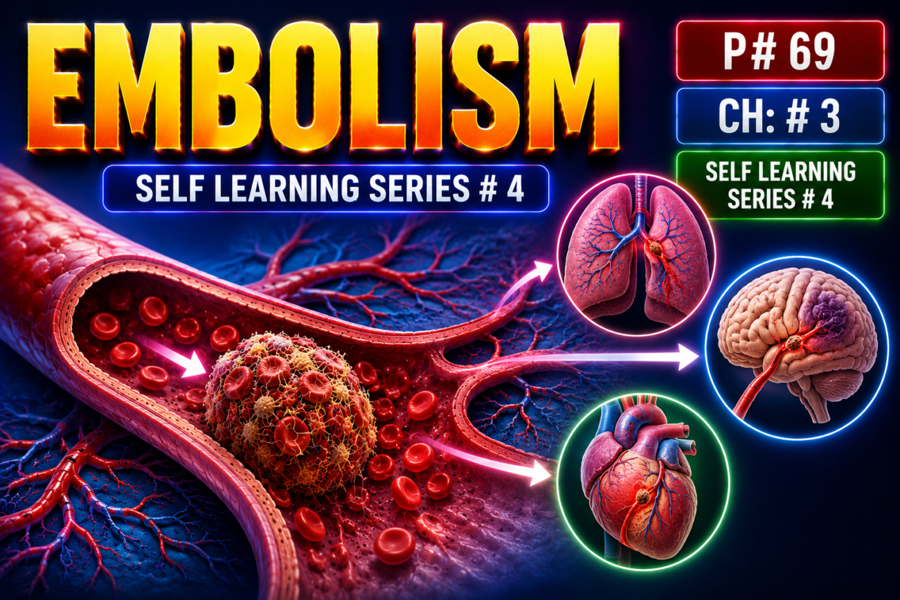
- An embolus = a detached solid, liquid, or gas inside blood vessels that travels from its origin to a distant site.
- It eventually lodges in a vessel too small to pass through → partial or complete vascular obstruction.
- This may cause tissue dysfunction or infarction.
- Most emboli come from dislodged thrombi → called thromboembolism.
- Less common emboli may consist of:
- fat droplets
- air or nitrogen bubbles
- atherosclerotic/cholesterol debris
- tumor fragments
- bone marrow
- amniotic fluid
- The final location of an embolus depends on its site of origin and direction of blood flow.
- Systemic embolism → blocks arterial blood supply → ischemia → infarction.
- Pulmonary embolism more commonly causes hypoxia, hypotension, and right-sided heart failure.
Pulmonary Thromboembolism
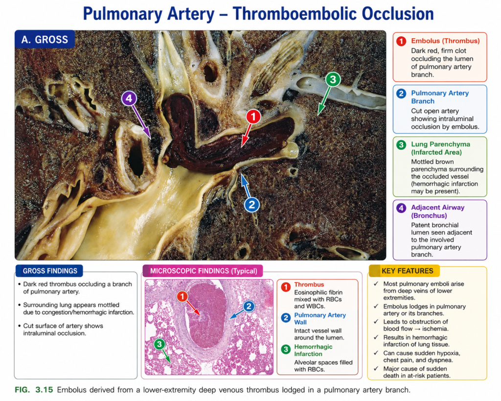
- Pulmonary emboli usually originate from deep venous thrombosis (DVT).
- More than 95% of venous emboli arise from deep leg veins proximal to the popliteal fossa.
- Therefore, risk factors for pulmonary embolism (PE) are essentially the same as those for DVT.
- A fragment from a DVT travels:
deep leg veins → larger veins → right side of heart → pulmonary arteries. - Depending on size, the embolus may:
- block the main pulmonary artery
- lodge at the bifurcation of right and left pulmonary arteries → saddle embolus
- travel into smaller pulmonary arteries/arterioles (Fig. 3.15)
- Pulmonary emboli may occur:
- one after another, or
- as multiple small emboli released from one large thrombus.
- A patient who has had one PE has an increased risk of another PE.
- Rarely, a venous embolus crosses an atrial or ventricular defect and enters systemic circulation → paradoxical embolism.
- Most pulmonary emboli (60%–80%) are small and clinically silent.
- Over time, they may organize and become incorporated into the vessel wall.
- Organization may sometimes leave fibrous webs within pulmonary vessels.
- A large embolus obstructing a major pulmonary artery → may cause sudden death.
- A medium-sized pulmonary artery embolus usually does not cause infarction because the lung has a dual blood supply, including bronchial arteries.
- However, with left-sided heart failure, reduced bronchial arterial perfusion may allow the same embolus to cause a pulmonary infarct.
- Emboli in small end-arteriolar branches usually cause infarction.
- Capillary rupture in the poorly oxygenated area may also produce hemorrhage.
- Repeated pulmonary emboli over time → pulmonary hypertension → right ventricular failure (cor pulmonale).
KEY CONCEPT
- Embolus = material traveling through blood that blocks a distant vessel.
- Most emboli are thromboemboli.
- DVT → right heart → pulmonary arteries = pulmonary embolism.
- Large PE → sudden death.
- Small peripheral PE → pulmonary infarction.
- Repeated PE → pulmonary hypertension → cor pulmonale.
- Paradoxical embolism = venous embolus enters systemic circulation through a cardiac defect.
CONCEPTUAL EXAMPLES
- Leg DVT breaks off → travels through right heart → blocks pulmonary artery → PE.
- Large embolus lodges at pulmonary artery bifurcation → saddle embolus.
- Small embolus blocks a terminal pulmonary branch → lung infarction.
- Repeated small emboli → progressively ↑ pulmonary pressure → right-sided heart failure.
Systemic Thromboembolism
- About 80% of systemic emboli arise from intracardiac mural thrombi.
- Of these:
- about 2/3 are associated with left ventricular infarction
- about 25% arise from a dilated left atrium, such as with mitral valve disease
- Other sources include:
- aortic aneurysms
- thrombi over ulcerated atherosclerotic plaques
- fragmented valvular vegetations
- venous thrombi entering systemic circulation → paradoxical emboli
- About 10%–15% have no identified source.
- Unlike venous emboli, which mainly travel to the lungs, systemic arterial emboli can travel almost anywhere.
- Their final site depends on:
- point of origin
- amount of blood flowing to downstream organs
- Common sites:
- Lower extremities → ~75%
- Central nervous system → ~10%
- Less commonly → intestines, kidneys, spleen
- The effect depends on:
- size of the blocked vessel
- presence of collateral circulation
- tissue sensitivity to lack of oxygen
- Infarction is common because arterial emboli often lodge in end arteries.
Fat Embolism
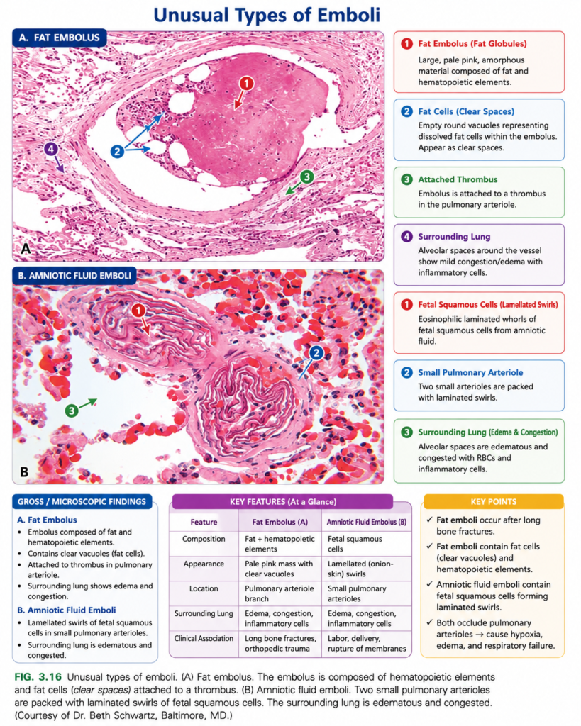
- Crush injury to soft tissue or rupture of bone-marrow vessels, especially after a long-bone fracture, can release:
- microscopic fat droplets
- marrow elements
into the circulation (Fig. 3.16A).
- Fat emboli may occur after vigorous cardiopulmonary resuscitation and are often clinically insignificant.
- A small number of patients with severe skeletal injury develop fat embolism syndrome.
- Major features:
- pulmonary insufficiency
- neurologic symptoms
- anemia
- thrombocytopenia
- diffuse petechial rash
- Symptoms usually appear 1–3 days after injury:
- rapid breathing
- dyspnea
- tachycardia
- irritability
- restlessness
- Severe cases may progress to delirium or coma.
- Death occurs in about 10% of cases.
- Thrombocytopenia occurs because platelets adhere to fat droplets → aggregate or become sequestered in the spleen.
- Anemia may result from red-cell aggregation and/or hemolysis.
- Petechial rash occurs in about 20%–50% of cases and is related to thrombocytopenia.
- Fat embolism syndrome develops through two main mechanisms:
- Mechanical obstruction → fat microemboli block pulmonary and cerebral microvessels and promote platelet aggregation.
- Biochemical injury → fatty acids released from fat droplets damage endothelium.
- Platelet activation and granulocyte recruitment further damage vessels through release of:
- free radicals
- proteases
- eicosanoids
- Routine tissue processing dissolves fat, so demonstrating isolated fat droplets microscopically requires special fat stains on frozen sections.
KEY CONCEPT
- Systemic embolus → usually from left-sided cardiac mural thrombus → travels to systemic arteries → commonly causes infarction.
- Fat embolism → usually follows severe skeletal injury or long-bone fracture.
- Fat embolism syndrome = respiratory distress + neurologic symptoms + anemia + thrombocytopenia + petechial rash.
- Mechanism = microvascular obstruction + fatty-acid–induced endothelial injury.
CONCEPTUAL EXAMPLES
- Left ventricular infarction → mural thrombus forms → fragment breaks off → travels to leg artery → systemic embolism and infarction.
- Long-bone fracture → marrow fat enters blood → reaches lung and brain microvessels → fat embolism syndrome.
- Fat droplets activate platelets → platelet count falls → thrombocytopenia + petechiae.
Amniotic Fluid Embolism
- Amniotic fluid embolism is a rare but very serious complication of labor or the immediate postpartum period.
- It occurs when amniotic fluid and fetal material enter the maternal circulation through tears in:
- placental membranes
- uterine veins
- It occurs in about 1 in 40,000 deliveries, but mortality approaches 80%.
- It causes about 5%–10% of maternal deaths in the United States.
- About 85% of survivors have some permanent neurologic deficit.
- Typical sudden presentation:
severe dyspnea → cyanosis → shock → seizures → coma. - If the patient survives the initial crisis:
- pulmonary edema usually develops.
- about half develop disseminated intravascular coagulation (DIC).
- DIC occurs because amniotic fluid contains thrombogenic substances that activate coagulation.
- The major injury is thought to result mainly from:
- activation of the coagulation system
- activation of the innate immune system
- Therefore, the severe effects are not mainly due to simple mechanical blockage of pulmonary vessels.
- In fatal cases, maternal pulmonary microcirculation may contain fetal material (Fig. 3.16B), including:
- fetal squamous cells
- lanugo hair
- fat from vernix caseosa
- fetal mucin
- Other pathologic findings include:
- marked pulmonary edema
- diffuse alveolar damage
- widespread fibrin thrombi caused by DIC
KEY CONCEPT
- Labor/postpartum → amniotic fluid enters maternal blood → coagulation + innate immune activation → sudden respiratory failure, shock, neurologic symptoms, and often DIC.
- Classic sequence:
sudden dyspnea + cyanosis + shock → seizures/coma → pulmonary edema ± DIC.
CONCEPTUAL EXAMPLES
- Tear in placental membranes → amniotic fluid enters maternal veins → sudden dyspnea and shock.
- Amniotic fluid activates coagulation → widespread fibrin formation → DIC.
- Fetal squamous cells, lanugo, fat, and mucin found in maternal lung vessels → supports amniotic fluid embolism.
Air Embolism
- Air/gas embolism occurs when gas bubbles enter the circulation, join together, and block blood flow → distal ischemia.
- Even a small amount of air can be dangerous if it enters:
- a coronary artery during bypass surgery → severe cardiac ischemia
- a cerebral artery during neurosurgery → severe brain ischemia
- Small venous gas emboli usually cause little harm.
- Larger amounts of venous air may enter during:
- obstetric procedures
- laparoscopic procedures
- chest-wall injury
- This air can reach pulmonary vessels → hypoxia.
- A very large venous gas embolus may become trapped in the heart → death.
- Decompression sickness is a special form of gas embolism caused by a rapid fall in atmospheric pressure.
- It can occur in:
- scuba divers
- underwater construction workers
- people in unpressurized aircraft after rapid ascent
- At high pressure, such as during deep diving:
↑ pressure → more nitrogen dissolves in blood and tissues. - If pressure falls too quickly:
dissolved nitrogen → expands → forms bubbles → gas emboli → tissue ischemia. - Nitrogen bubbles in muscles and tissues around joints cause severe pain called “the bends.”
- Gas bubbles in pulmonary vessels → pulmonary edema + hemorrhage + focal atelectasis/emphysema → respiratory distress.
- Gas bubbles in the central nervous system may cause:
- memory loss
- ataxia
- visual disturbances
- sudden coma
- Caisson disease is the chronic form of decompression sickness.
- Recurrent or persistent gas emboli in bones → multiple areas of ischemic bone necrosis.
- Commonly affected sites include the heads of the:
- femurs
- tibiae
- humeri
- Acute decompression sickness is treated by placing the patient in a high-pressure chamber → gas is forced back into solution.
- Then slow decompression allows gradual gas absorption and exhalation → prevents bubbles from forming again.
KEY CONCEPT
- Gas bubble → vessel obstruction → ischemia.
- Rapid decompression → dissolved nitrogen forms bubbles → decompression sickness.
- Muscle/joint bubbles → bends.
- Lung bubbles → respiratory distress.
- Brain bubbles → neurologic dysfunction/coma.
- Chronic bone involvement → caisson disease.
- Treatment → recompression followed by slow decompression.
CONCEPTUAL EXAMPLES
- Deep-sea diver ascends too quickly → nitrogen comes out of solution → gas emboli.
- Nitrogen bubbles around joints → severe pain → the bends.
- Gas bubbles enter pulmonary vessels → edema and hemorrhage → respiratory distress.
- Repeated decompression injury in bones → ischemic necrosis → caisson disease.
