- Thrombosis = formation of a clot inside the vascular system.
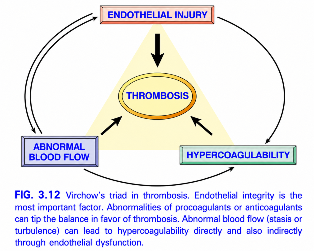
- The three major causes are the Virchow triad (Fig. 3.12):
- Endothelial injury
- Stasis or turbulent blood flow
- Hypercoagulability of blood
- Thrombosis is important because it underlies many serious and common cardiovascular diseases.
Endothelial Injury
- Endothelial injury → platelet activation → thrombus formation, especially in the heart and arteries.
- In arteries, rapid blood flow normally makes clot formation difficult.
- Therefore, platelet adhesion and activation are especially important for thrombus formation under high shear stress.
- Cardiac and arterial thrombi are therefore usually platelet-rich.
- This explains why aspirin and other platelet inhibitors are used in:
- coronary artery disease
- acute myocardial infarction
- Severe endothelial injury promotes thrombosis by exposing:
- vWF → promotes platelet adhesion
- tissue factor → activates coagulation
- Endothelium may also become activated or dysfunctional without major physical disruption.
- This changes endothelial activity from normally antithrombotic toward a prothrombotic state.
- Causes of endothelial activation/dysfunction include:
- physical injury
- infections
- abnormal blood flow
- cytokines and inflammatory mediators
- hypercholesterolemia
- homocystinemia
- toxins from cigarette smoke
- Endothelial activation is particularly important in arterial thrombosis.
- Procoagulant changes:
- Activated endothelium decreases normal coagulation inhibitors:
- thrombomodulin
- endothelial protein C receptor
- tissue factor pathway inhibitor
- It also increases tissue factor.
- Result → more coagulation.
- Activated endothelium decreases normal coagulation inhibitors:
- Antifibrinolytic effects:
- Activated endothelium increases plasminogen activator inhibitors (PAI).
- PAI inhibits t-PA and urokinase.
- Therefore → less fibrinolysis → clot persists more easily.
KEY CONCEPT
- Virchow triad = endothelial injury + abnormal blood flow + hypercoagulability.
- In arteries, endothelial injury and platelet activation are especially important.
- Activated endothelium becomes prothrombotic by:
↑ tissue factor + ↓ anticoagulant factors + ↑ PAI → ↓ fibrinolysis.
CONCEPTUAL EXAMPLES
- Arterial endothelial damage → vWF exposed → platelets adhere and activate → platelet-rich thrombus.
- Endothelial activation → ↓ thrombomodulin + ↑ tissue factor → coagulation increases.
- ↑ PAI → t-PA is inhibited → fibrin is removed less effectively → clot remains and can enlarge.
Abnormal Blood Flow
- Turbulence = chaotic blood flow → promotes arterial and cardiac thrombosis by:
- causing endothelial injury/dysfunction
- creating countercurrents and local areas of stasis
- Stasis = slowed or stopped blood flow → especially important in venous thrombosis.
- Normally, laminar blood flow keeps platelets and other blood cells mainly in the center of the vessel, separated from endothelium by a slower-moving plasma layer.
- Stasis and turbulence promote thrombosis because they:
- activate endothelium → ↑ procoagulant activity
- allow platelets and leukocytes to contact endothelium
- reduce washout of activated clotting factors
- reduce inflow of coagulation inhibitors
- Important clinical settings:
- Ulcerated atherosclerotic plaque → exposes subendothelial ECM + produces turbulence → thrombosis.
- Aneurysm → abnormal arterial dilation → local stasis → thrombus formation.
- Acute myocardial infarction → noncontracting myocardium → local stasis → mural thrombus.
- Previous infarction may cause ventricular aneurysm → more stasis → mural thrombus.
- Mitral stenosis → left atrial dilation.
- Left atrial dilation + atrial fibrillation → stasis + turbulence → thrombus formation.
- Hyperviscosity, such as polycythemia vera → ↑ resistance to flow → small-vessel stasis → thrombosis.
Hypercoagulability
- Hypercoagulability = abnormally increased tendency of blood to clot.
- It usually results from abnormalities in coagulation factors.
- It is especially important in venous thrombosis, but may also contribute to arterial or intracardiac thrombosis.
- Causes are divided into primary (genetic) and secondary (acquired) disorders (Table 3.2).
- Primary inherited hypercoagulability is most commonly caused by mutations involving factor V or prothrombin.
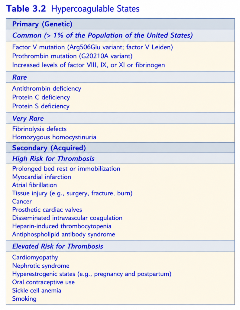
- Factor V Leiden mutation:
- Factor V becomes resistant to breakdown by protein C.
- Therefore, an important anticoagulant mechanism is lost.
- Heterozygotes → about 3–4× increased risk of venous thrombosis.
- Homozygotes → about 25–50× increased risk.
- It is common among patients with recurrent DVT.
- Prothrombin gene variant:
- Causes ↑ prothrombin production.
- Associated with about 3× increased risk of venous thrombosis.
- Less common inherited causes include deficiency of:
- antithrombin
- protein C
- protein S
- These patients often develop venous thrombosis and recurrent thromboembolism at a young age.
- Markedly ↑ homocysteine, such as with inherited cystathionine β-synthetase deficiency → increased risk of arterial and venous thrombosis.
- More modest elevations of homocysteine may also increase venous thromboembolism risk.
- Genetic risk becomes more important when combined with acquired risks such as:
- pregnancy
- prolonged bed rest
- long airplane flights
- Therefore, inherited hypercoagulability should be considered especially in patients younger than 50 years with thrombosis.
- Most patients with DVT have no known genetic abnormality, and most people with factor V Leiden never develop DVT.
- Genetic testing is therefore mainly considered in:
- strong family history of DVT
- DVT at a young age without an obvious acquired risk factor
- Secondary acquired hypercoagulability occurs in many conditions.
- Important mechanisms include:
- Trauma or cardiac failure → vascular injury or stasis.
- Oral contraceptives and pregnancy → ↑ hepatic coagulation-factor synthesis + ↓ antithrombin synthesis.
- Disseminated cancer → tumor products such as mucin promote coagulation.
- Increasing age → ↑ platelet aggregation + ↓ endothelial PGI₂.
- Smoking and obesity → promote hypercoagulability by uncertain mechanisms.
- Heparin-induced thrombocytopenia (HIT):
- May occur after unfractionated heparin treatment.
- Autoantibodies form against heparin–platelet factor 4 (PF4) complexes.
- Antibody complexes bind platelet Fc receptors → platelet activation + aggregation + removal from blood.
- Result → low platelets but increased thrombosis.
- Low-molecular-weight heparin causes this less often but can still cause thrombosis.
- Antiphospholipid antibody syndrome may cause:
- recurrent thrombosis
- repeated miscarriages
- cardiac valve vegetations
- thrombocytopenia
- Depending on the vessels involved, it may produce:
- DVT → pulmonary embolism
- recurrent pulmonary emboli → pulmonary hypertension
- stroke
- bowel infarction
- renovascular hypertension
- renal microangiopathy with renal failure
- Fetal loss appears to result mainly from antibody interference with trophoblast growth and placentation, rather than simply thrombosis.
- The name “antiphospholipid antibody” is somewhat misleading.
- The antibodies probably act against proteins associated with phospholipids, especially β₂-glycoprotein I.
- In the body, these antibodies promote a hypercoagulable state.
- In laboratory tests, however, they interfere with phospholipid-dependent clotting assays and therefore behave like “anticoagulants.”
- They may also cause a false-positive syphilis test because the test antigen contains the phospholipid cardiolipin.
- Antiphospholipid antibody syndrome may be:
- Secondary → associated with an autoimmune disease such as systemic lupus erythematosus.
- Primary → hypercoagulability occurs without another recognized autoimmune disease.
- Treatment involves anticoagulation and immunosuppression.
- Antiphospholipid antibodies may also occur in healthy people, so their presence alone is not sufficient to produce the full syndrome.
KEY CONCEPT
- Turbulence → mainly arterial/cardiac thrombosis.
- Stasis → mainly venous thrombosis.
- Abnormal flow promotes clotting by:
endothelial activation + platelet contact + retained clotting factors + reduced anticoagulant inflow. - Hypercoagulability = excessive tendency to clot.
- Major inherited causes → Factor V Leiden + prothrombin mutation.
- Important acquired causes → pregnancy, oral contraceptives, cancer, age, smoking, obesity, HIT, antiphospholipid syndrome.
- HIT = thrombocytopenia + paradoxical thrombosis.
- Antiphospholipid syndrome = thrombosis despite laboratory “anticoagulant” activity.
CONCEPTUAL EXAMPLES
- Atrial fibrillation → chaotic flow + atrial stasis → atrial thrombus.
- Aneurysm → blood stagnates inside dilated vessel → thrombus forms.
- Factor V Leiden → protein C cannot inactivate factor V normally → clotting persists longer.
- Heparin → anti-PF4 antibodies activate platelets → platelets fall but thrombosis increases.
- Antiphospholipid antibodies → hypercoagulable state → recurrent thrombosis or pregnancy loss.
MORPHOLOGY
- Thrombi can form anywhere in the cardiovascular system.
- Arterial/cardiac thrombi usually form where there is:
- endothelial injury, or
- turbulent blood flow.
- Venous thrombi usually form where there is stasis.
- Thrombi are attached to the vessel wall and tend to propagate toward the heart:
- Arterial thrombi → grow backward (retrograde) from the attachment point.
- Venous thrombi → grow in the direction of blood flow.
- The growing end of a thrombus is often poorly attached → may break off → travel in blood as an embolus.
- Thrombi may show visible layers called lines of Zahn:
- pale layers = platelets + fibrin
- dark layers = red blood cells
- Lines of Zahn form only in flowing blood → therefore indicate antemortem thrombosis.
- Postmortem clots are usually nonlaminated, although venous thrombi may sometimes resemble them.
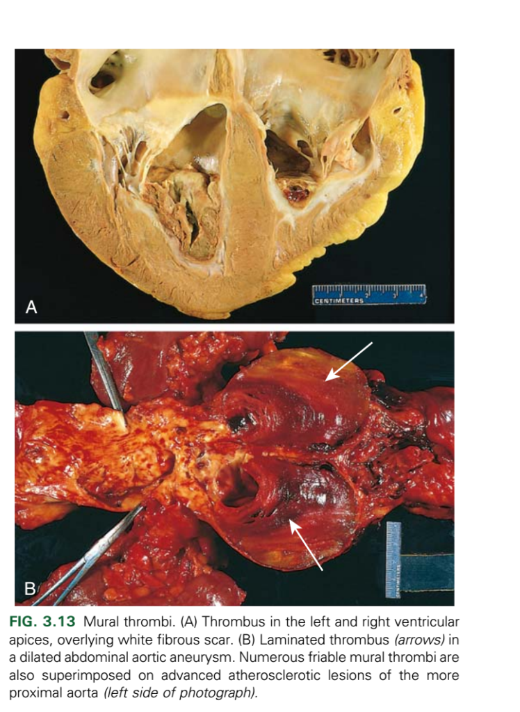
- Thrombi inside heart chambers or the aorta are called mural thrombi.
- Cardiac mural thrombi are promoted by:
- arrhythmias
- dilated cardiomyopathy
- myocardial infarction
- myocarditis
- catheter injury (Fig. 3.13A).
- Ulcerated atherosclerotic plaques and aneurysmal dilation promote aortic thrombosis (Fig. 3.13B).
- Arterial thrombi:
- are frequently occlusive.
- are usually platelet-rich because endothelial injury activates platelets.
- commonly form over a ruptured atherosclerotic plaque.
- may also follow vasculitis or trauma.
- Venous thrombi (phlebothrombosis):
- are almost always occlusive.
- often extend toward the heart as a long cast of the vessel.
- are prone to producing emboli.
- contain many trapped red cells because they form in sluggish blood → called red or stasis thrombi.
- About 90% of venous thromboses occur in the lower-extremity veins.
- They may also occur in upper-extremity veins, periprostatic plexus, ovarian and periuterine veins.
- In hypercoagulable states, thrombi may also occur in dural sinuses, portal vein, or hepatic vein.
- Postmortem clots may resemble venous thrombi but:
- are gelatinous
- are not attached to the vessel wall
- have a dark red dependent part from settled RBCs
- have a yellow upper “chicken fat” portion.
- In contrast, true red thrombi are:
- firm
- focally attached to the vessel wall
- contain gray strands of fibrin.
KEY CONCEPT
- Arterial/cardiac thrombus → injury or turbulence → platelet-rich.
- Venous thrombus → stasis → RBC-rich (“red thrombus”).
- Lines of Zahn → clot formed during life in flowing blood.
- Poorly attached thrombus tail → may detach → embolus.
- Postmortem clot = gelatinous + unattached; true thrombus = firm + attached.
CONCEPTUAL EXAMPLES
- Ruptured atherosclerotic plaque → platelet activation → arterial thrombus.
- Slow venous blood in the leg → RBC-rich clot → venous/stasis thrombus.
- Part of a venous thrombus breaks off → travels through blood → embolus.
- Firm attached clot with lines of Zahn → formed before death.
- Gelatinous unattached “chicken-fat” clot → formed after death.
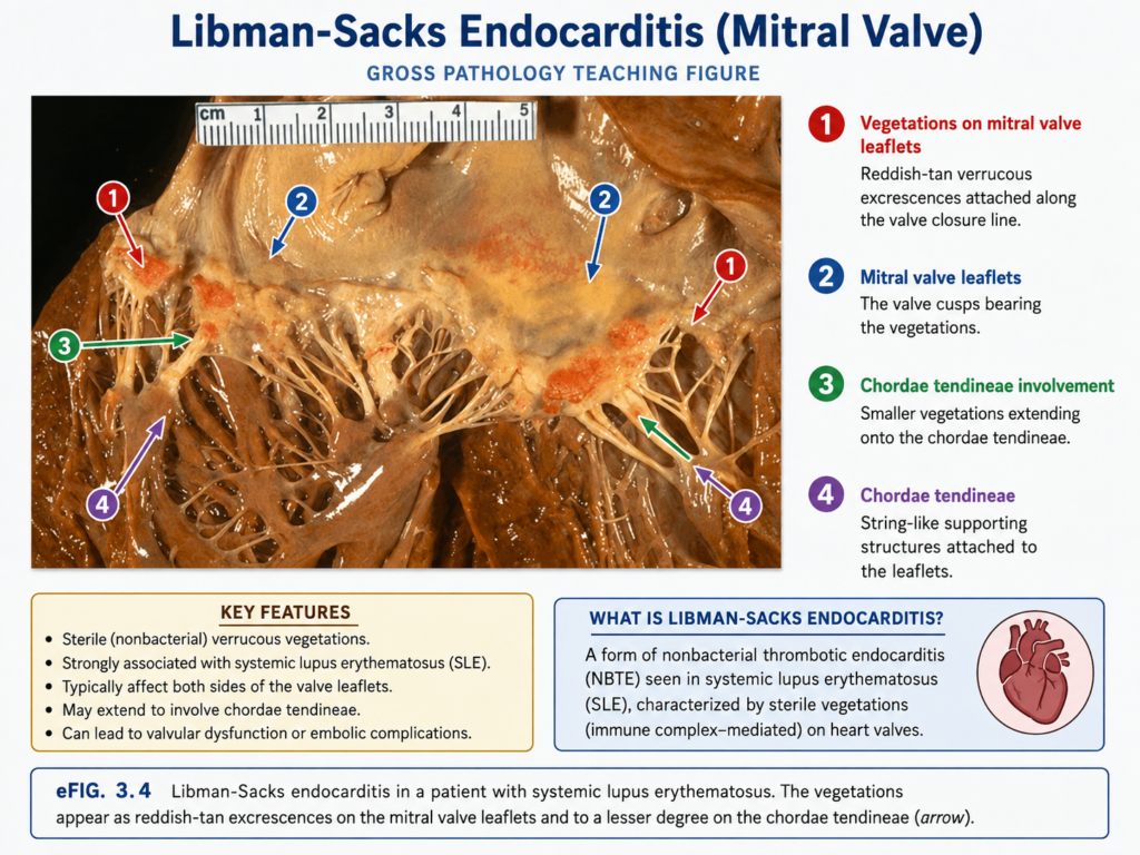
- Thrombi that form on heart valves are called vegetations.
- Bacterial or fungal bloodstream infection → valve damage → large thrombotic vegetations → infective endocarditis.
- Vegetations can also occur without infection.
- In hypercoagulable states:
- sterile vegetations may form on normal, noninfected valves.
- This is called nonbacterial thrombotic endocarditis.
- Less commonly, systemic lupus erythematosus (SLE) may cause sterile verrucous endocarditis (Libman-Sacks endocarditis) (eFig. 3.4).
KEY CONCEPT
- Valve thrombus = vegetation.
- Infection → infective endocarditis.
- Hypercoagulability → sterile nonbacterial thrombotic endocarditis.
- SLE → sterile Libman-Sacks endocarditis.
CONCEPTUAL EXAMPLES
- Bacteria in blood → valve injury + thrombotic mass → infective endocarditis.
- Hypercoagulable state + no infection → sterile valve vegetation → nonbacterial thrombotic endocarditis.
- SLE → sterile verrucous valve vegetations → Libman-Sacks endocarditis.
Fates of Thrombi
- If a patient survives the initial thrombotic event, the thrombus can undergo four possible changes over days to weeks:
- Propagation:
- More platelets + fibrin accumulate on the thrombus.
- Thrombus enlarges → ↑ chance of vessel occlusion and embolization.
- Embolization:
- Part or all of the thrombus breaks free.
- It travels through the circulation to another site.
- Dissolution:
- A newly formed thrombus can be rapidly broken down by activation of fibrinolytic factors.
- It may shrink or completely disappear.
- As a thrombus becomes older → extensive fibrin polymerization makes it increasingly resistant to plasmin-mediated breakdown.
- Therefore, fibrinolytic drugs such as t-PA are generally most effective when given within the first few hours of acute coronary thrombosis.
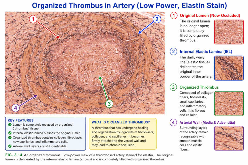
- Organization and recanalization:
- Older thrombi are invaded by:
- endothelial cells
- smooth muscle cells
- fibroblasts
- This process is called organization (Fig. 3.14).
- Small capillary channels then develop through the thrombus.
- These channels partially restore blood flow through the original vessel → recanalization.
- Further organization may convert the thrombus into vascularized connective tissue that becomes incorporated into the vessel wall.
- Occasionally, instead of organization, the center of the thrombus undergoes enzymatic digestion, probably from lysosomal enzymes released by trapped leukocytes.
- Older thrombi are invaded by:
KEY CONCEPT
- Remember the 4 fates of a thrombus:
Propagation → Embolization → Dissolution → Organization/Recanalization
- Propagation = clot gets bigger.
- Embolization = clot breaks off and travels.
- Dissolution = fresh clot is broken down.
- Organization/recanalization = old clot becomes incorporated into tissue and develops new channels for blood flow.
- Fresh thrombi respond better to fibrinolysis; older thrombi become resistant.
CONCEPTUAL EXAMPLES
- More fibrin and platelets collect → thrombus enlarges → propagation.
- Piece of thrombus breaks off → travels elsewhere → embolization.
- Fresh coronary thrombus + early t-PA → clot may dissolve → dissolution.
- Old thrombus → cells grow into it → small channels form through it → organization + recanalization.
Clinical Features
- Thrombi are dangerous mainly because they:
- obstruct arteries or veins
- may break off and form emboli
- Clinical effect depends mainly on the site of thrombosis.
- Venous thrombi:
- obstruct venous return → congestion + edema
- greatest danger → embolize to the lungs → potentially fatal pulmonary embolism
- Arterial thrombi:
- may embolize, but more importantly they block local blood flow
- obstruction of coronary or cerebral arteries → infarction
- Most venous thrombi occur in superficial or deep veins of the legs.
- Superficial venous thrombosis:
- commonly occurs in the saphenous veins, especially with varicose veins
- rarely embolizes
- may cause pain + local congestion + swelling
- impaired venous drainage may predispose to skin infection and varicose ulcers
- Deep venous thrombosis (DVT):
- commonly involves large veins at or above the knee:
- popliteal
- femoral
- iliac veins
- more serious because it can embolize to the lungs
- may cause pain and edema, but collateral veins can bypass the obstruction
- therefore, about 50% of DVTs may be asymptomatic until pulmonary embolization occurs
- commonly involves large veins at or above the knee:
- Lower-limb DVT is strongly associated with stasis + hypercoagulability (Table 3.2).
- Common predisposing factors:
- Congestive heart failure → venous stasis
- Bed rest/immobilization → ↓ leg-muscle pumping → ↓ venous return
- Trauma, surgery, burns → immobilization + vascular injury + increased procoagulant activity + ↓ t-PA
- Pregnancy → enlarged uterus/fetus compresses leg veins → stasis; late pregnancy and postpartum hormonal changes → hypercoagulability
- Disseminated cancer → tumor procoagulants → increased thrombosis
- Cancer-associated recurrent thrombosis in different venous sites is called migratory thrombophlebitis or Trousseau syndrome.
- DVT risk increases after 50 years of age and is greater in males than females.
- Atherosclerosis promotes arterial thrombosis through endothelial injury and abnormal blood flow (Fig. 3.13B).
- Myocardial infarction may cause abnormal ventricular contraction + endocardial injury → mural thrombus (Fig. 3.13A).
- Rheumatic heart disease may cause atrial dilation + atrial fibrillation → stasis → mural thrombus.
- Cardiac and aortic mural thrombi may embolize, especially to organs with rich blood flow:
- brain
- kidneys
- spleen
Disseminated Intravascular Coagulation (DIC)
- DIC = widespread formation of thrombi throughout the microcirculation.
- It may develop suddenly or gradually.
- It can occur in conditions ranging from obstetric complications to advanced malignancy.
- Widespread clotting consumes:
- platelets
- coagulation factors
- Therefore, DIC is also called consumptive coagulopathy.
- At the same time, fibrinolysis is activated.
- Final result:
widespread clotting + consumption of clotting components + fibrinolysis → thrombosis and bleeding can occur together.
KEY CONCEPT
- Venous thrombus → major danger = pulmonary embolism.
- Arterial thrombus → major danger = local obstruction → infarction.
- DVT may be silent in about 50% of patients.
- DVT is promoted mainly by stasis + hypercoagulability.
- Cancer-associated migratory thrombosis = Trousseau syndrome.
- DIC = widespread microthrombi + consumption of platelets/coagulation factors → simultaneous clotting and bleeding.
CONCEPTUAL EXAMPLES
- Bed rest → leg muscles stop pumping effectively → venous stasis → DVT.
- DVT breaks off → travels to lungs → pulmonary embolism.
- Coronary arterial thrombus → blocks blood flow → myocardial infarction.
- Cancer releases procoagulants → repeated thrombosis in different veins → Trousseau syndrome.
- DIC → many tiny clots use up platelets and clotting factors → patient can clot and bleed at the same time.