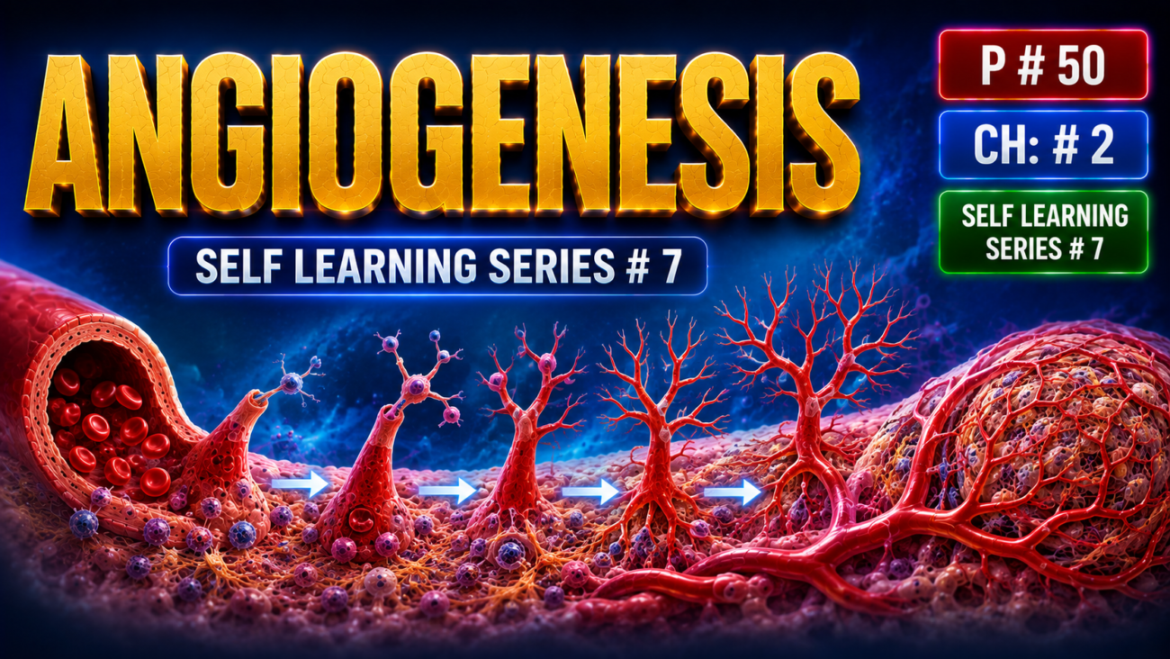
- Angiogenesis means formation of new blood vessels from existing vessels.
- It is important for:
- Healing at sites of injury
- Formation of collateral circulation in ischemic tissues
- Tumor growth
- Angiogenesis may be therapeutically:
- Increased to improve blood flow in ischemic heart disease
- Inhibited to reduce tumor growth or abnormal vessels, such as in wet macular degeneration
- New vessels develop by sprouting from existing vessels.
- Many factors regulate angiogenesis, but the most important is vascular endothelial growth factor (VEGF).
- VEGF → endothelial cell migration + proliferation
- At an injured site:Hypoxia → ↑ HIF → ↑ VEGF production
- HIF (hypoxia-inducible factor) is a transcription factor that increases VEGF production during hypoxia.
- VEGF causes nearby intact vessels to:
- Dilate
- Become more permeable
- Matrix metalloproteinases digest the basement membrane, allowing a new vascular sprout to form.
- Endothelial cells at the tip of the sprout migrate toward the injured tissue.
- Endothelial cells behind the tip proliferate and organize into vascular tubes.
- Pericytes are recruited around capillaries, while smooth muscle cells are recruited around larger vessels, producing a mature blood vessel.
- As healing continues, many newly formed vessels regress.
- Therefore, highly vascular granulation tissue gradually becomes a pale, largely avascular scar.
KEY CONCEPT
Injury → hypoxia → HIF ↑ → VEGF ↑ → endothelial migration + proliferation → new vascular sprouts → vascular tubes → mature vessels
Granulation tissue → vessel regression → pale scar
CONCEPTUAL EXAMPLES
- Injured tissue becomes hypoxic → VEGF increases → new vessels grow toward the wound
- Early granulation tissue → many vessels → pink/red appearance → healing progresses → vessels decrease → pale scar
Activation of Fibroblasts and Deposition of Connective Tissue
- Connective tissue deposition occurs in two main steps (Fig. 2.22):
- Fibroblasts migrate into the injured area and proliferate
- Fibroblasts produce and deposit ECM proteins
- These processes are stimulated by locally produced cytokines and growth factors, especially:
- PDGF
- FGF-2
- TGF-β
- Their major sources are inflammatory cells, especially macrophages at the injury site.
- Scar tissue mainly consists of connective tissue.
- Connective tissue contains:
- Fibroblasts
- Extracellular matrix (ECM)
- ECM mainly contains collagen and other glycoproteins.
- Cell–ECM interactions are essential for healing, normal tissue development, and maintenance of tissue architecture (Fig. 2.23).
- Major functions of ECM include:
- Providing mechanical support for cell attachment and migration
- Maintaining cell polarity
- Regulating cell proliferation
- Binding and storing growth factors
- Providing a scaffold for tissue regeneration
- An intact basement membrane or stromal scaffold is therefore essential for normal tissue regeneration.
- ECM exists in two major forms:
- Interstitial matrix → found between connective tissue cells and between epithelium and underlying supporting tissues.
- It contains mainly collagens, fibronectin, elastin, proteoglycans, and hyaluronate.
- Basement membrane → highly organized ECM surrounding epithelial, endothelial, and smooth muscle cells.
- Its major components are type IV collagen and laminin.
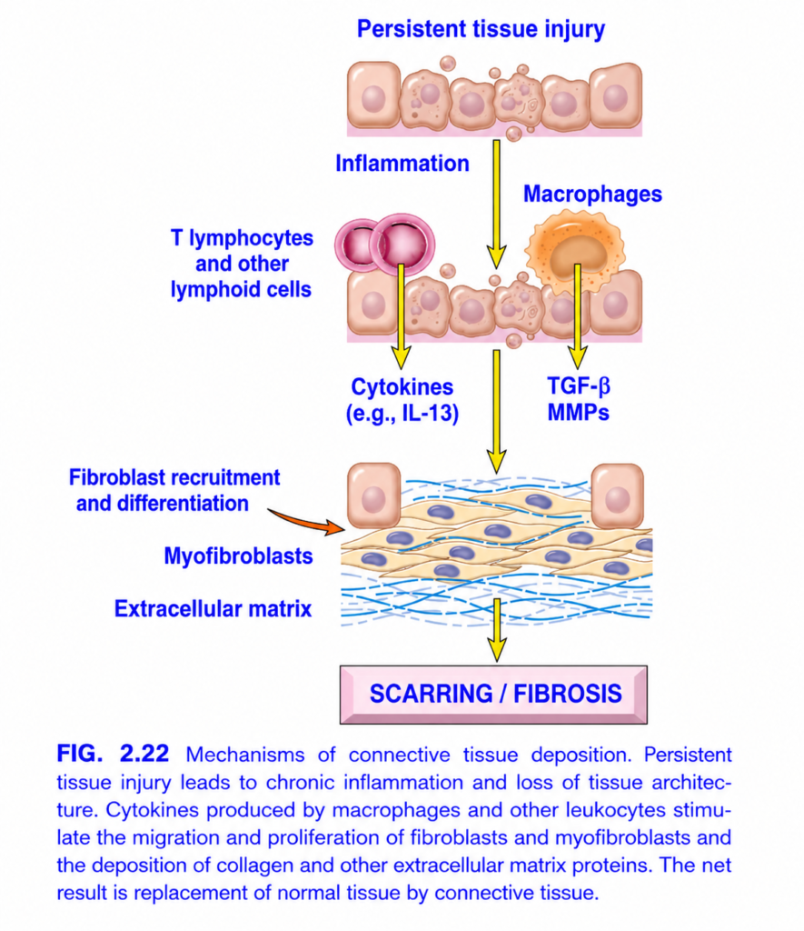
FIG. 2.22 — How Persistent Injury Causes Fibrosis
Think of the whole figure as one simple chain:
Persistent injury → chronic inflammation → fibroblasts activated → collagen/ECM deposited → SCAR / FIBROSIS
🔝 1. Persistent tissue injury
🟧 Peach cells = tissue cells.
Some are damaged and breaking apart.
⬇️ Yellow arrow = persistent damage keeps stimulating inflammation.
🔥 2. Inflammation
Two important inflammatory cells appear:
- 🩷 Pink round cells = T lymphocytes
- 🟠 Large orange cell = macrophage
These cells release signals that tell the tissue to start repair.
🩷 T lymphocytes → Cytokines
⬇️ Yellow arrow
T cells release cytokines, e.g. IL-13.
IL-13 → attracts/activates fibroblasts → promotes fibrosis.
🟠 Macrophage → TGF-β + MMPs
⬇️ Yellow arrow
TGF-β = MAIN fibrosis signal.
It tells fibroblasts:
“Multiply and make collagen.”
MMPs = enzymes that break down/remodel old extracellular matrix, allowing the scar tissue to be reorganized.
🟧 Orange arrow — Fibroblast recruitment & differentiation
Fibroblasts are brought into the damaged area and become activated.
➡️ Some become myofibroblasts.
Myofibroblast = fibroblast that makes lots of collagen + can contract the wound.
🟤 Beige spindle-shaped cells
= Fibroblasts / myofibroblasts
They produce the material needed for the scar.
🔵 Blue wavy lines
= Extracellular matrix (ECM), especially collagen
More fibroblast activity
→ more collagen/ECM
→ normal tissue gets replaced by fibrous tissue.
⬇️ Final yellow arrow
➡️ SCARRING / FIBROSIS
Fibrosis = excessive connective tissue/collagen replacing normal tissue.
🧠 Fastest exam memory
Persistent injury → Macrophage → TGF-β → Fibroblast → Collagen → FIBROSIS
⭐ Most important mediator of fibrosis = TGF-β.
Collagen
- Collagen is the major ECM protein of the interstitial matrix and scar tissue.
- More than 30 collagen types exist, and each collagen molecule contains three polypeptide chains arranged in a triple helix.
- Important fibrillar collagens are types I, II, III, and V.
- They are important in scar tissue, tendons, bone, and skin.
- Collagen gains tensile strength by cross-linking of its triple helices.
- This cross-linking requires vitamin C as a cofactor.
- Therefore:Vitamin C deficiency → defective collagen → poor wound healing + easy bleeding
Elastin
- Elastin allows tissues to stretch and return to their original shape.
- It is especially important in:
- Cardiac valves
- Large blood vessels
- Uterus
- Skin
- Ligaments
Proteoglycans and Hyaluronan
- Proteoglycans form highly hydrated gels that resist compressive forces.
- In joint cartilage, they also provide lubrication between bony surfaces.
- They can store growth factors such as FGF and HGF.
- Some proteoglycans also help regulate cell proliferation, migration, and adhesion.
Adhesive Glycoproteins and Adhesion Receptors
- These molecules help interactions between:
- Cell ↔ cell
- Cell ↔ ECM
- ECM ↔ ECM
- Two important adhesive glycoproteins are:
- Fibronectin → major component of interstitial ECM
- Laminin → major component of basement membrane
- Fibronectin is produced by fibroblasts, monocytes, endothelial cells, and other cells.
- In healing wounds, fibronectin provides a scaffold for:
- Further ECM deposition
- Angiogenesis
- Reepithelialization
- Laminin is the most abundant glycoprotein of the basement membrane.
- It helps in:
- Cell attachment
- Cell proliferation
- Differentiation
- Cell movement
- Cells attach to laminin and fibronectin through integrins (see Table 2.3).
- Integrins link the intracellular cytoskeleton to the ECM and can also mediate cell–cell adhesion.
- Integrin signaling influences:
- Cell migration
- Proliferation
- Shape
- Differentiation
- During scar formation, fibroblasts migrate from the wound edges toward the center in response to growth factors.
- Some fibroblasts become myofibroblasts.
- Myofibroblasts contain smooth muscle actin and contract strongly.
- Therefore:Myofibroblast contraction → wound margins pulled toward center → wound closes
- Activated fibroblasts and myofibroblasts produce connective tissue proteins, especially collagen and other ECM proteins.
- TGF-β is the most important cytokine for connective tissue synthesis and deposition.
- TGF-β is produced by many cells in granulation tissue, including activated macrophages.
- TGF-β causes:
- ↑ Fibroblast migration
- ↑ Fibroblast proliferation
- ↑ Collagen synthesis
- ↑ Fibronectin synthesis
- ↓ ECM degradation by inhibiting metalloproteinases
- Therefore:TGF-β → more ECM production + less ECM breakdown → scar formation
- TGF-β also contributes to fibrosis of the lung, liver, and kidneys during chronic inflammation.
- It also has an anti-inflammatory effect by inhibiting lymphocyte proliferation and reducing the activity of other leukocytes.
- As healing progresses, fibroblasts produce progressively more ECM.
- Collagen synthesis begins early, around days 3–5, and may continue for several weeks.
- Increasing collagen strengthens the healing wound.
- Final collagen accumulation depends on:
- Increased collagen synthesis
- Decreased collagen degradation
KEY CONCEPT
Macrophages → PDGF + FGF-2 + TGF-β → fibroblast migration/proliferation → collagen + ECM deposition → scar
TGF-β = major cytokine for fibrosis and scar formation
Fibroblast → myofibroblast → contraction → wound edges pulled together
Vitamin C → collagen cross-linking → strong wound
CONCEPTUAL EXAMPLES
- Macrophages enter wound → release growth factors → fibroblasts enter → collagen deposited → scar forms
- Myofibroblasts contract → wound edges move toward each other → wound becomes smaller
- Vitamin C deficiency → weak collagen → poor wound healing
- Chronic inflammation → persistent TGF-β activity → excess collagen deposition → fibrosis
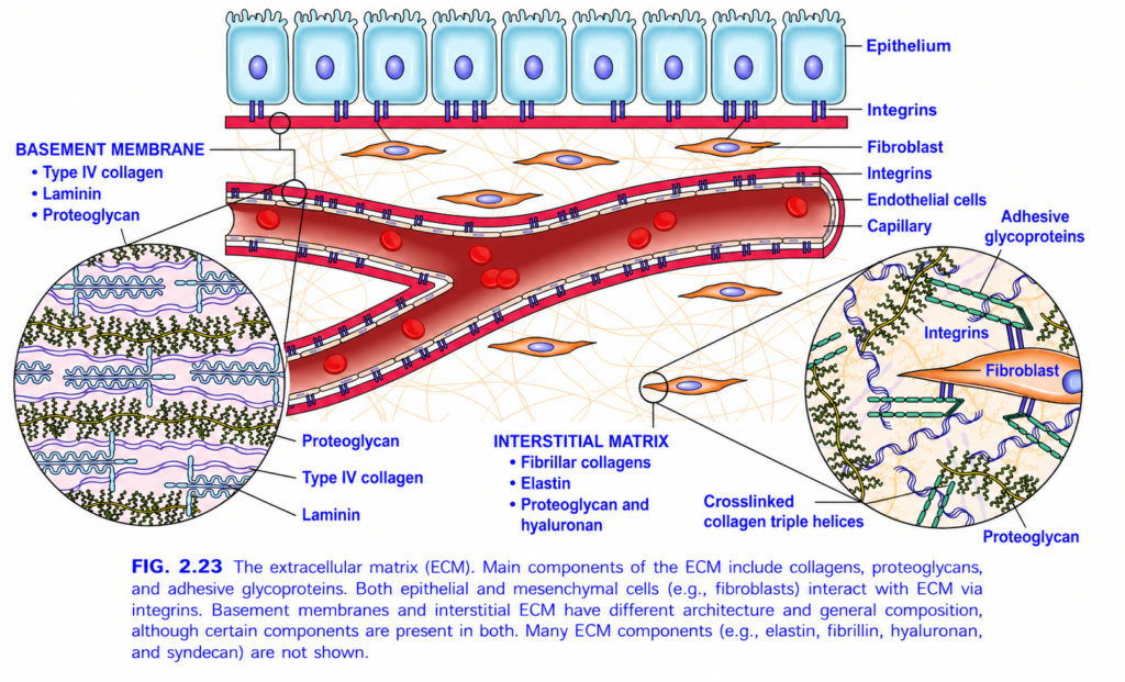
FIG. 2.23 — Extracellular Matrix (ECM)
Easiest concept: ECM = the supporting “scaffold” around cells
The whole image shows 2 types of ECM:
1. Basement membrane = thin sheet directly under cells
2. Interstitial matrix = loose 3-D network between cells
🔝 1️⃣ Top blue cells = EPITHELIUM
🔵 Blue rectangular cells = epithelial cells
🟣 Oval inside = nucleus
They sit on the basement membrane.
🟣 Small purple structures under the cells = INTEGRINS
Think:
Cell → integrin → ECM
Integrins are cell-surface attachment proteins.
They:
- anchor the cell to ECM
- also transmit signals between ECM ↔ cell
🟥 2️⃣ BASEMENT MEMBRANE
The thin red/pink horizontal layer directly below the epithelium represents the basement membrane.
Main components:
🔵 Type IV collagen
Forms a sheet-like supporting network.
🟣 Laminin
Acts like a connecting/adhesive protein.
It helps connect:
integrins on cells ↔ basement membrane
🟢 Proteoglycans
Highly hydrated molecules that:
- fill space
- bind water
- help filtration/support
🧠 Basement membrane memory:
“4-L-P”
Type IV collagen + Laminin + Proteoglycan
🔍 LEFT CIRCLE = Basement membrane magnified
This is a zoomed-in view.
- 🔵 Wavy network = Type IV collagen
- 🟣 connecting structures = laminin
- 🟢 branched structures = proteoglycans
Together they make a thin but strong supporting sheet.
Simple idea:
Epithelial cell
⬇️ integrin
Laminin
⬇️
Type IV collagen network + proteoglycan
🩸 3️⃣ CENTRAL RED TUBE = CAPILLARY
🔴 Inside = blood
🔴 Red round cells = red blood cells
The vessel wall is lined by:
⚪ Endothelial cells
Even endothelial cells have their own basement membrane outside them.
So basement membrane is found under:
- epithelium
- endothelium
🟧 4️⃣ Orange spindle-shaped cells = FIBROBLASTS
Fibroblasts are the main ECM-producing cells of connective tissue.
Think:
Fibroblast = ECM factory
They produce:
- collagen
- elastin
- proteoglycans
- adhesive glycoproteins
🕸️ 5️⃣ INTERSTITIAL MATRIX
The loose fibers filling the space around fibroblasts and vessels are the interstitial ECM.
It contains:
🧵 Fibrillar collagens
Give tensile strength.
🌀 Elastin
Allows stretch + recoil.
🌿 Proteoglycans + hyaluronan
Bind water and create a hydrated gel around cells.
🔍 RIGHT CIRCLE = Interstitial matrix magnified
This circle shows how a fibroblast attaches to the ECM.
🟧 Fibroblast
The orange cell.
🟣 Integrins
Integrins on the fibroblast membrane connect the cell to ECM.
🔗 Adhesive glycoproteins
Act like bridges between:
Integrin ↔ ECM proteins
🧵 Cross-linked collagen triple helices
Collagen molecules are organized and cross-linked.
This gives tissue strength.
🌿 Proteoglycans
Fill spaces, hold water and support the collagen network.
⭐ MAIN DIFFERENCE
| Basement membrane | Interstitial matrix |
|---|---|
| Thin sheet under cells | Network between cells |
| Type IV collagen | Fibrillar collagen |
| Laminin | Elastin |
| Proteoglycans | Proteoglycans + hyaluronan |
🧠 Whole figure in ONE FLOW
Cells
→ attach through integrins
→ to ECM
ECM has:
Basement membrane
→ Type IV collagen + laminin + proteoglycan
OR
Interstitial matrix
→ fibrillar collagen + elastin + proteoglycan/hyaluronan
🎯 Most important exam points
Basement membrane → Type IV collagen + laminin
Interstitial ECM → fibrillar collagen + elastin
Fibroblast → main producer of connective-tissue ECM
Integrins → connect cells to ECM and transmit signals.
Remodeling of Connective Tissue
- After a scar forms, continued remodeling makes it stronger and smaller.
- Scar strength increases because:
- Collagen fibers become cross-linked
- Collagen fibers become larger
- Early type III collagen is gradually replaced by stronger type I collagen
- In a well-sutured skin wound, tensile strength reaches about 70–80% of normal skin by 3 months.
- Over time, the scar becomes smaller because matrix metalloproteinases (MMPs) break down ECM components.
- MMPs require metal ions such as zinc for their activity.
- MMPs are produced by several cells, including:
- Fibroblasts
- Macrophages
- Neutrophils
- Synovial cells
- Some epithelial cells
- Their production is controlled by growth factors, cytokines, and other signals.
- Important MMP groups include:
- Interstitial collagenases → break down fibrillar collagen
- Gelatinases → degrade amorphous collagen and fibronectin
- Stromelysins → degrade several ECM components, including proteoglycans, laminin, fibronectin, and amorphous collagen
- TIMPs (tissue inhibitors of metalloproteinases) inhibit MMP activity.
- Therefore, the balance between MMPs and TIMPs determines the final size and composition of the scar.
KEY CONCEPT
Scar formed → collagen cross-linking + type III → type I collagen → scar becomes stronger
MMPs → break down ECM → scar becomes smaller
TIMPs → inhibit MMPs
MMP–TIMP balance → determines final scar size and structure
CONCEPTUAL EXAMPLES
- Early wound → more type III collagen → remodeling → stronger type I collagen
- Well-sutured skin wound → about 70–80% normal strength by 3 months
- More MMP activity → more ECM breakdown → smaller scar
- More TIMP activity → less MMP action → less ECM breakdown
MORPHOLOGY
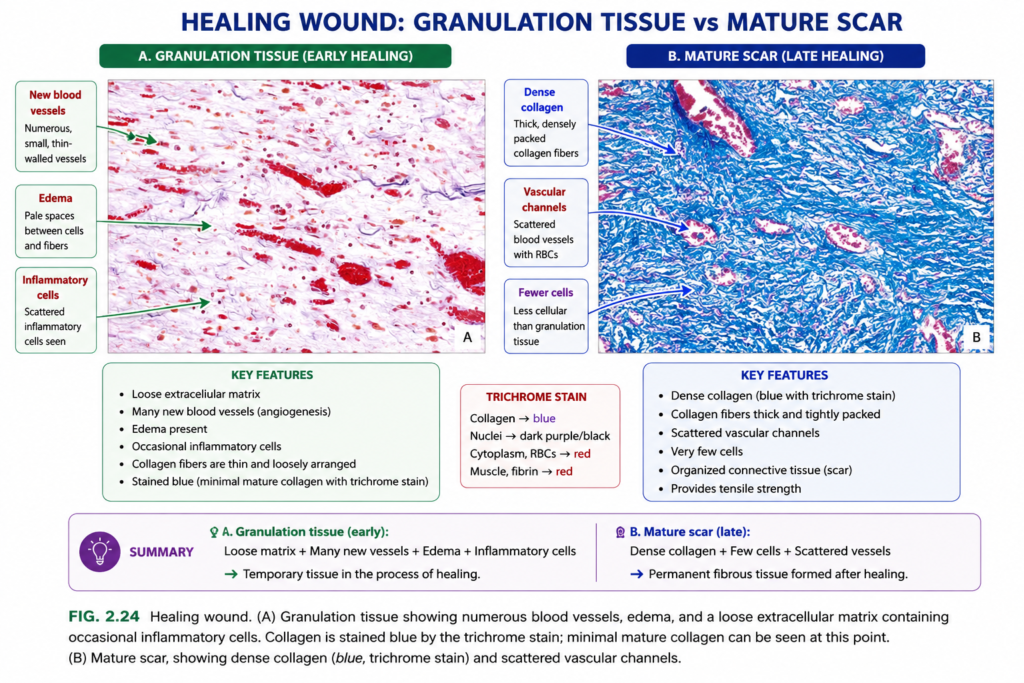
- Granulation tissue is made of:
- Proliferating fibroblasts
- Many thin-walled, delicate capillaries
- Loose extracellular matrix (ECM)
- Inflammatory cells, mainly macrophages (Fig. 2.24A)
- Granulation tissue is gradually deposited at the site of injury.
- The amount formed depends mainly on:
- Size of the tissue defect
- Intensity of inflammation
- A scar or fibrosis is composed of:
- Inactive, spindle-shaped fibroblasts
- Dense bundles of collagen
- Other ECM components (Fig. 2.24B)
- Special stains can identify different components of scars and fibrotic tissue.
- Trichrome stain detects collagen fibers.
- Red blood cells → orange
- Muscle → red
- Collagen → blue
- Elastin stain identifies delicate elastin fibers, which are the main component of pliable elastic tissue.
- Reticulin is another ECM protein present in:
- Connective tissue stroma of normal organs
- Early scars
- Reticulin is composed of type III collagen and can also be identified by a special stain.
KEY CONCEPT
Granulation tissue = fibroblasts + new capillaries + loose ECM + macrophages
Mature scar/fibrosis = inactive fibroblasts + dense collagen + ECM
Early repair → granulation tissue → increasing collagen → mature fibrous scar
CONCEPTUAL EXAMPLES
- Healing wound → many delicate new vessels + fibroblasts → granulation tissue
- Older healed wound → fewer active cells + dense collagen → scar
- Trichrome stain → collagen appears blue
- Early scar → reticulin/type III collagen is present
Factors That Interfere With Tissue Repair
- Factors that delay healing may be external or internal, and may act systemically or locally.
- Infection is one of the most important causes of delayed healing.
- It prolongs inflammation
- It may increase local tissue damage
- Diabetes is an important systemic cause of poor wound healing because it interferes with tissue repair in several ways.
- Poor nutrition delays healing.
- Protein deficiency → reduced repair
- Vitamin C deficiency → reduced collagen synthesis → delayed healing
- Glucocorticoids (steroids) suppress inflammation and inhibit TGF-β, which normally promotes collagen deposition.
- Therefore, after surgery: Glucocorticoids → ↓ TGF-β → ↓ collagen deposition → poor wound healing
- However, in some corneal infections, glucocorticoids may be given with antibiotics to reduce excessive collagen deposition and help prevent loss of vision.
- Mechanical stress, such as excessive pressure, twisting, or movement, may cause wound edges to pull apart.
- Poor blood perfusion also delays healing and may result from:
- Arteriosclerosis
- Diabetes
- Poor venous drainage, such as in varicose veins
- Foreign bodies, such as:
- Steel fragments
- Glass
- Bone fragments
can interfere with normal healing.
KEY CONCEPT
Delayed wound healing = infection + diabetes + poor nutrition + steroids + mechanical stress + poor perfusion + foreign bodies
Vitamin C deficiency → ↓ collagen synthesis → poor healing
Steroids → ↓ TGF-β → ↓ collagen deposition → delayed healing
CONCEPTUAL EXAMPLES
- Infected wound → inflammation continues → healing becomes slow
- Vitamin C deficiency → weak collagen formation → delayed wound repair
- Poor blood flow → less support to injured tissue → poor healing
- Excess movement at wound → wound edges separate → healing is delayed
Clinical Examples of Abnormal Wound Healing and Scarring
- Abnormal tissue repair may result from:
- Too little scar formation
- Excessive repair tissue deposition
- Contractures
Defects in Healing: Chronic Wounds
- Chronic wounds usually develop when local or systemic factors continuously interfere with healing.
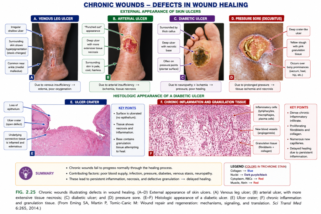
- Venous leg ulcers (Fig. 2.25A):
- Common in elderly people
- Caused by chronic venous hypertension
- May result from severe varicose veins or congestive heart failure
- Poor venous circulation leads to reduced oxygen delivery → poor healing
- Arterial ulcers (Fig. 2.25B):
- Occur with atherosclerosis of peripheral arteries
- Especially associated with diabetes
- Reduced arterial blood flow causes ischemia → poor repair + tissue necrosis
- These ulcers are often painful
- Diabetic ulcers (Fig. 2.25C):
- Mainly affect the lower limbs, especially the feet
- Poor healing results from:
- Small-vessel disease → ischemia
- Neuropathy
- Secondary infection
- Microscopically, they show epidermal ulceration (Fig. 2.25E) with extensive granulation tissue in the dermis (Fig. 2.25F).
- Pressure sores (Fig. 2.25D):
- Caused by prolonged compression of tissue against bone
- Common in bedridden patients
- Continuous pressure causes local ischemia → skin ulceration + underlying tissue necrosis
- Severe failure of healing may cause wound dehiscence, meaning the wound pulls apart or ruptures.
- Dehiscence occurs most commonly after abdominal surgery when vomiting, coughing, or ileus creates increased mechanical stress on the wound.
KEY CONCEPT
Poor blood flow / ischemia + neuropathy + infection + mechanical pressure → chronic wound → failure of normal healing
Venous ulcer → venous hypertension
Arterial ulcer → arterial ischemia
Diabetic ulcer → ischemia + neuropathy + infection
Pressure sore → prolonged pressure + ischemia
CONCEPTUAL EXAMPLES
- Severe varicose veins → venous hypertension → poor oxygen delivery → venous ulcer
- Peripheral atherosclerosis → ischemia → painful arterial ulcer
- Diabetic foot → ischemia + neuropathy + infection → chronic ulcer
- Bedridden patient → prolonged pressure over bone → ischemia → pressure sore
- Abdominal surgery + severe coughing/vomiting → mechanical stress → wound dehiscence
Excessive Scarring
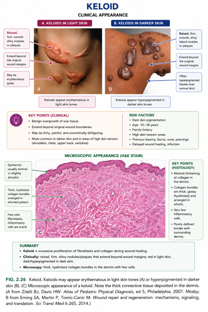
- Excessive repair tissue formation can produce hypertrophic scars and keloids.
- Hypertrophic scars:
- Contain many myofibroblasts
- Often grow rapidly
- Usually regress over several months
- Commonly develop after deep thermal or traumatic injury of the dermis
- A keloid forms when scar tissue:
- Grows beyond the original wound boundaries
- Does not regress (Fig. 2.26)
- Keloid formation appears to depend partly on individual predisposition.
- Normal wound healing includes wound contraction, which reduces wound size.
- Excessive contraction produces a contracture, causing deformity of the wound and surrounding tissues.
- Contractures commonly develop on:
- Palms
- Soles
- Anterior chest
- They are especially common after severe burns and may restrict joint movement.
KEY CONCEPT
Hypertrophic scar → excessive scar within wound → may regress
Keloid → scar extends beyond wound → does not regress
Excessive wound contraction → contracture → deformity + limited movement
CONCEPTUAL EXAMPLES
- Deep burn → excessive scar tissue within wound → hypertrophic scar
- Scar grows beyond original wound → keloid
- Severe burn near joint → excessive contraction → contracture → reduced joint movement
Fibrosis in Parenchymal Organs
- Fibrosis means excessive deposition of collagen and other ECM components in tissues.
- Although scar and fibrosis are often used interchangeably, fibrosis usually refers to abnormal collagen deposition in internal organs during chronic disease.
- The basic mechanism of fibrosis is similar to scar formation in skin.
- Fibrosis is caused by persistent injury, especially:
- Chronic infections
- Chronic immune reactions
- Persistent injury → prolonged repair signals → excessive ECM and collagen deposition → organ fibrosis
- Fibrosis may cause severe organ dysfunction and even organ failure.
- The most important cytokine promoting fibrosis is TGF-β.
- IL-13, produced by T lymphocytes, may also contribute (see Fig. 2.22).
- Important fibrotic disorders include:
- Liver cirrhosis
- Systemic sclerosis (scleroderma)
- Idiopathic pulmonary fibrosis
- Pneumoconioses
- Drug- and radiation-induced pulmonary fibrosis
- End-stage kidney disease
- Constrictive pericarditis
- Because fibrosis can severely impair organ function, there is major interest in developing antifibrotic drugs.
KEY CONCEPT
Persistent chronic injury → TGF-β ↑ → fibroblast activation → collagen + ECM deposition → fibrosis → organ dysfunction
CONCEPTUAL EXAMPLES
- Chronic liver injury → excess collagen → cirrhosis
- Chronic lung injury → fibrosis → reduced lung function
- Chronic kidney damage → fibrosis → end-stage kidney disease