- Repair (healing) means restoration of the normal structure and function of tissue after injury.
- Inflammation removes microbes and damaged tissue and also starts the process of tissue repair.
- Damaged tissues are repaired by two main processes: regeneration and scar formation (Fig. 2.20).
- Regeneration means damaged tissue is replaced by new cells, allowing the tissue to return almost to its normal state.
- Regeneration occurs when surviving cells multiply and are able to produce the mature cells of that tissue.
- These regenerating cells may be:
- Mature differentiated cells, or
- More commonly, tissue stem cells.
- Scar formation occurs when complete regeneration is not possible or when the supporting tissue structure is damaged.
- In this process, damaged tissue is replaced by connective (fibrous) tissue, producing a scar.
- The fibrous scar provides enough structural strength and stability for the injured tissue to continue functioning.
- When excessive scarring due to chronic inflammation occurs in organs such as the lungs, liver, or kidneys, it is called fibrosis.
- In many common injuries, both regeneration and scar formation occur together, but their contribution varies depending on the type and severity of injury.
KEY CONCEPT
Tissue injury → inflammation → repair
Repair occurs by:
Regeneration → damaged cells replaced → near-normal tissue
OR
Scar formation → fibrous connective tissue deposited → structural stability
- Many injuries heal by a combination of both processes.
CONCEPTUAL EXAMPLES
- If damaged cells can multiply and the tissue framework is preserved → regeneration is more likely.
- If tissue damage is severe or its supporting structure is destroyed → scar formation is more likely.
- Long-term scarring in organs such as the liver, lungs, or kidneys → fibrosis.
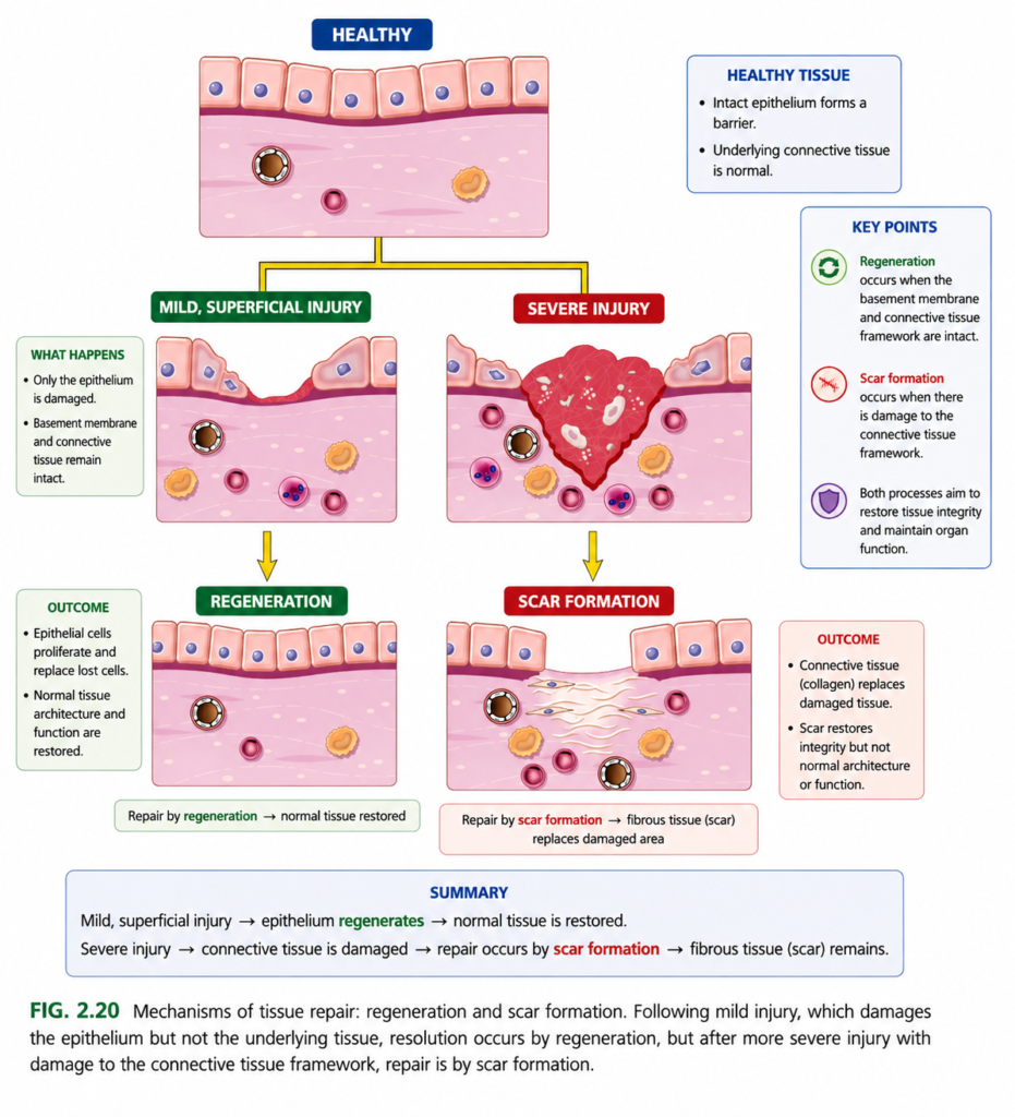
FIG. 2.20 — Tissue Repair: Regeneration vs Scar Formation
1️⃣ Top: HEALTHY
- 🩷 Peach/pink cells = normal surface epithelium
- 🔵 Blue-purple dots = nuclei
- 🟣 Dark purple line underneath = supporting basement membrane / tissue framework
- 🌸 Light pink area below = connective tissue
Everything is intact and normally organized.
🟨 Yellow branching arrow = “What happens after injury?”
It gives 2 possible pathways:
2️⃣ LEFT — Mild, superficial injury
Only the surface epithelial cells are damaged.
✅ Most important point: the underlying connective-tissue framework remains intact.
Mild injury → framework preserved → surviving cells multiply → wound closes
⬇️ Yellow arrow
3️⃣ REGENERATION
New epithelial cells replace the lost cells.
➡️ The tissue becomes almost exactly like normal again.
Regeneration = damaged cells replaced by the SAME type of cells.
4️⃣ RIGHT — Severe injury
The injury goes deep.
🔴 Dark red area = severely damaged/necrotic tissue + wound/clot
🟣/🔵 cells in the wound = inflammatory cells coming to clean the damage.
Now both are damaged:
Epithelium + underlying connective-tissue framework
So simple regeneration is not enough.
⬇️ Yellow arrow
5️⃣ SCAR FORMATION
🟡/cream wavy fibers = newly deposited collagen/fibrous connective tissue.
Severe injury → framework destroyed → collagen deposited → scar
The wound becomes strong, but the original architecture is not perfectly restored.
🧠 Easiest memory
SUPERFICIAL = SAME again → REGENERATION
SEVERE = SCAR
⭐ Whole figure in ONE line
Mild injury + intact framework → REGENERATION → normal tissue
Severe injury + destroyed framework → COLLAGEN → SCAR
🎯 Key exam point
The main deciding factor is whether the connective-tissue framework is intact or destroyed.
Cell and Tissue Regeneration
- The ability of a tissue to repair itself depends partly on its natural capacity for cell proliferation.
- In some tissues, cells are continuously lost and replaced by new cells derived from:
- Tissue stem cells
- Remaining mature tissue cells
- Examples include:
- Hematopoietic cells in bone marrow
- Basal cells of skin squamous epithelium
- Columnar epithelium of the gastrointestinal tract
- These tissues can regenerate easily after injury if their stem cell pool remains preserved.
- Other tissues contain cells that normally remain in the G0 stage of the cell cycle, meaning they are usually not dividing.
- However, these cells can start dividing after injury or loss of tissue mass.
- Examples include the parenchymal cells of most solid organs such as:
- Liver
- Kidney
- Pancreas
- Endothelial cells, fibroblasts, and smooth muscle cells are also normally inactive but can proliferate when stimulated by growth factors.
- Their proliferation is especially important in wound healing.
- Some tissues contain terminally differentiated cells that cannot divide, including most:
- Neurons
- Cardiac muscle cells
- Injury to these cells is usually irreversible, so healing mainly occurs by scar formation rather than regeneration.
KEY CONCEPT
- Continuously dividing tissues → regenerate easily
- G0/quiescent tissues → can divide after injury
- Terminally differentiated tissues → cannot regenerate → scar formation
CONCEPTUAL EXAMPLES
- Skin or intestinal epithelium injured → stem cells survive → rapid regeneration
- Liver tissue lost → resting cells enter the cell cycle → proliferation occurs
- Cardiac muscle or neurons severely injured → cells cannot divide → scar forms
Cell and Tissue Regeneration
- Cell proliferation is stimulated mainly by signals from:
- Growth factors
- Extracellular matrix (ECM)
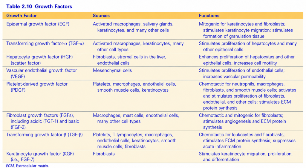
- Different growth factors may act on many cell types or only on specific cells (Table 2.10).
- Growth factors are usually produced by cells near the site of tissue injury.
- The most important source is macrophages activated by tissue damage, while epithelial and stromal cells also produce growth factors.
- Some growth factors bind to ECM proteins, so they become concentrated at the injured area.
- Growth factors activate signaling pathways that cause cell division and proliferation.
- Cells also attach to ECM proteins through integrins, and integrin signals can further stimulate cell proliferation.
- During regeneration, surviving cells proliferate, while stem cells also produce new mature cells.
- Embryonic stem cells (ES cells) can:
- Self-renew
- Produce all mature cell lineages, called totipotential ability
- Tissue stem cells are present in most adult tissues and usually produce the mature cells of the same tissue in which they live.
- Tissue stem cells have a more limited self-renewal capacity than embryonic stem cells.
- Stem cells undergo asymmetric cell division:
- One daughter cell remains a stem cell → maintains self-renewal.
- The other daughter cell differentiates → becomes a mature cell.
- Tissue stem cells live in specialized areas called stem cell niches.
- Injury stimulates these stem cells to proliferate and differentiate, helping to replace damaged cells.
- Stem cells are especially important when surviving mature cells have little or no ability to divide.
- The amount of regeneration depends on the type of tissue and severity of injury.
- In the intestinal epithelium and skin, damaged cells are rapidly replaced if the basement membrane remains intact.
- Repair occurs through:
- Proliferation of surviving cells
- Differentiation of tissue stem cells
- Parenchymal organs can regenerate if their mature cells can divide, but regeneration is usually limited except in the liver.
- The pancreas, adrenal, thyroid, and lung have some regenerative capacity.
- After surgical removal of one kidney, the remaining kidney undergoes a compensatory response:
- Hypertrophy → cells become larger
- Hyperplasia → proximal duct cells increase in number
- The liver has an exceptionally strong regenerative capacity and is an important model for studying regeneration.
- Complete restoration of normal tissue structure occurs only when the remaining tissue framework is intact, such as after partial surgical removal of the liver.
- If both the cells and their supporting framework are destroyed by infection or inflammation, regeneration becomes incomplete and scar formation occurs.
- For example, extensive liver destruction with collapse of the reticulin framework, as in a liver abscess, causes scarring even though surviving liver cells can regenerate.
KEY CONCEPT
- Growth factors + ECM signals → cell proliferation
- Stem cells → self-renew + produce mature cells
- Intact supporting framework → effective regeneration
- Destroyed supporting framework → incomplete regeneration + scar
- Liver → greatest regenerative capacity among major parenchymal organs
CONCEPTUAL EXAMPLES
- Skin injury + intact basement membrane → rapid regeneration
- Partial liver removal + preserved framework → liver cells regenerate
- Liver abscess + destroyed reticulin framework → regeneration cannot fully restore architecture → scar forms
- One kidney removed → remaining kidney enlarges by hypertrophy + hyperplasia
Liver Regeneration
- The liver has a remarkable ability to regenerate, especially after partial hepatectomy, which may be done for tumor removal or living-donor liver transplantation.
- Liver regeneration occurs by two main mechanisms:
- Proliferation of remaining hepatocytes
- Repopulation from stem cells
- Which mechanism is more important depends on the type of liver injury.
- After partial hepatectomy, the remaining hepatocytes can proliferate and regenerate up to 90% of the liver in humans.
- Hepatocyte proliferation is stimulated by the combined action of cytokines and polypeptide growth factors.
- First, cytokines such as IL-6, produced mainly by Kupffer cells, act on hepatocytes and make them ready to respond to growth-factor signals.
- Next, growth factors such as HGF and TGF-α, produced by several cell types (see Table 2.10), stimulate the prepared hepatocytes to proliferate.
- Therefore:Kupffer cells → IL-6 → hepatocytes become responsive → HGF + TGF-α → hepatocyte proliferation → liver regeneration
- If hepatocytes have a reduced ability to proliferate, such as during chronic liver injury or inflammation, liver stem cells help repopulate the tissue.
- Some liver stem cells are located in specialized niches called the canals of Hering, where bile canaliculi connect with larger bile ducts.
KEY CONCEPT
- Partial hepatectomy → remaining hepatocytes proliferate → liver regenerates
- IL-6 primes hepatocytes → HGF and TGF-α stimulate proliferation
- Chronic injury with poor hepatocyte proliferation → stem cells contribute to regeneration
CONCEPTUAL EXAMPLES
Part of liver surgically removed → remaining hepatocytes divide → liver mass is restoredChronic liver injury → hepatocytes cannot proliferate effectively → stem cells help replace liver cellsKupffer cell releases IL-6 → hepatocyte becomes ready → HGF/TGF-α stimulate cell division
Repair by Scarring
- When regeneration alone is not enough, injured cells are replaced by connective tissue, producing a scar.
- Scarring occurs especially when:
- Injury is severe or chronic
- Parenchymal cells, epithelium, and connective tissue framework are damaged
- Nondividing cells are injured
- Regeneration restores tissue, whereas a scar mainly patches the damaged area.
- Scar formation can occur in the skin or in internal organs when parenchymal cells are replaced by collagen.
- Example: after myocardial infarction, damaged heart muscle is replaced by a collagen scar.
Steps in Scar Formation
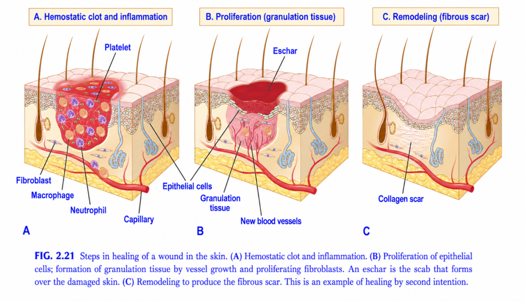
- Within minutes after injury, platelets form a hemostatic plug that:
- Stops bleeding
- Provides a scaffold for inflammatory cells
- Helps form a stable clot
- The later steps are shown in Fig. 2.21.
- Inflammation (6–48 hours):
- Neutrophils arrive first, followed by monocytes.
- They remove harmful agents and tissue debris.
- Macrophages are the central cells in repair.
- Macrophages clear microbes and necrotic tissue and release growth factors that stimulate later cell proliferation.
- When harmful agents and dead cells are removed, inflammation resolves.
- Cell proliferation (up to 10 days):
- Epithelial cells, endothelial/vascular cells, and fibroblasts proliferate and migrate into the cleaned wound.
- Epithelial cells migrate to cover the wound.
- Endothelial and other vascular cells form new blood vessels, called angiogenesis.
- Fibroblasts migrate into the wound and produce collagen fibers, which form the scar.
- Granulation tissue consists of:
- Proliferating fibroblasts
- ECM
- New blood vessels
- It has a characteristic pink, soft, granular appearance.
- Remodeling:
- Deposited connective tissue is reorganized into a stable fibrous scar.
- It begins about 2–3 weeks after injury.
- It may continue for months or years.
- Skin wounds heal by two patterns:
- First intention (primary union) → epithelial regeneration with minimal scarring, as in a well-apposed surgical incision.
- Second intention (secondary union) → larger wounds heal by a combination of regeneration and scarring.
- The main healing events are the same in both types.
KEY CONCEPT
Injury → platelet plug → inflammation → cell proliferation → angiogenesis + fibroblasts + collagen → granulation tissue → remodeling → stable scar
- Regeneration → restores tissue
- Scarring → patches tissue
- First intention → minimal scar
- Second intention → larger wound + regeneration + scar
CONCEPTUAL EXAMPLES
- Clean surgical incision → first intention → epithelial regeneration → minimal scar
- Large open wound → second intention → regeneration + scar formation
- Myocardial infarction → dead cardiac muscle replaced by collagen → fibrous scar
- Granulation tissue → remodeling over time → stable fibrous scar