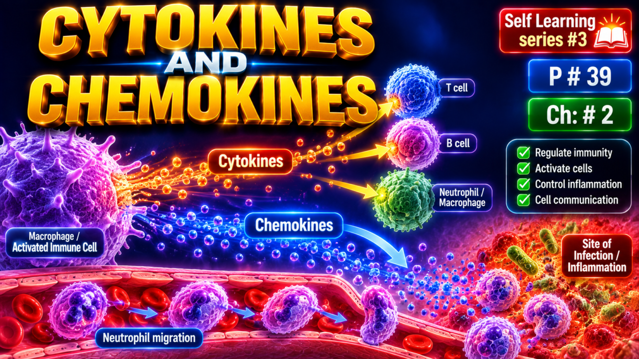
- Cytokines are proteins produced mainly by activated lymphocytes, macrophages, and dendritic cells, and also by endothelial, epithelial, and connective tissue cells.
- They control and regulate immune and inflammatory reactions.
- Growth factors acting mainly on epithelial and mesenchymal cells are not usually classified as cytokines.
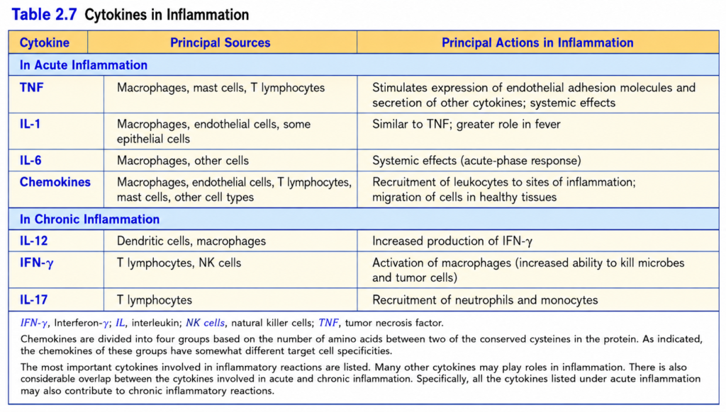
- Cytokines involved in acute inflammation are summarized in Table 2.7.
Tumor Necrosis Factor (TNF) and Interleukin-1 (IL-1)
- TNF and IL-1 are major inflammatory cytokines that help recruit leukocytes by increasing their adhesion to endothelium and movement through blood vessels.
- Both are produced mainly by activated macrophages and dendritic cells.
- TNF is also produced by T lymphocytes and mast cells, whereas IL-1 can also be produced by some epithelial cells.
- Their secretion is stimulated by microbial products, necrotic cells, and other inflammatory stimuli.
- TNF production is triggered through TLRs and other microbial sensors.
- IL-1 synthesis is stimulated by similar signals, but formation of its biologically active form requires inflammasome activation.
- TNF and IL-1 produce important local and systemic inflammatory effects (Fig. 2.7).
- Endothelial activation and leukocyte recruitment: TNF and IL-1 increase endothelial adhesion molecules, especially E-selectin, P-selectin, and ligands for leukocyte integrins.
- This allows leukocytes to attach to endothelium and reach the site of inflammation.
- They also stimulate production of other cytokines, chemokines, and eicosanoids and increase the procoagulant activity of endothelium.
- Activation of leukocytes and other cells: TNF increases neutrophil responses to stimuli such as bacterial endotoxin and enhances the microbicidal activity of macrophages.
- IL-1 activates fibroblasts to produce collagen and promotes proliferation of synovial and other mesenchymal cells.
- IL-1 also stimulates Th17 responses, which promote acute inflammation.
- Systemic acute-phase response: TNF and IL-1, together with IL-6, produce systemic responses to infection or injury, including fever.
- They also contribute to systemic inflammatory response syndrome (SIRS) in severe infections and other serious conditions.
- At high concentrations, TNF causes vasodilation and decreases myocardial contractility, contributing to the fall in blood pressure in SIRS.
- TNF also promotes lipid and protein mobilization and suppresses appetite.
- Therefore, prolonged TNF production can cause cachexia, characterized by weight loss and anorexia, especially in some chronic infections and cancers.
- TNF antagonists are highly effective in chronic inflammatory diseases such as rheumatoid arthritis, psoriasis, and some inflammatory bowel diseases.
- However, blocking TNF can increase susceptibility to mycobacterial infections because macrophages become less effective at killing intracellular microbes.
- Although TNF and IL-1 have many overlapping actions, IL-1 antagonists are generally less effective.
- Blocking either TNF or IL-1 does not improve the outcome of sepsis, probably because several other cytokines also contribute to this systemic inflammatory reaction.
KEY CONCEPT
- TNF + IL-1 → endothelial activation → leukocyte recruitment → inflammation.
- They also activate inflammatory cells and produce systemic effects such as fever and SIRS.
- Excess TNF → vasodilation + reduced cardiac contraction → low blood pressure, while prolonged TNF production may cause cachexia.
Conceptual Examples
- Local infection: Microbe activates macrophages → TNF and IL-1 released → endothelium becomes sticky → leukocytes enter the infected tissue.
- Severe systemic inflammation: Very high TNF → blood vessels dilate + heart contracts less strongly → blood pressure falls.
- Chronic TNF production: Increased breakdown of fat/protein + reduced appetite → weight loss and cachexia.
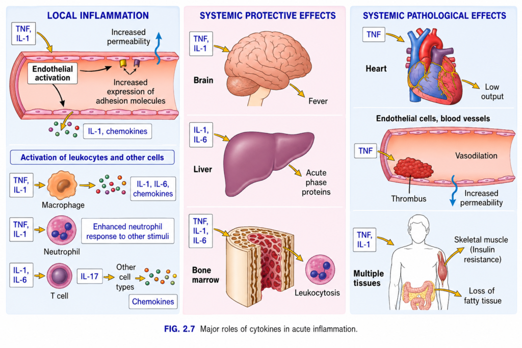
Fig. 2.7 — Major Roles of Cytokines in Acute Inflammation
First, understand the big idea
Cytokines = small chemical messengers made by immune cells.
Think of them as alarm messages sent during infection or tissue injury.
The most important cytokines in this figure are:
- TNF = Tumor Necrosis Factor
- IL-1 = Interleukin-1
- IL-6 = Interleukin-6
- IL-17 = Interleukin-17
- Chemokines = cytokines that mainly attract leukocytes to the site of inflammation
The figure shows that cytokines can have:
Local effects → help fight infection at the injured site
Systemic protective effects → help the whole body fight infection
Excessive systemic effects → become harmful and cause disease
1. LOCAL INFLAMMATION
This happens at the actual site of infection or tissue injury.
A. TNF + IL-1 → Endothelial activation
Endothelial cells = cells lining the inside of blood vessels.
Sequence
TNF + IL-1
→ activate endothelial cells
→ endothelial cells become more useful for inflammation.
Two important things happen:
1. Increased expression of adhesion molecules
Adhesion molecules = proteins that act like Velcro. ( A hook- and -loop fastening system made of two fabric strips that press together and stick.)
They allow circulating leukocytes to:
stick to endothelium → leave blood → enter infected tissue
So:
TNF/IL-1 → adhesion molecules ↑ → leukocyte attachment ↑ → leukocyte migration into tissue ↑
2. Increased vascular permeability
Permeability = how easily fluid/proteins can pass through the vessel wall.
TNF and IL-1 help increase permeability.
So:
Blood vessel becomes leaky → plasma proteins + fluid enter tissue
This helps defensive proteins reach the infected area.
But it also produces:
fluid accumulation → edema/swelling
Simple memory
TNF + IL-1 tell vessels:
“Become sticky and leaky.”
Sticky = leukocyte adhesion
Leaky = increased permeability
2. TNF + IL-1 → Macrophage Activation
A macrophage is a large immune cell that:
- eats microbes
- removes dead tissue
- produces inflammatory cytokines
Arrow sequence
TNF + IL-1
→ activate macrophage
→ macrophage produces more:
IL-1 + IL-6 + chemokines
These substances further increase inflammation.
So this creates an amplification loop:
Inflammation → cytokines → macrophage activation → more cytokines → stronger inflammation
3. TNF + IL-1 → Neutrophils
Neutrophils are the major early leukocytes in acute inflammation.
TNF and IL-1 act on neutrophils and cause:
Enhanced neutrophil response to other stimuli
Meaning the neutrophil becomes more ready and more powerful when it encounters microbes.
So:
TNF + IL-1 → neutrophil priming → stronger response to microbes
Important idea:
TNF/IL-1 do not necessarily make neutrophils attack randomly.
They make them more responsive when another activating signal appears.
4. IL-1 + IL-6 → T Cell → IL-17
Now follow the lower arrows carefully.
Step 1
IL-1 + IL-6
→ stimulate certain T cells
Step 2
Activated T cells produce:
IL-17
IL-17 is especially associated with Th17 cells.
Step 3
IL-17
→ acts on other cell types
Step 4
Those cells produce:
Chemokines
Step 5
Chemokines attract more leukocytes, especially neutrophils.
So the complete pathway is:
IL-1 + IL-6 → T cell → IL-17 → other tissue cells → chemokines → leukocyte recruitment
Memory trick
IL-17 = “Send neutrophils.”
5. SYSTEMIC PROTECTIVE EFFECTS
Now cytokines enter the circulation and act on distant organs.
These effects are usually helpful when controlled.
A. TNF + IL-1 → Brain → Fever
TNF and IL-1 act on the brain, especially temperature-regulating centers.
Result:
Body temperature rises → fever
So:
TNF + IL-1 → brain → fever
Why is fever useful?
Moderate fever may:
- slow growth of some microbes
- improve some immune responses
Easy memory
IL-1 = fever cytokine
Although TNF also contributes.
6. IL-1 + IL-6 → Liver → Acute-Phase Proteins
The next arrow shows:
IL-1 + IL-6 → liver
The liver responds by producing:
Acute-phase proteins
These are blood proteins whose levels rise during inflammation.
Examples include:
- C-reactive protein (CRP)
- fibrinogen
- serum amyloid A
They help the body’s inflammatory and antimicrobial responses.
So:
IL-1/IL-6 → liver → acute-phase proteins ↑
High-yield point
IL-6 is particularly important for acute-phase protein production.
Memory trick
IL-6 → Liver → CRP
7. TNF + IL-1 + IL-6 → Bone Marrow → Leukocytosis
Bone marrow produces blood cells.
TNF, IL-1, and IL-6 stimulate systemic responses that increase leukocyte production/release.
Result:
Leukocytosis
Leukocytosis = increased white blood cell count in blood.
So:
TNF + IL-1 + IL-6 → bone marrow → more circulating leukocytes
Why?
Because infection requires more immune soldiers.
Easy concept
Bone marrow receives the message:
“We have an infection—send more white cells.”
8. SYSTEMIC PATHOLOGICAL EFFECTS
This is extremely important.
Small/controlled amounts of cytokines are protective.
But very large amounts, especially during severe infection such as sepsis, can become dangerous.
Think:
Too much TNF/IL-1 → inflammation becomes harmful to the whole body.
9. TNF → Heart → Low Cardiac Output
TNF acts on the heart.
Result:
Low output
Meaning the heart pumps less blood effectively.
So:
Excess TNF → impaired cardiac function → cardiac output ↓
This contributes to poor tissue perfusion during severe systemic inflammation.
10. TNF → Endothelial Cells/Blood Vessels
Excess TNF causes several dangerous vascular effects.
A. Thrombus formation
Thrombus = blood clot formed inside a blood vessel.
Excess TNF activates endothelium and promotes coagulation.
So:
TNF ↑↑ → coagulation ↑ → small thrombi may form
If widespread, this may contribute to disseminated intravascular coagulation (DIC).
B. Vasodilation
Vasodilation = widening of blood vessels.
Blood vessels become wider.
Result:
vascular resistance ↓ → blood pressure ↓
So:
TNF ↑↑ → widespread vasodilation → hypotension
C. Increased permeability
Blood vessels also become excessively leaky.
So:
TNF ↑↑ → vascular permeability ↑ → fluid leaves circulation
Result:
circulating blood volume ↓ → blood pressure ↓ → tissue perfusion ↓
This can contribute to shock.
11. TNF + IL-1 → Multiple Tissues
Excess systemic cytokines also disturb normal metabolism.
Skeletal muscle → Insulin resistance
Insulin resistance means tissues do not respond normally to insulin.
Therefore:
glucose uptake by muscle decreases
So:
TNF/IL-1 → insulin action ↓ → insulin resistance
This helps explain metabolic disturbances in severe inflammation.
12. Loss of Fatty Tissue
Excess inflammatory cytokines, particularly TNF, promote breakdown of energy stores and suppress normal anabolic metabolism.
Result:
fat tissue decreases
With prolonged severe inflammation this contributes to:
Cachexia
Cachexia = severe loss of body weight, fat, and muscle associated with chronic disease/inflammation.
A classic older name for TNF is:
Cachectin
because of its association with cachexia.
Memory trick
TNF → “Thin”
Excess TNF can contribute to:
fat loss + muscle wasting
Whole-Figure Flow
Think of the entire figure as three levels:
Microbe/injury
→ TNF, IL-1, IL-6 and other cytokines released
Local level
→ endothelial activation
→ adhesion molecules ↑
→ permeability ↑
→ leukocyte recruitment
→ macrophage/neutrophil activation
→ stronger local defense
Controlled systemic level
→ brain → fever
→ liver → acute-phase proteins
→ bone marrow → leukocytosis
Excessive systemic level
→ heart → low cardiac output
→ vessels → vasodilation + leakage + thrombosis
→ metabolism → insulin resistance + wasting
→ severe cases → shock / organ dysfunction
Key Exam Points
TNF + IL-1
→ endothelial activation
→ adhesion molecules ↑
→ vascular permeability ↑
→ fever
IL-6
→ liver
→ acute-phase proteins, especially CRP
TNF + IL-1 + IL-6
→ bone marrow
→ leukocytosis
IL-1 + IL-6
→ T cells
→ IL-17
→ chemokines
→ leukocyte/neutrophil recruitment
Excess TNF
→ vasodilation + vascular leakage + thrombosis + low cardiac output
→ septic shock
One-line memory
“TNF/IL-1 make vessels STICKY and LEAKY; IL-6 tells the LIVER; IL-17 calls NEUTROPHILS.”
Chemokines
- Chemokines are small proteins (8–10 kD) that mainly act as chemoattractants, meaning they attract specific types of leukocytes.
- About 40 chemokines and 20 chemokine receptors have been identified.
- Different chemokines act on different cells according to the chemokine receptors present on those cells (Table 2.7).
- Chemokines bind to proteoglycans → become concentrated on endothelial surfaces and in the extracellular matrix (Fig. 2.3).
- Chemokines have two main functions:
- In inflammation: Microbes and other inflammatory stimuli → production of inflammatory chemokines.
- Chemokines bind to receptors on leukocytes → promote integrin-dependent attachment to endothelium → guide leukocytes by chemotaxis toward infection or tissue damage.
- Maintenance of tissue architecture: Some chemokines are continuously produced by stromal cells and are called homeostatic chemokines.
- Homeostatic chemokines guide different cells to their specific normal locations within tissues.
- For example, they help localize T and B lymphocytes to specific areas of the spleen and lymph nodes.
- Although chemokines have a well-established role in inflammation, it has been difficult to develop effective chemokine antagonists.
Other Cytokines in Acute Inflammation
- Many cytokines participate in inflammation; important additional cytokines include IL-6 and IL-17.
- IL-6 is produced by macrophages and other cells → participates in local and systemic inflammatory reactions.
- IL-17 is produced mainly by T lymphocytes → promotes neutrophil recruitment.
- Antagonists against IL-6 and IL-17 are effective in treating inflammatory diseases.
- Type I interferons normally inhibit viral replication and also contribute to some systemic effects of inflammation.
- Cytokines also play important roles in chronic inflammation.
KEY CONCEPT
- Inflammatory chemokines → leukocyte adhesion to endothelium → chemotaxis → leukocytes reach infection/tissue injury.
- Homeostatic chemokines → guide cells to their normal locations in tissues.
- IL-6 → local + systemic inflammation.
- IL-17 → neutrophil recruitment.
Conceptual Examples
- Infection: Microbe → chemokines released → leukocytes attach to endothelium → migrate toward the infected tissue.
- Normal lymphoid tissue: Homeostatic chemokines → guide T and B lymphocytes to specific regions of the spleen and lymph nodes.
- IL-17 response: T lymphocytes → IL-17 → neutrophil recruitment to the inflammatory site.
Complement System
- The complement system is a group of soluble proteins and membrane receptors that mainly provide defense against microbes and also participate in pathologic inflammation.
- It contains more than 20 proteins, including components C1–C9.
- Complement activation and functions are summarized in Fig. 2.8.
- Complement proteins normally exist as inactive proforms → inflammation activates them → an enzyme cascade begins with powerful amplification.
- The critical step is proteolytic cleavage of C3, the most abundant complement component.
- C3 can be activated through three pathways:
- Classical pathway: Antigen binds IgM or IgG → C1 binds the antibody → complement activation.
- Alternative pathway: Microbial surface molecules, such as LPS (endotoxin) and complex polysaccharides → complement activation without antibody.
- Lectin pathway: Mannose-binding lectin binds carbohydrates on microbes → activates complement without antibody.
- All three pathways → formation of C3 convertase → splits C3 into C3a + C3b.
- C3a is released, while C3b attaches to the surface where complement is activated.
- Additional C3b → formation of C5 convertase → splits C5 into C5a + C5b.
- C5a is released, while C5b remains attached to the cell surface.
- C5b binds C6–C9 → forms the membrane attack complex (MAC), containing multiple C9 molecules.
- The complement system has three major functions:
- Inflammation: C5a, and to a lesser extent C3a and C4a, recruit neutrophils and other leukocytes.
- They also stimulate mast-cell histamine release → increased vascular permeability + vasodilation.
- C3a, C4a, and C5a are called anaphylatoxins because their actions resemble mast-cell mediators involved in anaphylaxis.
- Opsonization and phagocytosis: C3b and iC3b attach to microbial surfaces and act as opsonins → neutrophils and macrophages recognize them → easier phagocytosis.
- Cell lysis: MAC forms holes in the cell membrane → water and ions move through the damaged membrane → cell lysis and death.
- MAC is especially important against thin-walled microbes such as Neisseria.
- Therefore, deficiency of terminal complement components or treatment with complement inhibitors → increased risk of disseminated Neisseria infections, including meningococci and gonococci.
- Complement activation is normally tightly controlled by regulatory proteins to protect normal host tissues.
- These regulators either prevent formation of active complement fragments or remove fragments deposited on cells.
- In autoimmune diseases, large amounts of complement may be deposited on host tissues → regulatory mechanisms can be overwhelmed → tissue injury.
- C1 inhibitor (C1 INH) blocks C1 activation in the classical pathway → inherited deficiency causes hereditary angioedema.
- Decay accelerating factor (DAF) prevents formation of C3 convertases.
- CD59 prevents formation of the MAC.
- DAF and CD59 are attached to cell membranes by a GPI anchor.
- Defective formation of GPI anchors → loss of DAF and CD59 → excessive complement activation → red blood cell lysis → paroxysmal nocturnal hemoglobinuria (PNH).
- Factor H promotes inactivation of C3 convertase → limits complement activation.
- Factor H deficiency → excessive complement activation.
- Factor H mutations are associated with hemolytic uremic syndrome and wet macular degeneration, which involves increased permeability of retinal vessels.
- Complement can cause disease when antibodies or antigen-antibody complexes activate complement on host cells and tissues → cell and tissue injury.
- Inherited deficiency of complement proteins → increased susceptibility to infections.
- Deficiency of complement regulatory proteins → excessive complement activation and disease.
- Excessive complement activity in disorders such as hemolytic uremic syndrome and PNH is also associated with an increased risk of thrombosis, although the mechanism is uncertain.
- Antibodies that block complement activation have been developed to treat several complement-related disorders.
KEY CONCEPT
- Classical / Alternative / Lectin pathway → C3 convertase → C3a + C3b → C5 convertase → C5a + C5b → C5b + C6–C9 → MAC.
- C3a + C4a + C5a → inflammation.
- C3b/iC3b → opsonization → phagocytosis.
- C5b–C9 → MAC → cell lysis.
- C1 INH deficiency → hereditary angioedema.
- Loss of DAF + CD59 → PNH.
Conceptual Examples
- Bacterial infection: Microbe → complement activated → C5a recruits leukocytes + C3b coats the microbe → easier killing and phagocytosis.
- Neisseria: MAC → holes in bacterial membrane → lysis; therefore terminal complement deficiency → high risk of Neisseria infection.
- PNH: Loss of GPI anchor → loss of DAF and CD59 → uncontrolled complement attack on RBCs → RBC lysis.
- Hereditary angioedema: C1 inhibitor deficiency → inadequate control of complement-related activation → hereditary angioedema.
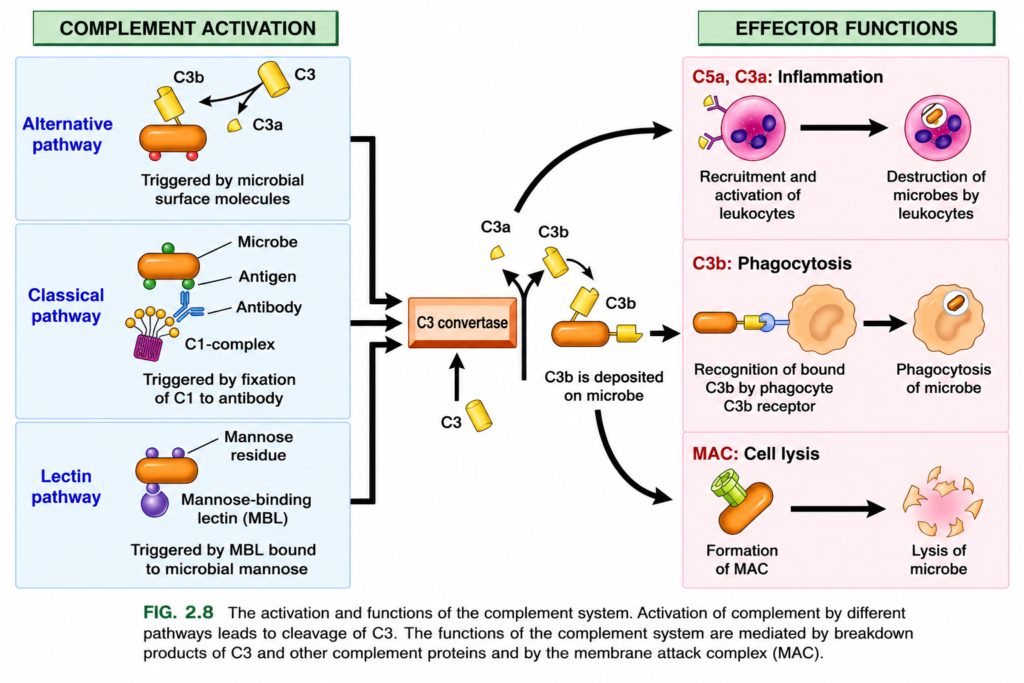
First: What is the “complement system”?
The complement system is a group of proteins normally present in our blood.
Most of these proteins are normally inactive.
When a microbe enters the body:
Microbe enters → complement proteins become activated one after another → microbe is attacked
It is called “complement” because these proteins help/complement the work of antibodies and immune cells.
What is C3?
This is the most important protein to understand in this figure.
C3 = Complement component 3
C3 is a large complement protein circulating in the blood.
Think of C3 as one large inactive defense protein waiting to be cut into useful pieces.
In the figure, C3 is shown as a large yellow cylinder.
So:
Yellow large cylinder = C3
C3 itself must be cleaved/cut before its major actions appear.
What cuts C3?
The orange box in the middle is:
C3 convertase
“Convertase” simply means an enzyme complex that converts/cuts C3.
So the most important central reaction is:
C3 convertase + C3 → C3a + C3b
Think:
C3 = one big yellow piece
✂️ C3 convertase cuts it
↓
small piece = C3a
large piece = C3b
This is the heart of the whole figure.
Now Start From the LEFT SIDE
There are 3 different ways to activate complement:
1. Alternative pathway
2. Classical pathway
3. Lectin pathway
They start differently.
But all 3 finally reach:
C3 convertase
Then:
C3 → C3a + C3b
1. Alternative Pathway — Top Blue Box
Look at the upper-left blue box.
You see an orange rod-shaped microbe.
The orange object represents a:
Microbe
On/near the microbial surface, complement becomes activated.
The figure says:
“Triggered by microbial surface molecules.”
Meaning:
Certain molecules on the microbe itself can start the alternative pathway.
Arrow sequence
Microbial surface
→ activates the alternative pathway
→ complement reactions occur
→ C3 convertase is formed
→ C3 is cleaved
Look at the yellow C3 in this box
You can see:
C3 → C3a + C3b
The large yellow C3 protein is split into:
- small C3a
- larger C3b
The C3b can attach to the microbial surface.
Very simple:
Microbe appears → complement recognizes its surface → C3 is activated → C3a + C3b are produced
Important
The alternative pathway does NOT require antibody.
Memory trick:
A = Alternative = Antibody Absent
2. Classical Pathway — Middle Blue Box
Now look at the middle-left box.
There is an orange microbe.
You can see:
- green dots
- a blue Y-shaped structure
- a purple structure
Let’s identify them.
Orange rod = microbe
Green dots = antigen on the microbe
An antigen is a foreign molecule that the immune system can recognize.
Blue Y = antibody
Antibodies bind specifically to antigens.
So:
Microbial antigen → antibody binds to it
What is the purple structure?
The purple structure is labeled:
C1 complex
C1 is the first complement component involved in the classical pathway.
Important:
C1 does not simply attach randomly to the microbe.
It recognizes the antibody that is already attached to the antigen.
So:
Microbe antigen
→ antibody binds antigen
→ C1 binds to the antibody
→ classical complement pathway starts
Arrow-by-arrow classical pathway
Arrow 1
Microbial antigen → antibody
Meaning antibody recognizes the antigen.
Arrow 2
Antibody → C1 complex
C1 attaches to the antigen-bound antibody.
This is called:
Fixation of C1
That is why the figure says:
“Triggered by fixation of C1 to antibody.”
Then:
C1 activation
→ activates more complement proteins
→ forms C3 convertase
So the complete sequence is:
Antigen on microbe
→ antibody binds
→ C1 binds antibody
→ classical pathway
→ C3 convertase
→ C3 cleavage
Important exam point
The major antibodies that activate the classical pathway are:
IgM and IgG
Memory:
Classical = antibody pathway
3. Lectin Pathway — Bottom Blue Box
Now look at the lower-left box.
Again:
Orange rod = microbe
There are small structures on its surface labeled:
Mannose residue
Mannose is simply a type of sugar/carbohydrate commonly found on microbial surfaces.
Purple structure = Mannose-binding lectin
The purple protein is:
MBL = Mannose-Binding Lectin
Its name tells you exactly what it does:
Mannose-binding lectin → binds mannose
So:
Mannose on microbe
→ MBL recognizes it
→ MBL binds it
→ lectin pathway becomes activated
→ C3 convertase is formedomplete lectin pathway
Microbial mannose
→ MBL binds
→ complement cascade
→ C3 convertase
→ C3 cleavage
Very important
The lectin pathway also:
does NOT require antibody
Memory trick:
Lectin Looks for sugar
Now Look at the 3 Thick Black Arrows
This part is extremely important.
You see:
Alternative pathway
↘
Classical pathway
→ C3 convertase
Lectin pathway
↗
Those three thick black arrows mean:
All three pathways start differently, but they come to the SAME central step.
That common step is:
Formation of C3 convertase
Now the Central Orange Box
The orange rectangular box says:
C3 convertase
Notice a yellow C3 molecule below it.
The black upward arrow means:
C3 comes to C3 convertase
Then C3 convertase cuts C3.
So:
C3 → C3a + C3b
Exactly How Does C3 Break Down?
Imagine C3 like a big protein made of connected amino acids.
C3 convertase acts like molecular scissors.
It cuts one specific peptide bond in C3.
So:
Large C3 molecule
↓ C3 convertase cuts it
Two pieces are produced:
Small piece → C3a
Large piece → C3b
In the picture:
- small yellow piece = C3a
- larger yellow piece = C3b
What Happens to C3a?
Look at the small yellow C3a.
It remains soluble and can move through fluid/tissue.
Then look at the large curved black arrow going upward/right.
That arrow takes us toward:
C3a and C5a → Inflammation
So:
C3 is cut
→ C3a released
→ inflammation increases
What Does C3a Do?
C3a is an inflammatory mediator.
It helps increase inflammation.
It can promote mast-cell activation and vascular changes.
In this figure, C3a is grouped with C5a because both promote inflammation.
So:
C3a/C5a
→ inflammation
→ recruitment of leukocytes
→ activation of leukocytes
→ leukocytes kill microbes
What is C5a?
C5 is another complement protein activated later in the cascade.
C5 is split into:
C5a + C5b
C5a is especially powerful for attracting and activating leukocytes.
C5a = very powerful chemotactic factor
Chemotaxis means:
movement of leukocytes toward the site of infection.
So:
C5a → “Come here, neutrophils!”
Easy memory:
C5a Calls leukocytes
Upper-Right Pink Box: Inflammation
The heading says:
C5a, C3a: Inflammation
Look at the pink round cell.
That is a leukocyte, especially representing a neutrophil.
The small receptor-like structures show that leukocytes can respond to complement signals.
The first black arrow means:
C3a/C5a
→ leukocytes are recruited and activated.
The next black arrow:
Activated leukocyte
→ attacks the microbe
→ destruction of microbes
So:
Complete arrow
C3a/C5a
→ leukocyte recruitment
→ leukocyte activation
→ microbial destruction
Now What Happens to C3b?
Return to the center.
Remember:
C3 → C3a + C3b
C3b is the large yellow fragment.
Unlike C3a, C3b can become attached to the microbial surface.
The figure specifically says:
“C3b is deposited on microbe.”
Meaning:
C3b sticks onto the microbe
Why Does C3b Stick to the Microbe?
C3b has a chemically reactive region after C3 is cleaved.
This allows C3b to bind to molecules on the microbial surface.
So now the microbe becomes:
C3b-coated microbe
This is extremely important.
What is Opsonization?
This C3b coating is called:
Opsonization
Opsonization means:
Coating a microbe so that a phagocyte can catch and eat it more easily.
Think of C3b as putting a big sticker on the bacterium saying:
“EAT ME!”
Middle-Right Pink Box: C3b → Phagocytosis
The heading says:
C3b: Phagocytosis
Look from left to right.
Orange object
= microbe
Yellow material attached to it
= C3b
Blue structure
= C3b receptor on the phagocyte
Large beige/orange cell
= phagocyte
A phagocyte can be:
- neutrophil
- macrophage
Arrow-by-arrow
Step 1
C3b attaches to microbe
↓
Microbe is now coated with C3b.
Step 2
Phagocyte has a:
C3b receptor
The receptor recognizes the bound C3b.
Step 3
C3b receptor binds C3b
So now the phagocyte grabs the microorganism very strongly.
Step 4
Look at the black arrow pointing to the right.
It means:
Recognition
→ engulfment
→ phagocytosis
The right-side cell contains the microorganism inside it.
That means:
Microbe has been eaten by the phagocyte.
Complete C3b pathway
C3
→ C3b
→ C3b coats microbe
→ phagocyte C3b receptor recognizes C3b
→ phagocyte attaches
→ phagocytosis
→ microbe destroyed
Memory:
C3b = coats the Bug
or:
C3b = “Eat me” tag
One More Important Job of C3b
C3b does more than opsonization.
Some C3b joins the complement enzyme complex and helps make:
C5 convertase
Then:
C5 → C5a + C5b
This takes us toward the final complement pathway:
MAC
Bottom-Right Pink Box — MAC: Cell Lysis
MAC means:
Membrane Attack Complex
This is the complement system’s hole-making machine.
The important complement proteins are:
C5b + C6 + C7 + C8 + C9
Together:
C5b–C9 = MAC
Look at the green tube-like structure
The green cylinder inserted into the orange microbe represents the:
MAC pore
The MAC becomes inserted into the microbial cell membrane.
Then it forms a hole/pore.
Arrow-by-arrow MAC pathway
Complement activation
↓
C5 is activated
↓
C5b
↓
C5b recruits:
C6 → C7 → C8 → C9
↓
MAC forms
↓
MAC inserts into microbial membrane
↓
Hole forms in membrane
↓
Water and ions move abnormally
↓
Microbial membrane loses integrity
↓
Microbe lyses
What Does “Lysis” Mean?
Lysis = breaking/bursting of a cell.
The broken orange pieces on the far right show:
microbe destroyed/broken apart
Memory trick:
MAC = Makes A hole in the Cell
Now Let’s Understand the COLORS
The colors are mainly used to help you visually separate structures. These are diagram colors; the actual molecules in the body do not literally have these colors.
| Color in figure | What it represents |
|---|---|
| 🟧 Orange rod | Microbe |
| 🟨 Yellow cylinders/pieces | Complement components, especially C3/C3b/C3a |
| 🟦 Blue Y | Antibody |
| 🟣 Purple structure in classical pathway | C1 complex |
| 🟣 Purple structure in lectin pathway | Mannose-binding lectin |
| 🟢 Green dots on classical-pathway microbe | Antigen |
| Small surface structures in lectin box | Mannose residues |
| 🟧 Orange central box | C3 convertase |
| ⚫ Thick black arrows | Direction of the complement pathway |
| 🩷 Pink boxes on right | Final/effect functions of complement |
| 🟦 Light-blue boxes on left | The 3 ways complement is activated |
| 🟩 Green headings | Main sections: activation vs functions |
The Most Important Sequence in the Whole Figure
Memorize this first:
3 pathways
⬇️
C3 convertase
⬇️
C3 → C3a + C3b
Now split your thinking into two branches:
C3a
C3a → inflammation
and with C5a:
C3a + C5a → leukocyte recruitment/activation
C3b
C3b → coats microbe → phagocytosis
And C3b also helps continue the cascade:
C3b → C5 activation → C5b–C9 → MAC → cell lysis
Whole Figure From Beginning to End
Route 1: Alternative
Microbial surface
→ Alternative pathway
→ C3 convertase
Route 2: Classical
Microbial antigen
→ antibody binds
→ C1 binds antibody
→ Classical pathway
→ C3 convertase
Route 3: Lectin
Mannose on microbe
→ MBL binds
→ Lectin pathway
→ C3 convertase
All three meet here:
C3 convertase
↓
C3
↓
C3a + C3b
Then:
C3a + C5a
→ inflammation
→ leukocytes come
→ leukocytes kill microbes
C3b
→ coats microbes
→ phagocyte recognizes it
→ phagocytosis
C5b–C9
→ MAC
→ hole in microbial membrane
→ lysis
⭐ Simplest Memory Trick
Think of complement as having 3 big jobs:
C3a/C5a = CALL
Call inflammatory cells.
C3b = COAT
Coat the bacterium so phagocytes can eat it.
C5b–9 = KILL
Make a hole in the membrane and lyse the microbe.
So remember:
CALL → COAT → KILL
C3a/C5a → CALL
C3b → COAT
C5b–9 → KILL
One-line exam recall
All 3 complement pathways form C3 convertase → C3 is split into C3a and C3b; C3a/C5a cause inflammation, C3b causes opsonization, and C5b–C9 forms MAC causing cell lysis.
Other Mediators of Inflammation
- Platelet-activating factor (PAF) is a phospholipid-derived mediator originally discovered because it causes platelet aggregation.
- PAF can be produced by platelets, basophils, mast cells, neutrophils, macrophages, and endothelial cells.
- PAF → platelet aggregation + vasoconstriction + bronchoconstriction.
- At low concentrations, PAF → vasodilation + increased venular permeability.
- However, its exact role in acute inflammation remains unclear.
- Coagulation and inflammation are closely linked because tissue injury commonly activates both processes.
- This relationship was supported by the discovery of protease-activated receptors (PARs), which are activated by thrombin and are present on platelets and leukocytes.
- The major role of PARs is probably platelet activation during clotting.
- Tissue injury → clotting + inflammation, while inflammation alters endothelial cells → increases the chance of abnormal clotting (thrombosis).
- However, whether coagulation products themselves directly produce significant inflammation is still uncertain.
- Kinins are vasoactive peptides formed from plasma proteins called kininogens by enzymes called kallikreins.
- Kallikrein acts on high-molecular-weight kininogen → produces bradykinin.
- Bradykinin → increased vascular permeability + smooth-muscle contraction + vasodilation + pain.
- These effects are similar to those produced by histamine.
- Bradykinin acts only briefly because it is rapidly broken down by kininase.
- Bradykinin may contribute to some allergic reactions, including anaphylaxis.
- Neuropeptides are small peptides released by sensory nerves and various leukocytes and may help initiate and regulate inflammation.
- Important examples include substance P and neurokinin A, which are produced in the central and peripheral nervous systems.
- Substance P → transmission of pain signals + increased vascular permeability.
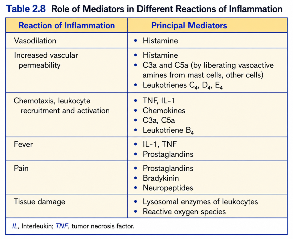
- Many different inflammatory mediators have now been identified, but only some appear to be especially important in acute inflammation in vivo (Table 2.8).
- Different mediators often have overlapping and synergistic actions, helping maintain a strong and reliable inflammatory response.
KEY CONCEPT
- PAF → platelet aggregation + vascular/bronchial effects.
- Tissue injury → clotting + inflammation, and inflammation can increase the risk of thrombosis.
- Kallikrein → kininogen → bradykinin → vasodilation + ↑ vascular permeability + pain.
- Substance P → pain transmission + ↑ vascular permeability.
- Multiple inflammatory mediators work together → strong, effective inflammatory response.
Conceptual Examples
- PAF: PAF released during inflammation → affects platelets and blood vessels → contributes to vascular changes.
- Bradykinin: Tissue injury → kallikrein activation → bradykinin formation → blood vessels dilate + become more permeable + pain occurs.
- Substance P: Sensory nerves activated during injury → substance P released → pain signals + increased vascular permeability.
- Inflammation and clotting: Tissue injury → inflammation and clot formation occur together → endothelial changes may increase the risk of thrombosis.