


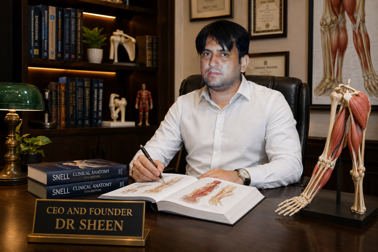
- The upper limb is a freely movable structure with many joints.
- It is attached to the trunk at the shoulder joint.
- Its main function is to move the hand into positions where it can handle and manipulate objects.
- The hand is a highly developed organ.
- It can grasp objects in both coarse (strong) and fine (precise) ways.
- The thumb is especially important because it can move opposite to the other fingers like a pincer.
- This opposable thumb allows the tip of the thumb to touch the tips of the other fingers.
- The upper limb is divided into the shoulder region, the arm, the cubital fossa, the forearm, the wrist, and the hand.
- The arm, forearm, and hand are divided into compartments that work as separate units.
- Each compartment has its own muscles.
- These muscles perform both group actions and individual actions.
- Each compartment has its own nerve supply.
- Each compartment has its own blood supply.
- Physicians commonly see pain in the upper limb.
- Fractures are common injuries of the upper limb.
- Dislocations are common injuries of the upper limb.
- Nerve injuries are also common in the upper limb.
- Wrist injuries need special attention.
- Hand injuries also need special attention.
- Preserving as much thumb function as possible is very important.
KEY CONCEPT
- The upper limb mainly positions the hand to perform different tasks.
- The hand can perform both powerful and delicate movements.
- The opposable thumb is the key feature that makes the hand highly functional.
- The upper limb is organized into different regions and compartments, each with its own muscles, nerves, and blood supply.
- Thumb function should be preserved as much as possible after wrist or hand injuries.
Conceptual Examples
- Holding a hammer = coarse (strong) grasp.
- Holding a pen = fine (precise) grasp.
- Picking up a small coin by touching the thumb to the index finger = opposable thumb (pincer action).
OSTEOLOGY
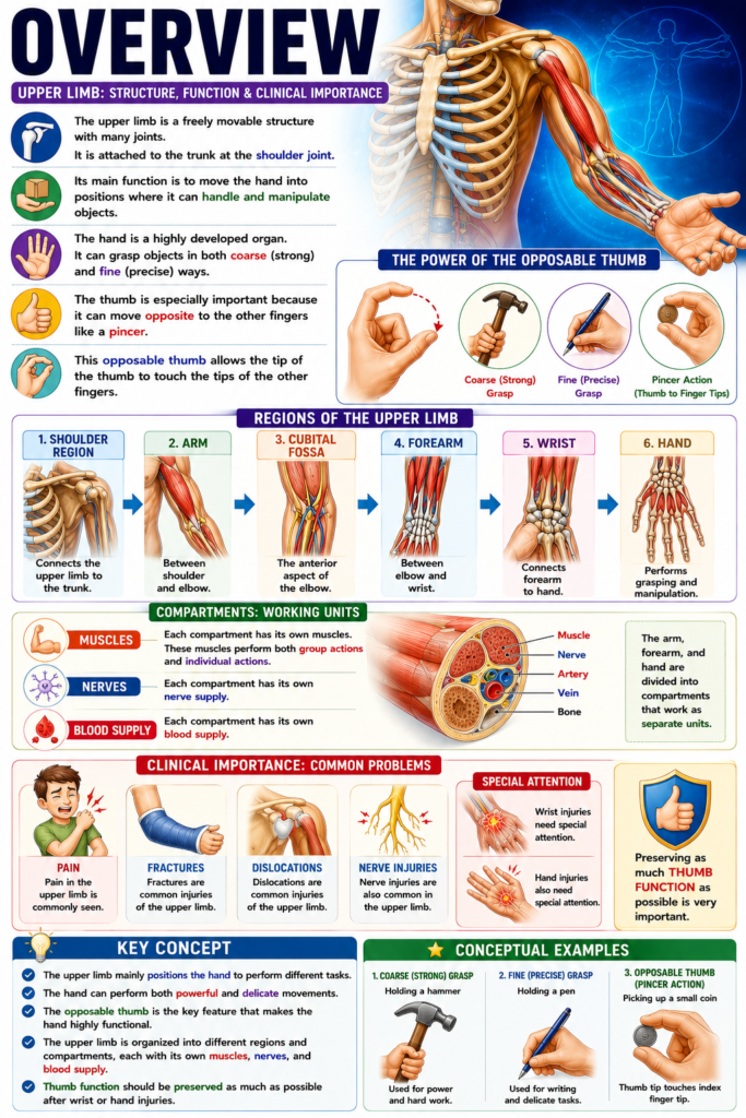
- The upper limb is part of the appendicular skeleton.
- The bones of the upper limb are the clavicle, scapula, humerus, ulna, radius, carpal bones, metacarpal bones, and phalanges.
- The clavicle and scapula together form the shoulder girdle.
- The humerus forms the arm.
- The radius and ulna form the forearm.
- The carpal bones form the wrist.
- The metacarpals and phalanges form the hand.
- This section gives a complete description of the bones of the upper limb and their important features.
- Do not learn these bones by only memorizing difficult words.
- Try to understand the meaning of the anatomical terms.
- For example, understand the difference between a tubercle and a tuberosity.
- Understanding the terms helps you better understand the anatomy.
- Most importantly, ask functional questions while examining the bones.
- Ask yourself whether the bone is from the right side or the left side.
- Ask which bone or structure articulates with this area.
- Ask which muscle, ligament, or other structure attaches to this area.
- Ask whether this structure can be felt (palpated).
- Ask whether this structure can be identified on a standard radiographic image.
- Ask whether there are any important nerves or blood vessels related to this region or structure.
KEY CONCEPT
- The upper limb is made up of the clavicle, scapula, humerus, ulna, radius, carpal bones, metacarpals, and phalanges.
- The shoulder girdle is formed by the clavicle and scapula.
- The humerus forms the arm, the radius and ulna form the forearm, the carpals form the wrist, and the metacarpals and phalanges form the hand.
- Learning the meaning of anatomical terms is better than only memorizing names.
- Always study bones by asking functional questions about side, articulations, attachments, palpation, X-ray appearance, and neurovascular relations.
Conceptual Examples
- Right or left bone: Identify whether a humerus belongs to the right or left upper limb.
- Articulation: The humerus articulates with the scapula at the shoulder joint.
- Attachment: Muscles attach to different bony prominences.
- Palpation: The clavicle can be easily felt under the skin.
- Radiograph: The humerus can be identified on a standard X-ray.
- Neurovascular relation: Important nerves and blood vessels pass close to some upper limb bones.
Clavicle
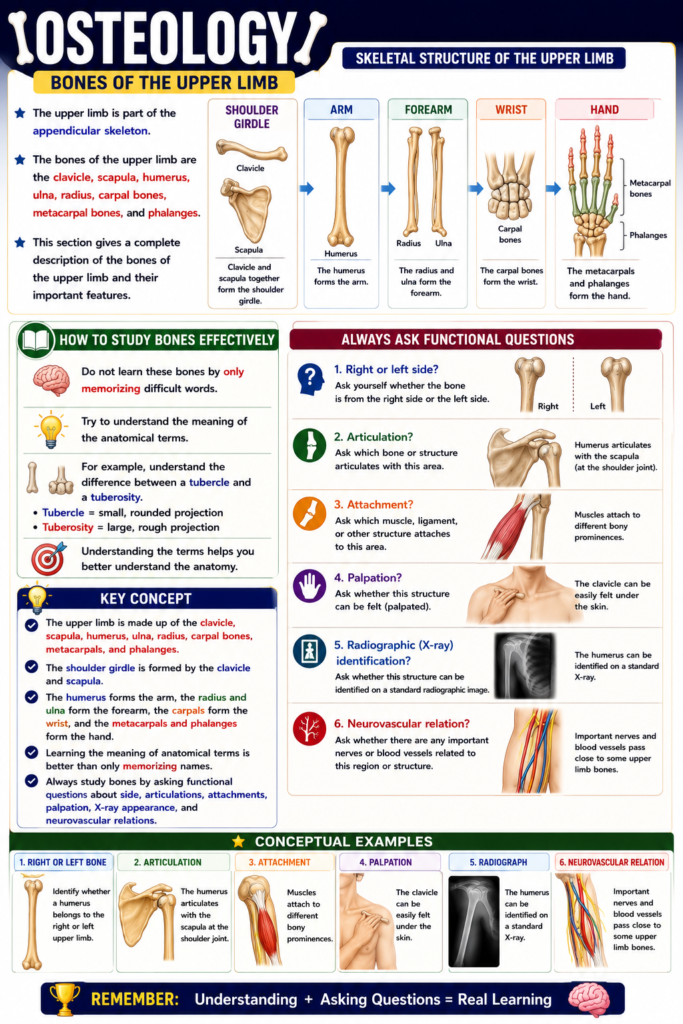
- The clavicle is also called the collar bone.
- The word clavicul- is Latin for “key.”
- The clavicle is located between the sternum and the scapula.
- It lies horizontally across the root of the neck.
- It is roughly S-shaped.
- It looks like a large, old-style key.
- The clavicle acts as a light strut.
- It connects the upper limb to the thorax.
- It allows the upper limb to move freely away from the trunk.
- The clavicle is the first bone to begin ossification.
- The clavicle is just under the skin (subcutaneous).
- It can be easily felt (palpated) along its entire length.
- The sternal extremity (Fig. 3.1) is the blunt, thick, proximal (medial) end of the clavicle.
- It articulates with the clavicular notch of the sternum.
- This joint is a compound synovial joint.
- The joint contains an articular disc.
- The acromial extremity is the flattened distal (lateral) end of the clavicle.
- It articulates with the acromion process of the scapula.
- The conoid tubercle is a small, rough elevation on the inferior surface near the acromial end.
- The word cono- is Greek for “pine cone.”
- The conoid tubercle provides attachment for the conoid ligament.
- The conoid ligament is part of the coracoclavicular ligament.
- The important muscles and ligaments attached to the clavicle are shown in Figures 3.1 and 3.2.
KEY CONCEPT
- The clavicle is the collar bone connecting the upper limb to the thorax.
- It is S-shaped and lies between the sternum and the scapula.
- The sternal extremity (Fig. 3.1) forms a compound synovial joint with the sternum.
- The acromial extremity articulates with the acromion of the scapula.
- The conoid tubercle provides attachment for the conoid ligament of the coracoclavicular ligament.
- The muscles and ligaments attached to the clavicle are shown in Figures 3.1 and 3.2.
Conceptual Examples
- The sternal extremity is the end of the clavicle that joins the sternum.
- The acromial extremity is the end of the clavicle that joins the scapula.
- The conoid tubercle acts as an attachment point where the conoid ligament is fixed.
Figure Number: Fig. 3.1 (mentioned with the sternal extremity), Figures 3.1 and 3.2 (mentioned for muscle and ligament attachments).
Clinical Notes
Clavicle Fracture
- The clavicle acts as a strut that keeps the arm away from the trunk.
- This allows the arm to move freely.
- The clavicle is the only bony connection between the upper limb and the axial skeleton.
- It transfers all forces from the upper limb to the trunk.
- Because of its position, the clavicle is easily injured by trauma.
- It is the most commonly fractured bone in the body.
- A clavicle fracture usually happens after a fall on the shoulder.
- It can also occur after a fall on an outstretched hand.
- The force travels along the clavicle.
- The clavicle usually breaks at its weakest point.
- The weakest point is the junction of the middle one-third and outer one-third.
- After the fracture, the lateral (outer) fragment moves downward because of the weight of the arm.
- The lateral fragment is also pulled medially (toward the body) and forward.
- This pull is produced by the strong adductor muscles of the shoulder joint.
- The pectoralis major is the main muscle responsible for this pull.
- The medial (inner) fragment is pulled upward.
- The sternocleidomastoid muscle causes this upward tilt.
- The supraclavicular nerves lie very close to the clavicle.
- After a clavicle fracture, these nerves may become involved in callus formation.
- This nerve involvement may cause persistent pain over the side of the neck.
KEY CONCEPT
- The clavicle keeps the arm away from the trunk and transfers forces from the upper limb to the axial skeleton.
- It is the most commonly fractured bone because it is easily exposed to trauma.
- Fractures usually occur at the junction of the middle one-third and outer one-third.
- After a fracture:
- The lateral fragment moves downward, medially, and forward.
- The medial fragment moves upward.
- Involvement of the supraclavicular nerves during healing may cause long-lasting pain on the side of the neck.
Conceptual Examples
- Falling directly on the shoulder → Force travels through the clavicle → Fracture occurs at its weakest point.
- After the fracture, the arm hangs down because the lateral fragment is pulled downward by the weight of the arm.
- The sternocleidomastoid muscle pulls the medial fragment upward.
- If the supraclavicular nerves become trapped in the healing callus, pain may continue on the side of the neck.
Clinical Notes
Clavicle Fracture
- The clavicle acts as a strut that keeps the arm away from the trunk.
- This allows the arm to move freely.
- The clavicle is the only bony connection between the upper limb and the axial skeleton.
- It transfers all forces from the upper limb to the trunk.
- Because of its position, the clavicle is easily injured by trauma.
- It is the most commonly fractured bone in the body.
- A clavicle fracture usually occurs after a fall on the shoulder.
- It can also occur after a fall on an outstretched hand.
- The force travels through the clavicle.
- The clavicle usually breaks at its weakest point.
- The weakest point is the junction of the middle one-third and outer one-third.
- After the fracture, the lateral (outer) fragment moves downward because of the weight of the arm.
- The lateral fragment is also pulled medially (toward the body) and forward.
- This pull is caused by the strong adductor muscles of the shoulder joint.
- The pectoralis major is the main muscle responsible for this pull.
- The medial (inner) fragment is pulled upward.
- The sternocleidomastoid muscle causes this upward movement.
- The supraclavicular nerves lie very close to the clavicle.
- After a clavicle fracture, these nerves may become involved in callus formation.
- This nerve involvement may cause persistent pain on the side of the neck.
KEY CONCEPT
- The clavicle keeps the arm away from the trunk and transfers forces from the upper limb to the axial skeleton.
- It is the most commonly fractured bone because it is easily exposed to trauma.
- Most fractures occur at the junction of the middle one-third and outer one-third.
- After a fracture:
- The lateral fragment moves downward, medially, and forward.
- The medial fragment moves upward.
- During healing, involvement of the supraclavicular nerves may cause long-lasting pain on the side of the neck.
Conceptual Examples
- Fall on the shoulder → Force passes through the clavicle → Fracture occurs at the weakest point.
- Weight of the arm → Pulls the lateral fragment downward.
- Pectoralis major muscle → Pulls the lateral fragment medially and forward.
- Sternocleidomastoid muscle → Pulls the medial fragment upward.
- Supraclavicular nerves trapped in healing callus → Persistent pain on the side of the neck.
Scapula
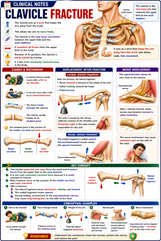
- The scapula is also called the shoulder bone.
- The word scapul- is Latin for “shoulder blade.”
- The scapula is a large, flat, triangular bone.
- It lies on the posterior chest wall between the 2nd and 7th ribs.
- It articulates with the acromial end of the clavicle.
- It also articulates with the head of the humerus.
- The main features of the scapula are:
- Three borders – superior, medial, and lateral.
- Three angles – superior, inferior, and lateral.
- Two surfaces – dorsal and costal.
- Three bony processes – spine, acromion, and coracoid (Fig. 3.3).
- The superior border is the short, thin upper edge of the scapula.
- The scapular notch is present on the lateral part of the superior border near the base of the coracoid process.
- The superior transverse scapular ligament bridges the scapular notch.
- The suprascapular artery passes above the ligament.
- The suprascapular nerve passes below the ligament.
- Remember: Army goes over the bridge; Navy goes under the bridge.
- The medial (vertebral) border is the long edge nearest the vertebral column.
- The lateral (axillary) border is the thick edge nearest the axilla.
- The junction of the superior and medial borders forms the superior angle.
- The junction of the medial and lateral borders forms the inferior angle.
- The inferior angle can be easily felt (palpated) in a living person.
- It marks the level of the 7th rib.
- It also marks the level of the spine of the 7th thoracic vertebra.
- The junction of the superior and lateral borders forms the lateral angle.
- The lateral angle is the thickest and most complex part of the scapula.
- It mainly consists of the head of the scapula.
- The head is connected to the rest of the scapula by the neck of the scapula.
- The lateral surface of the head forms the glenoid cavity (glenoid fossa).
- The word glen- is Greek for “pit” or “socket.”
- The glenoid cavity articulates with the head of the humerus.
- The glenoid labrum is a fibrocartilage ring around the glenoid cavity.
- It broadens and deepens the joint cavity.
- The supraglenoid tubercle is located above the glenoid cavity near the base of the coracoid process.
- The infraglenoid tubercle is located immediately below the glenoid cavity.
- The dorsal (posterior) surface is divided into two unequal regions by the spine of the scapula.
- The area above the spine is the supraspinous fossa.
- The area below the spine is the infraspinous fossa.
- The spine is a large triangular ridge.
- It extends laterally from the medial border to the acromion process.
- The lateral end of the spine joins the neck of the scapula.
- This forms the spinoglenoid (greater scapular) notch.
- The notch connects the supraspinous and infraspinous fossae.
- The suprascapular nerve and vessels pass through this notch between the two fossae.
- The acromion is the broad, flat lateral extension of the spine.
- The word acromi- is Greek for “point of the shoulder.”
- The acromion forms the easily felt tip of the shoulder.
- It partly covers the glenoid cavity.
- It articulates with the clavicle at the acromioclavicular joint.
- The costal (ventral, anterior) surface lies against the posterior surface of the rib cage.
- Most of this surface forms the subscapular fossa.
- The coracoid process is a thick, beak-shaped projection.
- The word coraco- is Greek for “like a crow’s beak.”
- It projects anterolaterally from the junction of the neck and the lateral end of the superior border.
- The coracoid process can be felt by applying deep pressure through the anterior part of the deltoid muscle below the lateral end of the clavicle.
- The main muscles and ligaments attached to the scapula are shown in Figures 3.1 and 3.3.
KEY CONCEPT
- The scapula is a flat triangular bone located on the posterior chest wall.
- It articulates with the clavicle and the humerus.
- It has 3 borders, 3 angles, 2 surfaces, and 3 processes (Fig. 3.3).
- The glenoid cavity forms the socket for the head of the humerus.
- The glenoid labrum makes the socket broader and deeper.
- The spine divides the dorsal surface into the supraspinous and infraspinous fossae.
- The acromion forms the tip of the shoulder and articulates with the clavicle.
- The coracoid process is a beak-like projection for important muscle and ligament attachments.
- Remember: Suprascapular artery passes over the superior transverse scapular ligament, while the suprascapular nerve passes under it.
Conceptual Examples
- Feel the tip of your shoulder → You are touching the acromion.
- Feel the lower point of your shoulder blade on your back → This is the inferior angle.
- The glenoid cavity acts like a shallow socket, while the head of the humerus acts like a ball, together forming the shoulder joint.
- Think of the superior transverse scapular ligament as a bridge:
- Army (artery) goes over the bridge.
- Navy (nerve) goes under the bridge.
Figure Numbers: Fig. 3.3 (main features of the scapula); Figures 3.1 and 3.3 (muscle and ligament attachments).
Clinical Notes
Scapular Fractures
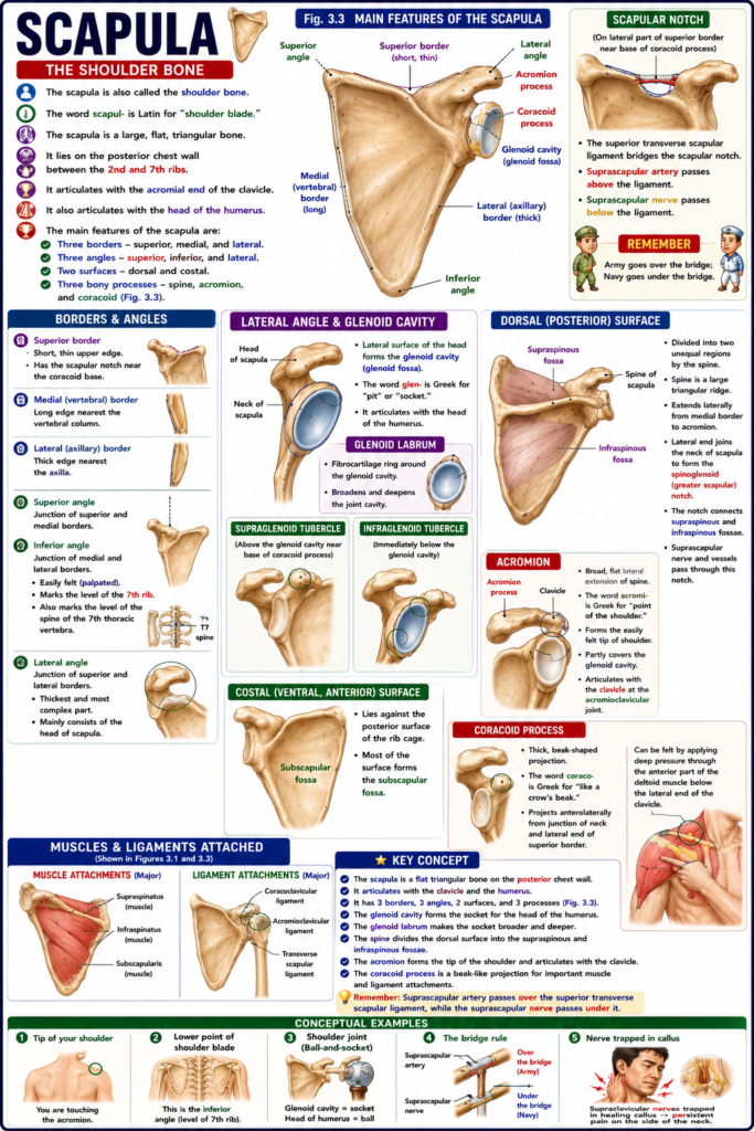
- Scapular fractures usually occur because of severe trauma.
- They are commonly seen in run-over accident victims.
- They are also common in people involved in motor vehicle crashes.
- Scapular fractures are usually associated with fractured ribs.
- Most scapular fractures need little direct treatment.
- The muscles on the anterior and posterior surfaces of the scapula hold the broken pieces in place (splint the fragments).
Dropped Shoulder and Winged Scapula
- The scapula is kept in its normal position on the posterior chest wall by the tone and balance of its attached muscles.
- If one of these muscles becomes paralyzed, this balance is disturbed.
- Dropped shoulder occurs due to paralysis of the trapezius muscle.
- Winged scapula (Fig. 3.4) occurs due to paralysis of the serratus anterior muscle.
- In winged scapula, the scapula projects backward like a wing.
- These muscle imbalances can be detected by a careful physical examination.
KEY CONCEPT
- Scapular fractures are usually caused by severe trauma.
- They are commonly associated with rib fractures.
- Most scapular fractures heal with minimal direct treatment because surrounding muscles stabilize the bone fragments.
- The normal position of the scapula depends on the balance of its muscles.
- Trapezius paralysis → Dropped shoulder.
- Serratus anterior paralysis → Winged scapula (Fig. 3.4).
- Careful physical examination helps identify these muscle imbalances.
Conceptual Examples
- High-speed road traffic accident → Severe trauma → Scapular fracture, often with rib fractures.
- Trapezius muscle paralysis → Shoulder hangs lower than normal → Dropped shoulder.
- Serratus anterior paralysis → Medial border of the scapula sticks out like a wing when pushing against a wall → Winged scapula (Fig. 3.4).
Figure Number: Fig. 3.4 (Winged scapula).
Humerus
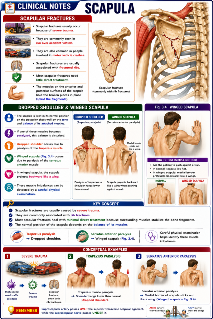
- The humerus is the bone of the arm (brachium).
- The word humer- is Latin for “shoulder.”
- It is the longest bone of the upper limb.
- Proximally, it articulates with the glenoid cavity of the scapula at the glenohumeral (shoulder) joint.
- Distally, it articulates with the head of the radius and the trochlear notch of the ulna at the elbow joint.
- The humerus has three main regions:
- Proximal extremity
- Body (shaft)
- Distal extremity
- The main muscles and ligaments attached to the humerus are shown in Figures 3.1 and 3.5.
Proximal Extremity
- The head is the round, smooth upper end of the humerus.
- It forms about one-third of a sphere.
- It faces medially, superiorly, and slightly posteriorly.
- It articulates with the glenoid cavity of the scapula to form the glenohumeral joint.
- The greater tubercle is a large rough projection on the lateral side of the proximal humerus.
- It lies lateral to the head.
- The lesser tubercle is a small rough projection on the anterior side of the proximal humerus.
- It lies below the head and medial to the greater tubercle.
- The anatomical neck is the slightly narrowed region around the articular surface of the head.
- The articular capsule of the glenohumeral joint attaches along its lower border.
- Fractures of the anatomical neck are rare.
- They are more common in older people.
- The surgical neck is the narrowed area below the greater and lesser tubercles.
- It forms the junction between the proximal end and the shaft.
- It is closely related to the axillary nerve.
- It is also related to the anterior and posterior circumflex humeral vessels.
- Fractures of the surgical neck are common.
- The intertubercular (bicipital) groove is a deep groove on the front of the humerus.
- It lies between the greater and lesser tubercles.
- It contains the tendon of the long head of the biceps brachii muscle.
- It extends into the upper one-third of the shaft.
Body (Shaft)
- The deltoid tuberosity is a rough triangular area on the anterolateral surface of the middle of the shaft.
- The word delt- comes from the triangular Greek letter “delta.”
- It is the attachment site for the deltoid muscle.
- The posterior border of the deltoid tuberosity forms the radial nerve groove.
- The radial groove (spiral groove) is a shallow groove that winds around the back and side of the shaft.
- It is most prominent between the deltoid tuberosity and the upper end of the lateral supracondylar ridge.
- It is closely related to the radial nerve.
- It is also related to the profunda brachii vessels.
- Fractures of the middle shaft are common.
- These fractures often occur below the deltoid tuberosity.
- They may injure the radial nerve and the structures in the radial groove.
- The medial supracondylar ridge extends upward from the medial epicondyle.
- It forms the lower medial border of the humerus.
- The lateral supracondylar ridge extends upward from the lateral epicondyle.
- It forms the lower lateral border of the humerus.
Distal Extremity
- The lateral epicondyle is a small rough projection on the outer side of the distal humerus.
- It lies above the capitulum.
- It can be easily felt (palpated).
- The common extensor tendon attaches here.
- Inflammation of this tendon is called lateral epicondylitis (tennis elbow).
- The medial epicondyle is a large rounded projection on the inner side of the distal humerus.
- It lies above the trochlea.
- It is easily felt and is an important surface landmark.
- The ulnar nerve passes behind it in the ulnar sulcus.
- The ulnar nerve can be injured by trauma or fractures.
- The nerve can be felt behind the medial epicondyle.
- Striking this nerve causes the “funny bone” tingling sensation.
- The tingling is felt along the medial side of the hand and the little finger (fifth digit).
- The capitulum is a rounded articular surface on the outer side of the distal humerus.
- The word capit- is Latin for “little head.”
- It lies lateral to the trochlea.
- It articulates with the head of the radius.
- This joint allows flexion, extension, and rotation.
- The trochlea is a pulley-shaped articular surface on the inner side of the distal humerus.
- The word trochle- is Greek for “pulley.”
- It lies medial to the capitulum.
- It articulates with the trochlear notch of the ulna.
- This joint mainly allows hinge movement.
- The coronoid fossa is a depression above the trochlea on the front of the humerus.
- It receives the coronoid process of the ulna during full elbow flexion.
- The radial fossa is a shallow depression above the capitulum on the front of the humerus.
- It receives the head of the radius during full elbow flexion.
- The olecranon fossa is a deep depression above the trochlea on the back of the humerus.
- It receives the olecranon process of the ulna during elbow extension.
KEY CONCEPT
- The humerus is the longest bone of the upper limb.
- It consists of three regions: proximal extremity, shaft, and distal extremity.
- The head articulates with the glenoid cavity to form the shoulder joint.
- The surgical neck is a common fracture site and is closely related to the axillary nerve.
- The intertubercular groove contains the long head of the biceps tendon.
- The radial groove contains the radial nerve and profunda brachii vessels.
- The lateral epicondyle is the attachment site of the common extensor tendon and is affected in tennis elbow.
- The medial epicondyle is closely related to the ulnar nerve, producing the funny bone sensation when struck.
- The capitulum articulates with the radius, while the trochlea articulates with the ulna.
- The coronoid, radial, and olecranon fossae receive their corresponding bony structures during elbow movements.
Conceptual Examples
- Shoulder joint → Head of the humerus fits into the glenoid cavity like a ball in a shallow socket.
- Surgical neck fracture → May damage the axillary nerve.
- Midshaft fracture → May injure the radial nerve in the radial groove.
- Tennis elbow → Pain occurs where the common extensor tendon attaches to the lateral epicondyle.
- Hit the inside of your elbow → The ulnar nerve is stimulated behind the medial epicondyle, causing the funny bone tingling sensation.
- Bending the elbow fully → The coronoid process and head of the radius fit into the coronoid and radial fossae.
- Straightening the elbow fully → The olecranon process fits into the olecranon fossa.
Figure Numbers: Figures 3.1 and 3.5 (muscle and ligament attachments).
Clinical Notes
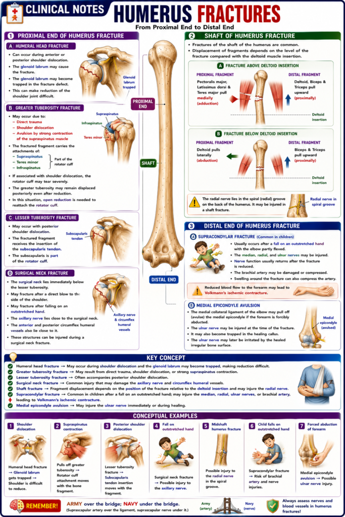
Proximal End of Humerus Fracture
Humeral Head Fracture
- A humeral head fracture can occur during anterior or posterior shoulder dislocation.
- The glenoid labrum may cause the fracture.
- The glenoid labrum may become trapped in the fracture defect.
- This can make reduction of the shoulder joint difficult.
Greater Tuberosity Fracture
- The greater tuberosity may fracture due to direct trauma.
- It may also be displaced by the glenoid labrum during shoulder dislocation.
- It can also be avulsed by a strong contraction of the supraspinatus muscle.
- The fractured bone fragment carries the attachments of the:
- Supraspinatus
- Teres minor
- Infraspinatus
- These tendons form part of the rotator cuff.
- If associated with shoulder dislocation, the rotator cuff may tear severely.
- The greater tuberosity may remain displaced posteriorly even after the shoulder joint is reduced.
- In this situation, open reduction is needed to reattach the rotator cuff.
Lesser Tuberosity Fracture
- A lesser tuberosity fracture may occur with posterior shoulder dislocation.
- The fractured fragment receives the insertion of the subscapularis tendon.
- The subscapularis is part of the rotator cuff.
Surgical Neck Fracture
- The surgical neck lies immediately below the lesser tuberosity.
- It may fracture after a direct blow to the side of the shoulder.
- It may also fracture after falling on an outstretched hand.
- The axillary nerve lies close to the surgical neck.
- The anterior and posterior circumflex humeral vessels also lie close to it.
- These structures can be injured during a surgical neck fracture.
Shaft of Humerus Fracture
- Fractures of the shaft of the humerus are common.
- The displacement of the fragments depends on the level of the fracture compared with the deltoid muscle insertion.
- If the fracture is above the deltoid insertion:
- The pectoralis major, latissimus dorsi, and teres major pull the proximal fragment medially (adduction).
- The deltoid, biceps, and triceps pull the distal fragment upward (proximally).
- If the fracture is below the deltoid insertion:
- The deltoid pulls the proximal fragment laterally (abduction).
- The biceps and triceps pull the distal fragment upward (proximally).
- The radial nerve lies in the spiral (radial) groove on the back of the humerus.
- It may be injured in a shaft fracture.
Distal End of Humerus Fracture
- Supracondylar fractures are common in children.
- They usually occur after a fall on an outstretched hand with the elbow partly flexed.
- The median, radial, and ulnar nerves may be injured.
- Nerve function usually returns after the fracture is reduced.
- The brachial artery may be damaged or compressed.
- Swelling around the fracture can also compress the artery.
- Reduced blood flow to the forearm may lead to Volkmann’s ischemic contracture.
- The medial collateral ligament of the elbow may pull off (avulse) the medial epicondyle if the forearm is forcibly abducted.
- The ulnar nerve may be injured at the time of the fracture.
- It may also become trapped in the healing callus.
- The ulnar nerve may later be irritated by the healed irregular bone surface.
KEY CONCEPT
- Humeral head fracture → May occur during shoulder dislocation and the glenoid labrum may become trapped, making reduction difficult.
- Greater tuberosity fracture → May result from direct trauma, shoulder dislocation, or strong supraspinatus contraction.
- Lesser tuberosity fracture → Often accompanies posterior shoulder dislocation.
- Surgical neck fracture → Common injury that may damage the axillary nerve and circumflex humeral vessels.
- Shaft fracture → Fragment displacement depends on the position of the fracture relative to the deltoid insertion and may injure the radial nerve.
- Supracondylar fracture → Common in children after a fall on an outstretched hand; may injure the median, radial, ulnar nerves, or brachial artery, leading to Volkmann’s ischemic contracture.
- Medial epicondyle avulsion → May injure the ulnar nerve immediately or during healing.
Conceptual Examples
- Shoulder dislocation → Humeral head fracture → Glenoid labrum gets trapped → Shoulder is difficult to reduce.
- Strong contraction of the supraspinatus → Pulls off the greater tuberosity → Rotator cuff attachment moves with the bone fragment.
- Fall on an outstretched hand → Surgical neck fracture → Possible injury to the axillary nerve.
- Midshaft humerus fracture → Possible injury to the radial nerve in the spiral groove.
- Child falls on an outstretched hand → Supracondylar fracture → Risk of brachial artery and nerve injuries.
- Forced abduction of the forearm → Medial epicondyle avulsion → Possible ulnar nerve injury.
MADE BY EASIEST SELF LEARNING CEO AND FOUNDER DR SHEEN
