


FILLING
- The walls of the ureters contain smooth muscle.
- The smooth muscle is arranged in:
- Spiral bundles
- Longitudinal bundles
- Circular bundles
- The muscle is not organized into distinct layers.
- The ureters produce regular peristaltic contractions.
- These contractions occur about 1–5 times per minute.
- Peristaltic contractions push urine from the renal pelvis to the bladder.
- Urine enters the bladder in small spurts.
- Each spurt occurs at the same time as a peristaltic wave.
- The ureters pass obliquely through the wall of the bladder.
- There are no true ureteral sphincters.
- The oblique course of the ureters through the bladder wall keeps the ureters closed most of the time.
- The ureters open only during peristaltic waves to allow urine to enter the bladder.
- This arrangement prevents the backflow (reflux) of urine from the bladder into the ureters.
KEY CONCEPT
- Urine is transported from the kidneys to the bladder by regular peristaltic contractions of the ureters. The ureters enter the bladder obliquely, creating a valve-like mechanism that prevents urine from flowing backward (vesicoureteral reflux) while allowing urine to enter the bladder during each peristaltic wave.
THE BLADDER
EMPTYING
- The bladder wall contains smooth muscle arranged in:
- Spiral bundles
- Longitudinal bundles
- Circular bundles
- The circular smooth muscle is called the detrusor muscle.
- The detrusor muscle is mainly responsible for emptying the bladder during urination (micturition).
- Some smooth muscle bundles pass on both sides of the urethra.
- These muscle bundles are sometimes called the internal urethral sphincter.
- However, they do not completely surround the urethra.
- Further along the urethra is the external urethral sphincter.
- The external urethral sphincter is made of skeletal muscle.
- The bladder lining (epithelium) consists of:
- A superficial layer of flat cells.
- A deep layer of cuboidal cells.
- The nerve supply of the bladder is shown in Figure 37–19.
- Micturition is mainly a spinal reflex.
- This spinal reflex is controlled by higher brain centres.
- Urination can be voluntarily started or delayed.
- As urine enters the bladder, the bladder can store a large volume without a significant increase in pressure.
- The bladder muscle has the property of plasticity.
- When the bladder is stretched, the initial increase in muscle tension gradually decreases.
- Bladder pressure can be measured by cystometry.
- In cystometry, the bladder is emptied with a catheter and then filled in 50-mL steps with water or air while pressure is recorded.
- A graph showing bladder pressure against bladder volume is called a cystometrogram.
- The cystometrogram has three parts:
- An initial slight rise in pressure.
- A long, nearly flat phase while the bladder continues to fill.
- A sudden sharp rise in pressure when the micturition reflex begins.
- These three parts are called:
- Segment Ia
- Segment Ib
- Segment II
- The first desire to urinate is usually felt when the bladder contains about 150 mL of urine.
- A strong feeling of bladder fullness is usually felt at about 400 mL.
- The nearly flat part of the cystometrogram (Segment Ib) follows the Law of Laplace.
- According to the Law of Laplace: Pressure = (2 × Wall Tension) ÷ Radius
- As the bladder fills:
- Wall tension increases.
- Bladder radius also increases.
- Therefore, bladder pressure rises only slightly until the bladder is almost full.
- During normal urination:
- The pelvic floor muscles relax.
- The external urethral sphincter relaxes.
- The detrusor muscle contracts.
- Urine flows through the urethra.
- The smooth muscle bundles beside the urethra have little role in urination.
- In males, their main function is to prevent semen from flowing backward into the bladder during ejaculation.
- The exact mechanism that starts voluntary urination is not completely understood.
- One of the first events is relaxation of the pelvic floor muscles.
- This relaxation may help trigger detrusor muscle contraction.
- The external urethral sphincter and perineal muscles can be voluntarily contracted.
- This can prevent urination or stop urine flow after urination has started.
- Adults learn to keep the external urethral sphincter contracted until an appropriate time to urinate.
- After urination:
- In females, the urethra empties by gravity.
- In males, the bulbocavernosus muscle contracts several times to expel the remaining urine from the urethra.
Figure: Figure 37–19, Figure 37–20
KEY CONCEPT
- Bladder emptying (micturition) is a spinal reflex controlled by higher brain centres. The detrusor muscle contracts while the pelvic floor muscles and external urethral sphincter relax, allowing urine to pass. The bladder stores urine with very little increase in pressure because of its plasticity and the Law of Laplace. Adults voluntarily delay urination by keeping the external urethral sphincter contracted until an appropriate time to void.
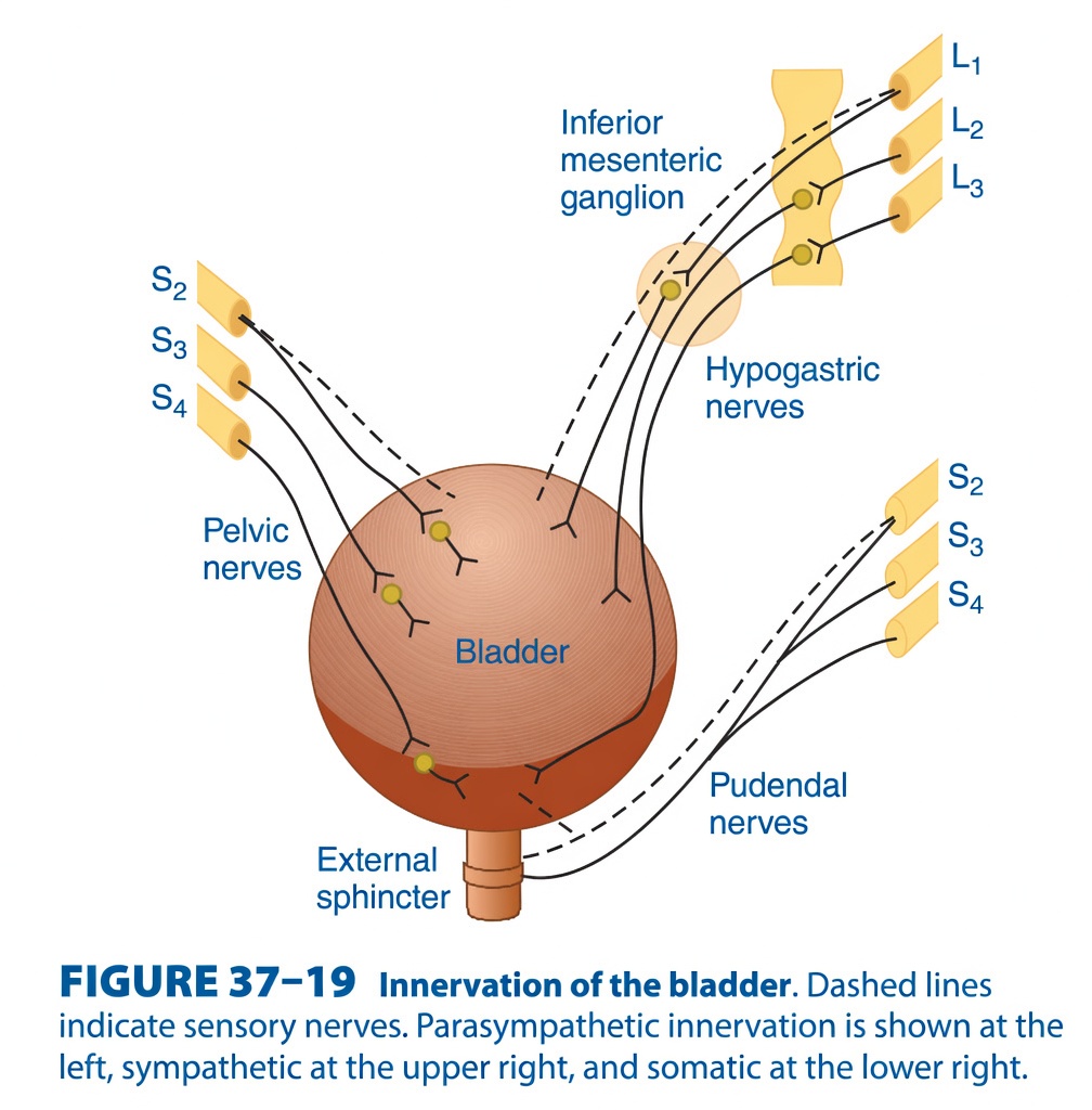
Figure 37-19: Innervation of the Bladder
Easiest & Most Conceptual Explanation for self learners
This figure explains which nerves control the bladder and urination (micturition).
The bladder is controlled by three different nerve systems, and each has one simple job.
Think of the bladder as a water tank with one door.
- One nerve fills the tank.
- One nerve empties the tank.
- One nerve locks or unlocks the door.
⭐ One-Line Concept
Parasympathetic = Empty the bladder
Sympathetic = Fill the bladder
Somatic = Voluntary control of urine
First Understand the Picture
The figure shows:
        Brain & Spinal Cord

        │
        │
   -----------------
   |               |
Sympathetic    Parasympathetic
(L1–L3)          (S2–S4)

        ↓
      Bladder

        ↓
External Sphincter
        ↑
   Pudendal Nerve
     (S2–S4)
There are three separate nerve pathways going to the bladder.
Three Main Nerves
| Nerve | Spinal Level | Function |
|---|---|---|
| Pelvic nerve | S2–S4 | Empty bladder |
| Hypogastric nerve | L1–L3 | Fill bladder |
| Pudendal nerve | S2–S4 | Control external sphincter |
The Dashed Lines
Notice the dashed lines.
These represent:
Sensory (Afferent) nerves
They carry information:
Bladder
    ↓
Spinal cord
They tell the brain:
“The bladder is filling.”
The Solid Lines
The solid lines are:
Motor (Efferent) nerves
They carry commands:
Brain/Spinal Cord
        ↓
Bladder
They tell the bladder what to do.
1. Parasympathetic Nerves (Pelvic Nerves)
Look at the left side.
They arise from:
S2
S3
S4
These join to form:
Pelvic nerves
They go directly to the bladder.
Main Job
They cause:
✅ Bladder contraction
AND
✅ Internal sphincter relaxation
Result:
Urination occurs.
Easy Memory
Pelvic nerve = Pee nerve
Easy Analogy
Imagine squeezing a water balloon.
The bladder muscle contracts.
Urine comes out.
Parasympathetic Summary
S2–S4
     ↓
Pelvic nerve
     ↓
Detrusor contracts
     ↓
Bladder empties
2. Sympathetic Nerves (Hypogastric Nerves)
Look at the upper right side.
Origin:
L1
L2
L3
They first synapse in:
Inferior Mesenteric Ganglion
Then travel through:
Hypogastric nerves
to the bladder.
Main Job
They help store urine.
They cause:
- Relaxation of detrusor muscle
- Contraction of internal urethral sphincter
Result:
The bladder fills.
Easy Memory
Hypogastric nerve = Hold urine
Easy Analogy
Imagine tying the mouth of a balloon.
Water cannot escape.
Sympathetic Summary
L1–L3
      ↓
Hypogastric nerve
      ↓
Bladder relaxes
      ↓
Internal sphincter closes
      ↓
Urine stored
3. Somatic Nerves (Pudendal Nerve)
Look at the lower right side.
Origin:
S2
S3
S4
These form:
Pudendal nerve
It goes to the:
External urethral sphincter
Main Job
This is voluntary control.
It allows you to:
- Hold urine
- Start urination when appropriate
Easy Concept
This is the only muscle you consciously control.
Easy Analogy
Think of the external sphincter as a door lock.
You decide:
- Lock it → Hold urine
- Unlock it → Pass urine
Pudendal Summary
S2–S4
      ↓
Pudendal nerve
      ↓
External sphincter
      ↓
Voluntary control
What Happens While the Bladder Is Filling?
Sympathetic dominates.
Hypogastric nerve

↓

Bladder relaxes

↓

Internal sphincter closes
At the same time
Pudendal nerve

↓

External sphincter contracts
Result:
Urine is stored.
What Happens During Urination?
Now the bladder becomes full.
Stretch receptors are activated.
Sensory signals travel to:
S2–S4The spinal cord activates:
Parasympathetic nerves.
Now:
Pelvic nerve

↓

Detrusor contracts
Meanwhile
Internal sphincter relaxesThen
You voluntarily relax
External sphincterUrine flows out.
Complete Story of Micturition
Bladder fills
      ↓
Stretch receptors activated
      ↓
Sensory impulses
      ↓
S2–S4 spinal cord
      ↓
Parasympathetic activated
      ↓
Detrusor contracts
      ↓
Internal sphincter relaxes
      ↓
External sphincter voluntarily relaxes
      ↓
Urination
Everyday Analogy
Imagine a water tank with a tap.
During Filling
- Tank expands
- Tap remains closed
- Door is locked
No water comes out.
During Emptying
- Tank is squeezed
- Tap opens
- Door unlocks
Water flows out.
Exactly how the bladder works.
Easy Memory Trick
Three Ps
Pelvic = Pee
S2–S4

Contracts bladder
Pudendal = Permission
S2–S4

You decide when to urinate.
Hypogastric = Hold
L1–L3

Stores urine.
Comparison Table
| Nerve | Origin | Type | Main Action | Result |
|---|---|---|---|---|
| Pelvic nerve | S2–S4 | Parasympathetic | Contracts detrusor, relaxes internal sphincter | Urination |
| Hypogastric nerve | L1–L3 | Sympathetic | Relaxes detrusor, contracts internal sphincter | Urine storage |
| Pudendal nerve | S2–S4 | Somatic | Contracts external sphincter | Voluntary continence |
High-Yield Exam Points
- The bladder receives parasympathetic, sympathetic, and somatic innervation.
- Pelvic nerves (S2–S4) provide parasympathetic innervation and stimulate detrusor muscle contraction, promoting bladder emptying.
- Hypogastric nerves (L1–L3) provide sympathetic innervation, causing detrusor relaxation and internal urethral sphincter contraction, facilitating urine storage.
- Pudendal nerves (S2–S4) provide somatic innervation to the external urethral sphincter, allowing voluntary control of micturition.
- The dashed lines in the figure represent sensory (afferent) fibers, which carry bladder stretch information from the bladder to the spinal cord.
- Normal micturition requires coordinated activation of the parasympathetic pathway and inhibition of the sympathetic and somatic pathways.
KEY CONCEPT (Figure 37-19)
Figure 37-19 demonstrates the three neural pathways controlling bladder function. The pelvic nerves (S2–S4) carry parasympathetic fibers that contract the detrusor muscle and promote bladder emptying. The hypogastric nerves (L1–L3) carry sympathetic fibers that relax the detrusor muscle and contract the internal urethral sphincter, allowing urine storage. The pudendal nerves (S2–S4) provide somatic innervation to the external urethral sphincter, enabling voluntary control of urination. Sensory fibers from the bladder travel to the sacral spinal cord and initiate the micturition reflex when bladder filling reaches an appropriate level.
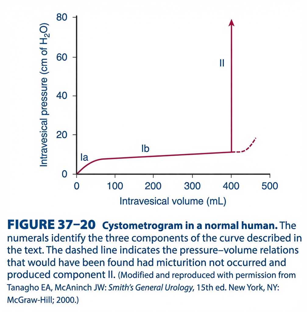
Figure 37-20 – Cystometrogram (Pressure–Volume Curve of the Urinary Bladder)
Easy Conceptual Summary for self learners
This figure shows how the pressure inside the urinary bladder changes as the bladder fills with urine.
It is called a cystometrogram.
The graph demonstrates an important property of the bladder:
The bladder can store a large volume of urine with only a very small increase in pressure because it is highly compliant (stretchable).
Basic Concept
Imagine an empty balloon.
When you first start filling it with water:
- The pressure rises slightly.
- Then the balloon stretches easily.
- A lot more water can enter without much increase in pressure.
- Finally, when it becomes almost full, it can no longer stretch, and the pressure rises rapidly.
The urinary bladder behaves almost exactly like this balloon.
Understanding the Axes
X-axis
Intravesical Volume (mL)
This shows:
How much urine is inside the bladder.
Moving to the right means:
➡️ The bladder is filling.
Y-axis
Intravesical Pressure (cm H₂O)
This shows:
The pressure inside the bladder.
Higher on the graph means:
➡️ Higher bladder pressure.
The Graph Has Three Components
The graph is divided into:
- Component Ia
- Component Ib
- Component II
Each represents a different stage of bladder filling.
Component Ia – Initial Rise in Pressure
This is the first small upward curve.
What happens?
When the first urine enters the empty bladder,
pressure rises quickly from:
0 → about 5–7 cm H₂O
Why?
Initially,
the bladder wall is relaxed and folded.
When urine first enters,
these folds begin to unfold.
This causes a small increase in pressure.
Easy Concept
Think of opening a folded shopping bag.
At first,
it takes a little effort.
Once opened,
it expands much more easily.
Key Point
Small amount of urine
↓
Initial stretching
↓
Small pressure rise
Component Ib – Plateau (High Compliance Phase)
This is the long, almost horizontal part of the graph.
What happens?
The bladder volume increases from about:
50 mL → 400 mL
Yet,
pressure increases only slightly
(from about 7 → 10 cm H₂O).
Why?
This is due to:
Stress Relaxation (Plasticity)
The bladder smooth muscle gradually relaxes after being stretched.
Therefore,
even though more urine enters,
the muscle adapts,
so pressure hardly increases.
This property is called:
Bladder Compliance
High compliance means:
Large increase in volume
↓
Very small increase in pressure
Easy Concept
Imagine a new rubber balloon.
You keep adding water,
but the pressure hardly changes because the balloon stretches easily.
Clinical Importance
Because of this high compliance,
we can comfortably store:
300–400 mL
without continuously feeling pain or urgency. Point
Large increase in volume
↓
Only small increase in pressure
↓
Normal urine storageomponent II – Sudden Vertical Rise
This is the tall vertical arrow.
What happens?
At around:
350–400 mL
the pressure suddenly rises dramatically.
Why?
The bladder has reached its functional limit.
Stretch receptors become strongly activated.
These receptors send signals to the spinal cord.
This triggers the:
Micturition Reflex
What happens during the reflex?
- Detrusor muscle contracts.
- Internal urethral sphincter relaxes.
- External urethral sphincter is voluntarily relaxed.
- Urination begins.
Easy Concept
Imagine filling a water balloon until it can no longer stretch.
Suddenly,
the pressure rises sharply,
and the water is forced out.
Key Point
Bladder almost full
↓
Stretch receptors activated
↓
Micturition reflex begins
↓
Pressure rises suddenly
↓
Urination occurs
What Does the Dashed Line Mean?
The graph also shows a dashed line after 400 mL.
What does it represent?
It shows what would happen
if urination did not occur.
Normally,
the micturition reflex empties the bladder.
But if urine were not passed,
the bladder would continue filling.
Eventually,
pressure would rise gradually because the bladder can no longer stretch efficiently.
Easy Concept
Imagine continuing to inflate a balloon after it is already full.
Now,
every small amount of extra air causes a big increase in pressure.
Understanding Bladder Compliance
Compliance tells us:
How easily the bladder stretches.
Formula:Compliance=ΔPΔV
Where:
- ΔV = Change in volume
- ΔP = Change in pressure
High Compliance
Large volume increase
Small pressure increase
Normal bladder
Low Compliance
Small volume increase
Large pressure increase
Diseased bladder
Clinical Importance
1. Normal Bladder
- High compliance
- Stores 400–500 mL comfortably
- Low pressure during filling
2. Neurogenic Bladder
Compliance decreases.
Pressure rises rapidly.
Patient develops:
- Frequency
- Urgency
- Incontinence
3. Fibrotic Bladder
The bladder becomes stiff.
It cannot stretch well.
Even small amounts of urine produce high pressure.
4. Bladder Outlet Obstruction
Over time,
the bladder wall hypertrophies.
Compliance decreases,
and pressure rises earlier during filling.
Comparison of the Three Components
| Component | What Happens? | Pressure Change |
|---|---|---|
| Ia | Initial filling | Small rise |
| Ib | Bladder stretches easily (high compliance) | Almost constant |
| II | Micturition reflex starts | Sudden large rise |
Easy Memory Trick
Ia = “Initial Stretch” 🎈
Small pressure increase.
Ib = “Big Storage” 🫙
Large urine storage.
Very little pressure increase.
II = “It’s Time to Pee!” 🚽
Stretch receptors activated.
Pressure shoots up.
Urination begins.
Flow Diagram
Empty bladder
      ↓
Urine begins entering
      ↓
Small rise in pressure (Ia)
More urine enters
↓
Bladder stretches easily
↓
Pressure remains nearly constant (Ib)Bladder nearly full (≈350–400 mL)
↓
Stretch receptors activated
↓
Micturition reflex starts
↓
Detrusor contracts
↓
Pressure rises sharply (II)
↓
UrinationQuick Summary Table
| Bladder Volume | Bladder Pressure | Reason |
|---|---|---|
| 0–50 mL | Slight increase | Initial stretching |
| 50–400 mL | Almost constant | High compliance and stress relaxation |
| 400–500 mL | Sudden increase | Micturition reflex activation |
Key Concept
A cystometrogram illustrates the relationship between bladder volume and intravesical pressure during bladder filling. Initially (Component Ia), the pressure rises slightly as the bladder begins to stretch. During Component Ib, the bladder exhibits high compliance (stress relaxation), allowing it to store 300–400 mL of urine with only a minimal increase in pressure, making urine storage comfortable. When the bladder approaches its capacity (about 350–400 mL), stretch receptors are strongly activated, triggering the micturition reflex. This produces Component II, in which intravesical pressure rises sharply because the detrusor muscle contracts, initiating urination. The dashed line indicates the pressure-volume relationship that would occur if micturition did not take place and the bladder continued to fill, leading to a further gradual increase in pressure.
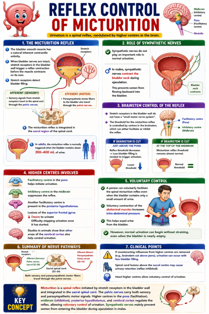
REFLEX CONTROL
- The bladder smooth muscle can contract on its own (inherent contractile activity).
- When the bladder nerves are intact, stretch receptors in the bladder wall trigger a reflex contraction before the muscle contracts on its own.
- Stretch receptors detect bladder filling.
- The sensory (afferent) signals travel through the pelvic nerves.
- The motor (efferent) parasympathetic fibers to the bladder also travel through the pelvic nerves.
- The micturition reflex is integrated in the sacral region of the spinal cord.
- In adults, the micturition reflex is normally triggered when the bladder contains about 300–400 mL of urine.
- Sympathetic nerves do not play an important role in normal urination.
- In males, sympathetic nerves contract the bladder neck during ejaculation.
- This prevents semen from flowing backward into the bladder.
- The stretch receptors in the bladder wall do not have a small motor nerve system.
- The threshold for the micturition reflex is controlled by centres in the brainstem.
- These brainstem centres can either facilitate or inhibit the reflex.
- A facilitatory centre is located in the pons.
- An inhibitory centre is located in the midbrain.
- If the brainstem is cut just above the pons, the reflex threshold decreases.
- As a result, less bladder filling is needed to trigger urination.
- If the brainstem is cut at the top of the midbrain, the micturition reflex threshold remains almost normal.
- Another facilitatory centre is present in the posterior hypothalamus.
- People with lesions of the superior frontal gyrus have a reduced desire to urinate.
- They also have difficulty stopping urination once it has started.
- Studies in animals show that other areas of the cerebral cortex also help control urination.
- A person can voluntarily facilitate the spinal micturition reflex even when the bladder contains only a small amount of urine.
- Voluntary contraction of the abdominal muscles increases intra-abdominal pressure.
- This helps expel urine from the bladder.
- However, normal urination can begin without straining, even when the bladder is nearly empty.
KEY CONCEPT
- Micturition is a spinal reflex initiated by stretch receptors in the bladder wall and integrated in the sacral spinal cord. The pelvic nerves carry both sensory and parasympathetic motor signals. Higher centres in the pons, midbrain, hypothalamus, and cerebral cortex regulate the reflex, allowing voluntary control of urination. Sympathetic nerves mainly prevent semen from entering the bladder during ejaculation in males.
EFFECTS OF DEAFFERENTATION
- Deafferentation means the sensory (afferent) nerves from the bladder are interrupted.
- This may occur when the sacral dorsal roots are cut in experimental animals.
- It can also occur in diseases affecting the dorsal roots, such as tabes dorsalis.
- When the sensory nerves are interrupted, all reflex contractions of the bladder are lost.
- The bladder becomes greatly distended (overfilled).
- The bladder wall becomes thin.
- The bladder becomes hypotonic (reduced muscle tone).
- Despite the loss of reflexes, some bladder contractions still occur.
- These contractions are produced by the bladder smooth muscle’s own response to stretching.
EFFECTS OF DENERVATION
- Denervation means both the sensory (afferent) and motor (efferent) nerves of the bladder are destroyed.
- This may occur due to tumors affecting the cauda equina or the filum terminale.
- Immediately after denervation, the bladder becomes flaccid (soft) and distended.
- Gradually, the bladder muscle becomes active again without nerve control.
- The decentralized bladder develops many spontaneous contraction waves.
- These contractions expel only small dribbles of urine through the urethra.
- Over time, the bladder becomes small (shrunken).
- The bladder wall becomes thickened (hypertrophied).
- The exact reason why a denervated bladder becomes small and hypertrophied, while a deafferented bladder becomes large and hypotonic, is not fully understood.
- The increased activity of the denervated bladder suggests the development of denervation hypersensitivity.
- This occurs even though the interrupted nerve fibers are preganglionic neurons.
KEY CONCEPT
- Deafferentation removes only the sensory nerve supply, abolishing the micturition reflex and producing a large, thin-walled, hypotonic bladder with only weak stretch-induced contractions. Denervation removes both sensory and motor nerves, initially causing a flaccid bladder, but later the bladder develops spontaneous contractions, becomes shrunken, and its wall becomes hypertrophied because of denervation hypersensitivity.
EFFECTS OF SPINAL CORD TRANSECTION
- During spinal shock, the bladder is flaccid and unresponsive.
- It becomes overfilled, and urine dribbles through the sphincters (overflow incontinence).
- After spinal shock has passed, the voiding reflex returns.
- There is no voluntary control and no inhibition or facilitation from higher centers when the spinal cord is transected.
- Some paraplegic patients train themselves to initiate voiding by pinching or stroking their thighs, provoking a mild mass reflex (see Chapter 12).
- In some instances, the voiding reflex becomes hyperactive.
- Bladder capacity is reduced, and the wall becomes hypertrophied.
- This type of bladder is sometimes called the spastic neurogenic bladder.
- The reflex hyperactivity is made worse by, and may be caused by, infection in the bladder wall.
KEY CONCEPT Spinal cord transection first causes a flaccid, overfilled bladder with overflow incontinence during spinal shock; later the voiding reflex returns without voluntary control and may become hyperactive, leading to a small, thickened spastic neurogenic bladder.
CLINICAL BOX 37–4
Abnormalities of Micturition
- Three major types of bladder dysfunction are caused by damage to the nervous system.
- Type 1: Interruption of the sensory (afferent) nerves from the bladder.
- Type 2: Interruption of both the sensory (afferent) and motor (efferent) nerves to the bladder.
- Type 3: Interruption of the facilitatory and inhibitory nerve pathways descending from the brain to the spinal cord.
- In all three types of bladder dysfunction, the bladder is still able to contract.
- However, these contractions are usually too weak to empty the bladder completely.
- As a result, some urine remains in the bladder after urination.
- This remaining urine is called residual urine.
KEY CONCEPT
- Neural lesions can cause three major types of bladder dysfunction: interruption of sensory nerves, interruption of both sensory and motor nerves, or interruption of brain pathways controlling micturition. In all three conditions, bladder contractions are usually incomplete, leading to residual urine remaining in the bladder after voiding.
Made by self learning CEO and founder Dr sheen
