


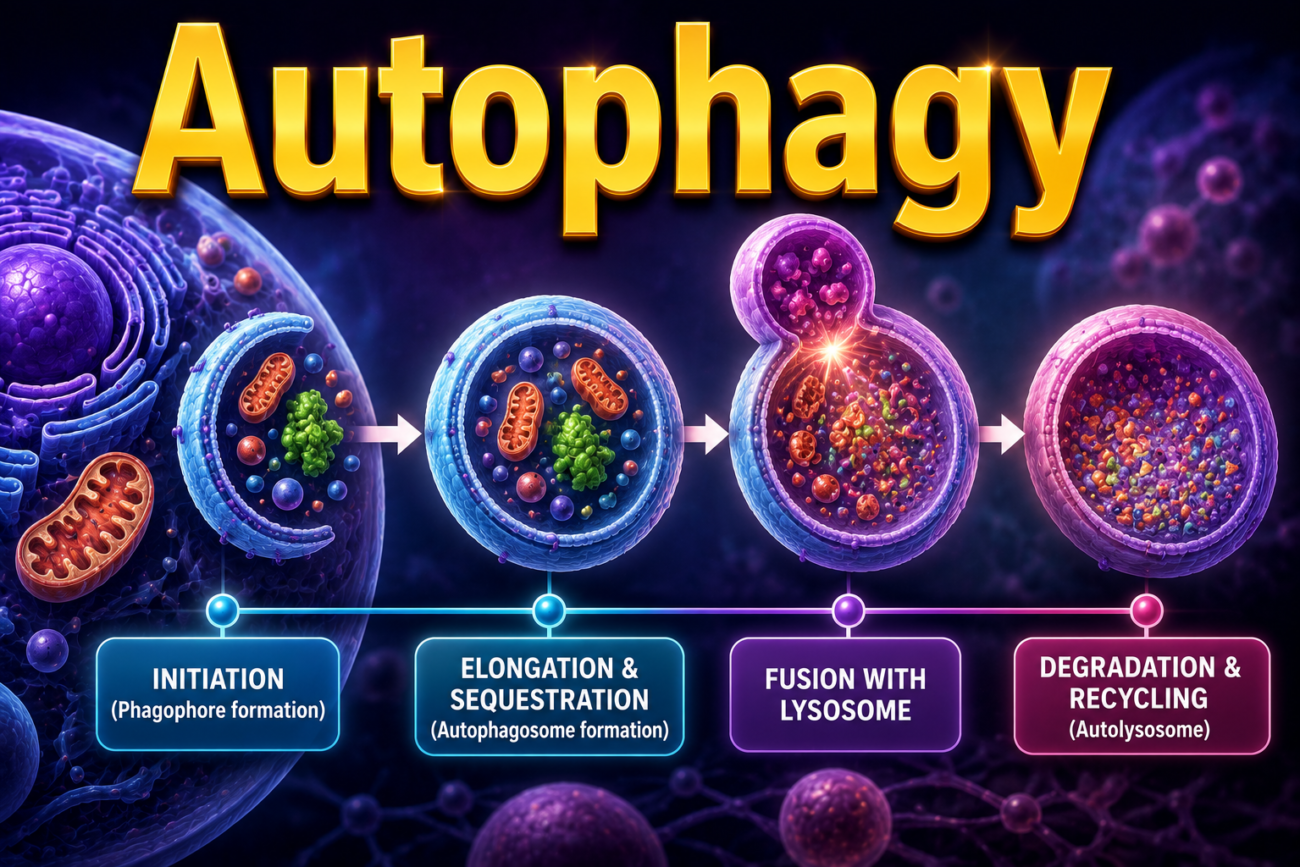
- Autophagy means “self-eating.”
- It is the process in which lysosomes digest the cell’s own components.
- Autophagy is a survival mechanism during nutrient deprivation (starvation).
- It helps the cell stay alive by:
- Breaking down its own contents.
- Recycling them to produce nutrients and energy.
- During autophagy:
- Cell organelles and parts of the cytoplasm are enclosed by a double membrane derived from the endoplasmic reticulum (ER).
- This membrane is called the phagophore.
- The phagophore develops into an autophagic vacuole (autophagosome).
- Formation of the autophagosome is started by cytosolic proteins that detect nutrient deprivation (Fig. 1.14).
- The autophagosome then fuses with lysosomes.
- This forms an autophagolysosome.
- Lysosomal enzymes digest the enclosed cellular components.
- In some situations, autophagy is associated with tissue atrophy.
- In these cases, it acts as an adaptation that helps cells survive during periods of limited nutrients.
- If starvation continues and the cell can no longer survive by digesting its own contents:
- Autophagy may lead to apoptotic cell death.
- Extensive autophagy is seen in:
- Ischemic injury.
- Some types of myopathies.
- In infected cells:
- Autophagic vacuoles can surround microbes.
- This helps destroy the infectious organisms.
- Cancer cells can survive under stressful conditions without depending on autophagy.
- Autophagy, once thought to be a minor survival process, is now recognized as having important roles in many human diseases.
KEY CONCEPT
- Autophagy (“self-eating”) is the lysosomal digestion of the cell’s own components.
- It helps cells survive starvation by recycling nutrients and energy.
- Autophagosomes form from the ER and later fuse with lysosomes to digest cell contents (Fig. 1.14).
- Autophagy may:
- Help cells survive during nutrient deficiency.
- Contribute to apoptosis if starvation is prolonged.
- It is commonly seen in:
- Ischemic injury.
- Some myopathies.
- Destruction of microbes inside infected cells.
- Autophagy also plays an important role in human diseases, including cancer.
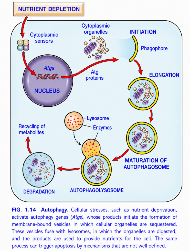
Autophagy (Figure 1.14) – The Cell’s Self-Recycling System
This figure explains Autophagy, which literally means:
“Auto” = Self
“Phagy” = Eating
So,
Autophagy = Self-Eating
But don’t misunderstand it.
The cell is not committing suicide.
Instead,
it is recycling its old or damaged parts to survive during starvation or stress.
Main Concept
When the cell has little food or damaged organelles, it “eats” and recycles its own old components to produce energy and survive.
Think of it as the cell’s recycling factory.
The Whole Story in One Sentence
No Food
   ↓
Cell Detects Starvation
   ↓
Collects Old Organelles
   ↓
Wraps Them in a Membrane
   ↓
Fuses with Lysosome
   ↓
Digests Them
   ↓
Recycles Nutrients
   ↓
Cell Survives
Think of the Cell as a House
Imagine your house has:
- Old furniture 🪑
- Broken TV 📺
- Damaged refrigerator ❄️
You don’t throw away the entire house.
Instead,
you recycle the old items
and use the materials again.
The cell does exactly the same thing.
Step-by-Step Explanation of the Figure
STEP 1 – Nutrient Depletion
At the top,
the figure shows:
Nutrient Depletion
This means
the cell has very little food.
Examples
- Starvation
- Fasting
- Severe illness
- Lack of oxygen
- Cellular stress
Easy Concept
Imagine
your wallet becomes empty.
You cannot buy food.
So you start using old things in your house.
The cell does exactly this.
STEP 2 – Cytoplasmic Sensors
The small colored boxes represent
cytoplasmic sensors.
These sensors constantly monitor:
- Nutrient level
- Energy level
- Stress
Their Job
They ask:
“Do we have enough food?”
If the answer is:
❌ No
they activate autophagy.
Easy Concept
They are like
food detectors inside the cell.
STEP 3 – Activation of Atg Genes (Autophagy Genes)
The signal reaches the nucleus.
Inside the nucleus are
Atg genes (Autophagy-related genes).
What do these genes do?
They produce
Atg proteins.
These proteins build the autophagy machinery.
Easy Concept
The nucleus is like
the factory manager.
Sensors report:
“We are starving.”
The manager orders:
“Start recycling!”
STEP 4 – Atg Proteins are Produced
The Atg genes make
Atg proteins.
These proteins are the
construction workers
that build the recycling vesicle.
Easy Concept
Atg proteins are like
workers who build a garbage collection bag.
STEP 5 – Initiation (Formation of the Phagophore)
The figure now shows
Initiation
with a structure called the
Phagophore.
What is a Phagophore?
It is
a small cup-shaped membrane.
This is the beginning of the recycling bag.
Easy Concept
Imagine opening
a plastic shopping bag.
Initially,
it is only partly open.
That is the phagophore.
STEP 6 – Elongation
The membrane grows larger.
It surrounds:
- Old mitochondria
- Damaged ER
- Protein aggregates
- Old organelles
Easy Concept
The garbage bag becomes bigger
until all the waste fits inside.
STEP 7 – Maturation of Autophagosome
The membrane completely closes.
Now it forms
a double-membrane vesicle
called the
Autophagosome.
Important Point
The autophagosome
does NOT digest anything.
It only stores the material.
Easy Concept
It is like
a sealed garbage bag.
The garbage is inside,
but nothing has been destroyed yet.
STEP 8 – Fusion with Lysosome
Now the autophagosome
joins with
the lysosome.
The figure labels this new structure
Autophagolysosome.
What is the Lysosome?
The lysosome is the
digestive stomach of the cell.
It contains many powerful digestive enzymes.
Easy Concept
Imagine taking the garbage bag
to a recycling factory.TEP 9 – Enzymes Digest the Contents
Lysosomal enzymes digest
everything inside.
Examples:
- Proteins
- Lipids
- Old mitochondria
- Damaged organelles
Everything is broken into small molecules.
Easy Concept
Like crushing old furniture
into small reusable pieces.
STEP 10 – Recycling of Metabolites
The small molecules are released.
Examples:
- Amino acids
- Fatty acids
- Sugars
The cell uses them again.
Why?
Because the cell has no food.
So it recycles its own materials.
Easy Concept
Instead of buying new bricks,
you recycle bricks from an old building.
STEP 11 – Cell Survives
Thanks to autophagy,
the cell now has:
- Energy
- Nutrients
- Building materials
The cell survives starvation.
Complete Flow of the Figure
Nutrient Depletion
        ↓
Cytoplasmic Sensors
        ↓
Activation of Atg Genes
        ↓
Production of Atg Proteins
        ↓
Initiation
(Phagophore)
        ↓
Elongation
        ↓
Autophagosome Formation
        ↓
Fusion with Lysosome
        ↓
Autophagolysosome
        ↓
Enzymatic Digestion
        ↓
Recycling of Nutrients
        ↓
Cell Survival
Easy House Analogy
Imagine your family loses electricity and money.
You cannot buy new things.
Instead,
you:
- Break old furniture
- Melt old metal
- Reuse wood
- Reuse nails
Nothing is wasted.
The cell does exactly the same.
Autophagy vs Apoptosis
Many students confuse these two.
| Autophagy | Apoptosis |
|---|---|
| Cell survives | Cell dies |
| Recycling | Suicide |
| Triggered by starvation or damaged organelles | Triggered by severe or irreparable damage |
| Protective process | Programmed cell death |
Easy Memory
Autophagy = Repair & Recycle
Apoptosis = Remove the whole cell
Easy Memory Trick
Think:
“4 R Rule”
Recognize Starvation
        ↓
Recycle Old Organelles
        ↓
Recover Nutrients
        ↓
Remain Alive
Important Points from Figure 1.14
- Autophagy means “self-eating”, a protective mechanism that helps cells survive during nutrient deprivation or cellular stress.
- Nutrient depletion is detected by cytoplasmic sensors, which activate autophagy-related genes (Atg genes) in the nucleus.
- Atg proteins initiate the formation of a cup-shaped membrane called the phagophore.
- The phagophore elongates and surrounds damaged organelles and portions of cytoplasm, forming a double-membrane autophagosome.
- The autophagosome fuses with a lysosome, forming an autophagolysosome.
- Lysosomal enzymes digest the enclosed cellular components into amino acids, fatty acids, sugars, and other small metabolites.
- These metabolites are recycled and reused for energy production and biosynthesis, allowing the cell to survive during stress.
- If cellular stress is severe or prolonged, autophagy may eventually contribute to apoptosis (programmed cell death).
KEY CONCEPT (Figure 1.14)
Figure 1.14 illustrates autophagy, the cell’s internal recycling mechanism. During nutrient depletion or cellular stress, cytoplasmic sensors activate Atg genes, leading to the production of Atg proteins that form a phagophore. This membrane expands around damaged organelles and cytoplasm to create an autophagosome, which subsequently fuses with a lysosome to form an autophagolysosome. Lysosomal enzymes digest the enclosed material, and the resulting metabolites are recycled to provide energy and building blocks, enabling the cell to survive until normal nutrient availability is restored.
MECHANISMS OF CELL INJURY AND CELL DEATH
- Cell injury and cell death have many different external causes.
- Therefore, cells also have many different internal biochemical pathways that lead to injury and death.
- Before learning the individual mechanisms, some general principles should be understood.
- The cellular response depends on:
- The type of injury.
- The duration of the injury.
- The severity of the injury.
- Examples:
- A small amount of toxin or short period of ischemia usually causes reversible cell injury.
- A large amount of toxin or prolonged ischemia usually causes irreversible injury and necrosis.
- The effect of an injurious stimulus also depends on:
- The type of cell.
- The cell’s metabolic state.
- The cell’s ability to adapt.
- The cell’s genetic makeup.
- Example:
- Skeletal muscle can survive complete ischemia for 2–3 hours.
- Cardiac muscle is more metabolically active and usually dies after 20–30 minutes of ischemia.
- Genetic differences also affect how cells respond to injury.
- Example:
- People with different cytochrome P-450 gene variants break down the same toxin at different rates.
- As a result, the same toxin may produce different effects in different individuals.
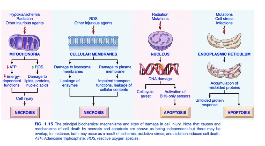
- Cell injury usually occurs because of functional and biochemical abnormalities in one or more essential cell components (Fig. 1.15).
- Examples:
- Hypoxia and ischemia mainly damage energy-dependent cellular functions.
- Protein damage and DNA damage mainly trigger apoptosis.
- A single harmful stimulus can activate multiple overlapping biochemical pathways.
- Because of this, blocking only one pathway usually cannot completely prevent cell injury.
- The following sections explain the different mechanisms that cause cell injury and cell death.
- Although each mechanism mainly causes either necrosis or apoptosis, the two pathways can overlap.
- Example:
- Ischemia and free radical production usually cause necrosis.
- They can also trigger apoptosis.
KEY CONCEPT
- Cell injury occurs through multiple biochemical pathways.
- The severity of injury depends on:
- Type of injury
- Duration
- Severity
- The response also depends on:
- Cell type
- Metabolic activity
- Adaptability
- Genetic makeup
- Hypoxia and ischemia mainly impair energy production.
- DNA and protein damage mainly induce apoptosis (Fig. 1.15).
- One harmful stimulus can activate several overlapping pathways, leading to necrosis, apoptosis, or both.
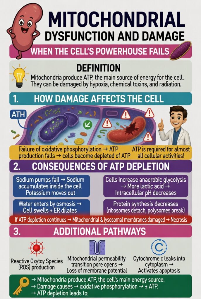
Mitochondrial Dysfunction and Damage
- Mitochondria produce ATP, the main source of energy for the cell.
- Mitochondria can be damaged by:
- Hypoxia
- Chemical toxins
- Radiation
- Mitochondrial damage has two major consequences.
- Failure of oxidative phosphorylation
- Oxidative phosphorylation decreases.
- ATP production falls.
- Cells become depleted of ATP.
- ATP is required for almost all cellular activities.
- Therefore, ATP depletion affects many cell functions.
- ATP loss commonly occurs during ischemia.
- Reduced ATP causes:
- ATP-dependent sodium pumps in the plasma membrane to fail.
- Sodium accumulates inside the cell.
- Potassium moves out of the cell.
- Water enters the cell by osmosis.
- The cell swells.
- The endoplasmic reticulum (ER) becomes dilated.
- To compensate for low ATP:
- Cells increase anaerobic glycolysis.
- More lactic acid is produced.
- Intracellular pH decreases.
- Many cellular enzymes become less active.
- If ATP depletion continues:
- The protein-making machinery is damaged.
- Ribosomes detach from the rough ER.
- Polysomes break apart.
- Protein synthesis decreases.
- With prolonged ATP depletion:
- Mitochondrial membranes become permanently damaged.
- Lysosomal membranes also become damaged.
- The cell undergoes necrosis.
- Although necrosis is the main form of cell death caused by hypoxia:
- The mitochondrial pathway of apoptosis may also contribute.
- Abnormal oxidative phosphorylation also produces reactive oxygen species (ROS).
- Mitochondrial damage is often associated with opening of the mitochondrial permeability transition pore.
- Opening of this pore causes:
- Loss of mitochondrial membrane potential.
- Changes in mitochondrial pH.
- Further reduction in oxidative phosphorylation.
- Mitochondria contain proteins such as cytochrome c.
- When mitochondria are damaged:
- Cytochrome c leaks into the cytoplasm.
- This signals internal cell injury.
- It activates apoptosis.
- Release of cytochrome c is controlled by regulatory proteins.
- It occurs in response to:
- Loss of survival signals.
- Other proapoptotic stimuli.
- Healthy mitochondria support cell survival.
- Damaged mitochondria can activate both protective and cell death pathways.
KEY CONCEPT
- Mitochondria produce ATP, the cell’s main energy source.
- Damage to mitochondria causes:
- ↓ Oxidative phosphorylation
- ↓ ATP production
- ATP depletion leads to:
- Failure of sodium pumps
- Sodium and water accumulation
- Cell swelling
- ER dilation
- Increased anaerobic glycolysis
- Lactic acid accumulation
- Decreased intracellular pH
- Reduced protein synthesis
- Severe ATP depletion damages mitochondria and lysosomes, causing necrosis.
- Damaged mitochondria also:
- Produce reactive oxygen species (ROS).
- Open the mitochondrial permeability transition pore.
- Release cytochrome c, which activates apoptosis.
Oxidative Stress
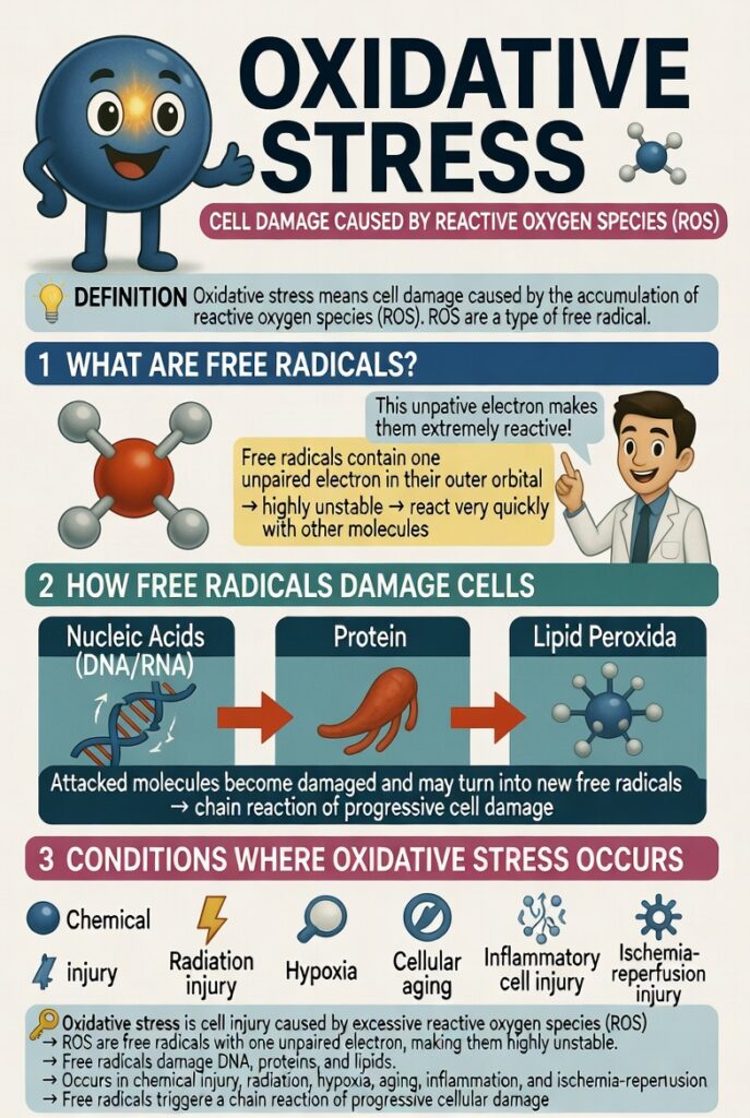
- Oxidative stress means cell damage caused by the accumulation of reactive oxygen species (ROS).
- Reactive oxygen species (ROS) are a type of free radical.
- Free radicals damage cells in many conditions, including:
- Chemical injury
- Radiation injury
- Hypoxia
- Cellular aging
- Tissue injury caused by inflammatory cells
- Ischemia-reperfusion injury
- Free radicals are chemical molecules that contain one unpaired electron in their outer orbital.
- Because of this unpaired electron:
- Free radicals are highly unstable.
- They react very quickly with other molecules.
- Free radicals can react with:
- Nucleic acids (DNA and RNA)
- Proteins
- Lipids
- During these reactions:
- The attacked molecules become damaged.
- They may also change into new free radicals.
- These newly formed free radicals continue attacking other molecules.
- This creates a chain reaction that spreads cell damage.
KEY CONCEPT
- Oxidative stress is cell injury caused by excessive reactive oxygen species (ROS).
- ROS are free radicals with one unpaired electron, making them highly unstable.
- Free radicals damage:
- DNA (nucleic acids)
- Proteins
- Lipids
- Cell injury due to ROS occurs in:
- Chemical injury
- Radiation injury
- Hypoxia
- Aging
- Inflammation
- Ischemia-reperfusion injury
- Free radicals trigger a chain reaction, causing progressive cellular damage.
Generation and Removal of Reactive Oxygen Species
- The amount of reactive oxygen species (ROS) in a cell depends on:
- How much ROS is produced.
- How much ROS is removed (Fig. 1.16).
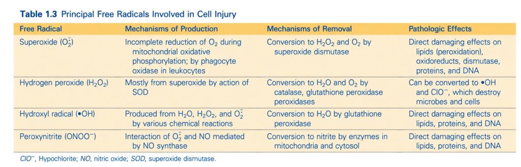
- The major properties and harmful effects of ROS are summarized in Table 1.3.
- ROS are normally produced by two major pathways.
- ROS production during normal energy generation
- Small amounts of ROS are produced in all cells during reduction-oxidation (redox) reactions.
- These reactions occur while mitochondria produce energy.
- Normally, oxygen receives four electrons and is completely converted into water.
- Sometimes oxygen is only partially reduced.
- This produces highly reactive and short-lived ROS.
- The sequence is:
- Oxygen → Superoxide (O₂•⁻)
- Superoxide → Hydrogen peroxide (H₂O₂)
- This occurs naturally and with the help of the enzyme superoxide dismutase (SOD).
- Hydrogen peroxide → Hydroxyl radical (•OH)
- This occurs in the presence of metals such as Fe²⁺ (iron).
- Hydrogen peroxide (H₂O₂) is:
- More stable than superoxide.
- Able to cross biological membranes.
- Hydroxyl radical (•OH) is the most highly reactive and damaging ROS.
- Ionizing radiation and high doses of ultraviolet (UV) light increase ROS production.
- They do this by breaking water into:
- Hydroxyl radicals (•OH)
- Hydrogen free radicals (H•)
- ROS production by phagocytic leukocytes
- Phagocytic leukocytes, especially neutrophils, produce ROS.
- They use ROS to destroy:
- Ingested microbes.
- Other foreign substances during inflammation.
- ROS are produced inside phagolysosomes.
- This process is called:
- Respiratory burst
- Oxidative burst
- During the respiratory burst:
- Phagocyte oxidase produces superoxide (O₂•⁻).
- Superoxide is converted into hydrogen peroxide (H₂O₂).
- Myeloperoxidase, an enzyme abundant in neutrophils, converts H₂O₂ into hypochlorite (OCl⁻).
- Hypochlorite is the major active component of household bleach.
- It is a very powerful substance for killing microbes.
- ROS released from neutrophils can also damage normal body tissues.
KEY CONCEPT
- The level of ROS depends on the balance between production and removal (Fig. 1.16).
- Major ROS production pathways:
- Normal mitochondrial redox reactions
- Respiratory burst in neutrophils
- ROS formation occurs in the following sequence:
- O₂ → Superoxide (O₂•⁻) → Hydrogen peroxide (H₂O₂) → Hydroxyl radical (•OH)
- Superoxide dismutase (SOD) converts superoxide into H₂O₂.
- Fe²⁺ converts H₂O₂ into the highly damaging hydroxyl radical (•OH).
- Myeloperoxidase converts H₂O₂ into hypochlorite (OCl⁻) during the respiratory burst.
- ROS help destroy microbes but can also injure normal tissues (Table 1.3).
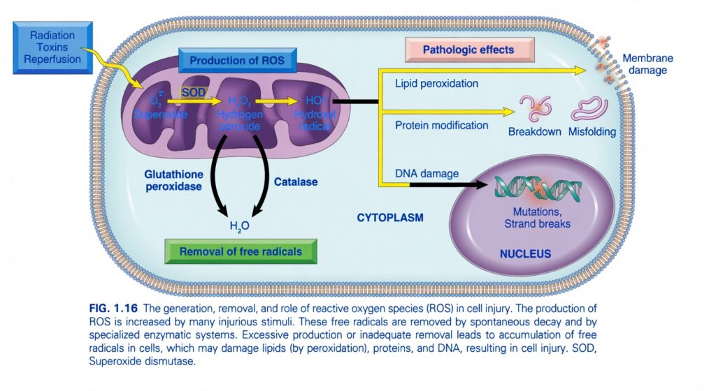
Reactive Oxygen Species (ROS) in Cell Injury (Figure 1.16)
This figure explains how harmful oxygen molecules (Reactive Oxygen Species, ROS) are:
- Produced
- Neutralized (Removed)
- Cause cell injury when they become excessive
Main Concept
ROS are highly reactive oxygen molecules. In small amounts they are normal, but when they become excessive, they damage the cell membrane, proteins, and DNA, leading to cell injury or death.
Think of ROS as sparks of fire inside the cell.
- 🔥 Small sparks = Normal and controlled.
- 🔥🔥🔥 Too many sparks = Fire spreads and damages the whole house.
Overall View of the Figure
The figure has three major parts:
1. ROS Production
        ↓
2. ROS Removal
        ↓
3. Pathologic Effects (Cell Injury)
PART 1 – Production of ROS
The left side of the figure shows how ROS are formed.
Step 1 – Cell Injury Starts
The blue box lists causes:
- Radiation
- Toxins
- Reperfusion injury
These injure the cell and especially the mitochondria.
Easy Concept
Imagine a factory’s power plant becomes damaged.
Instead of producing only electricity,
it starts producing dangerous sparks.
The mitochondria behave the same way.
Step 2 – Formation of Superoxide (O₂•⁻)
The first ROS produced is:
Superoxide (O₂•⁻)
It is the first free radical.
Easy Memory
Think of it as
The first spark.
Step 3 – Superoxide → Hydrogen Peroxide
The enzyme
Superoxide Dismutase (SOD)
changes
Superoxide (O₂•⁻)
        ↓
Hydrogen peroxide (H₂O₂)
Important Point
Hydrogen peroxide is not a free radical, but it is still harmful.
Easy Concept
SOD acts like a firefighter trying to control the first spark.
Step 4 – Hydrogen Peroxide → Hydroxyl Radical
Hydrogen peroxide can be converted into
Hydroxyl Radical (•OH)
This is the most dangerous ROS.
Easy Memory
Superoxide
      ↓
Hydrogen peroxide
      ↓
Hydroxyl radical
The danger increases at each step.
Easy Concept
Think of it as
Spark
     ↓
Small Fire
     ↓
Explosion
The hydroxyl radical is the explosion.
PART 2 – Removal of ROS
The middle of the figure explains how the cell protects itself.
Fortunately,
the cell has antioxidant enzymes.
1. Superoxide Dismutase (SOD)
Function:
Superoxide
      ↓
Hydrogen peroxide
Easy Memory
SOD controls the first spark.
2. Catalase
Catalase converts
Hydrogen peroxide
        ↓
Water (H₂O)
Water is harmless.
Easy Concept
Catalase is like a firefighter that completely extinguishes the fire.
3. Glutathione Peroxidase
This enzyme also converts
Hydrogen peroxide
        ↓
Water
It works together with glutathione to detoxify peroxides.
Easy Concept
Think of Catalase and Glutathione Peroxidase as two cleanup teams.
Both convert dangerous peroxide into harmless water.
Simple Removal Flow
Superoxide
      ↓
SOD
      ↓
Hydrogen peroxide
      ↓
Catalase
OR
Glutathione peroxidase
      ↓
Water
PART 3 – Pathologic Effects of ROS
If too many ROS are produced,
or too few are removed,
they begin attacking the cell.
The figure shows three major targets.
1. Lipid Peroxidation → Membrane Damage
ROS attack membrane lipids.
This process is called
Lipid Peroxidation
Result
The cell membrane becomes:
- Weak
- Leaky
- Damaged
Eventually,
the membrane may rupture.
Easy Concept
Imagine rust slowly destroying the metal body of a car.
ROS similarly “rust” the cell membrane.
2. Protein Modification
ROS attack proteins.
The figure shows:
- Protein breakdown
- Protein misfolding
Result
Enzymes stop working properly.
Structural proteins lose their normal shape.Easy Concept
Imagine bending a key.
It no longer fits the lock.
Damaged proteins can no longer perform their normal functions.
3. DNA Damage
ROS attack DNA in the nucleus.
The figure shows:
- Mutations
- DNA strand breaks
Result
The cell may:
- Stop dividing
- Become cancerous
- Undergo apoptosis
- Die
Easy Concept
DNA is the instruction manual.
ROS tear pages from the manual.
Now the cell cannot read the instructions correctly.
Complete Story of the Figure
Radiation
Toxins
Reperfusion
        ↓
Mitochondria
        ↓
Superoxide
        ↓
SOD
        ↓
Hydrogen Peroxide
        ↓
Hydroxyl Radical
        ↓
ROS Accumulation
        ↓
Damage Cell Membrane
Damage Proteins
Damage DNA
        ↓
Cell Injury
The Cell’s Defense System
ROS Produced
      ↓
SOD
      ↓
Hydrogen Peroxide
      ↓
Catalase
OR
Glutathione Peroxidase
      ↓
Water
      ↓
Cell Protected
Easy Real-Life Analogy
Imagine your kitchen.
Cooking naturally produces a little smoke.
A chimney removes the smoke,
so everything is fine.
Now imagine:
- Smoke becomes excessive.
- The chimney stops working.
Soon,
the smoke fills the room,
damages the walls,
burns the furniture,
and ruins the house.
In the same way:
- ROS = Smoke/fire
- Antioxidant enzymes = Chimney/firefighters
- Cell = House
Easy Memory Trick
3 ROS
O₂•⁻
   ↓
H₂O₂
   ↓
•OH
Danger increases from top to bottom.
3 Antioxidant Enzymes
SOD
Catalase
Glutathione Peroxidase
Remember:
“SCG saves the Cell.”
3 Major Targets of ROS
ROS attack:
- Lipids → Membrane damage
- Proteins → Misfolding and breakdown
- DNA → Mutations and strand breaks
Important Points from Figure 1.16
- Reactive oxygen species (ROS) are chemically reactive oxygen-containing molecules produced mainly in the mitochondria.
- Cell injury caused by radiation, toxins, and reperfusion increases ROS production.
- Superoxide (O₂•⁻) is the first ROS formed.
- Superoxide dismutase (SOD) converts superoxide into hydrogen peroxide (H₂O₂).
- Hydrogen peroxide is detoxified to water (H₂O) by catalase or glutathione peroxidase.
- If ROS accumulate, hydroxyl radicals (•OH) and other ROS damage cellular components.
- Lipid peroxidation damages cell membranes.
- Protein oxidation causes protein modification, misfolding, and breakdown.
- DNA damage leads to mutations and DNA strand breaks.
- Excessive ROS production or inadequate antioxidant defenses results in oxidative stress, which contributes to cell injury, necrosis, apoptosis, aging, and many diseases.
KEY CONCEPT (Figure 1.16)
Figure 1.16 illustrates the balance between ROS generation and antioxidant defense. Harmful stimuli such as radiation, toxins, and reperfusion increase mitochondrial production of superoxide, which is converted to hydrogen peroxide by superoxide dismutase (SOD). Hydrogen peroxide is then detoxified to water by catalase or glutathione peroxidase. When ROS production exceeds the cell’s antioxidant capacity, oxidative stress develops. Excess ROS attack membrane lipids (lipid peroxidation), proteins (oxidation, misfolding, breakdown), and DNA (mutations and strand breaks), ultimately leading to cellular dysfunction, injury, and possibly cell death.
Generation and Removal of Reactive Oxygen Species
- Cells have protective mechanisms that remove free radicals.
- These mechanisms reduce the harmful effects of reactive oxygen species (ROS).
- Free radicals are naturally unstable.
- They can disappear on their own.
- Cells also contain free radical scavengers.
- These include:
- Nonenzymatic systems.
- Enzymatic systems (Fig. 1.16).
- Superoxide dismutase (SOD)
- Found in many types of cells.
- Converts superoxide (O₂•⁻) into hydrogen peroxide (H₂O₂).
- The H₂O₂ is then broken down by catalase.
- Glutathione peroxidase
- A family of enzymes that protects cells from oxidative damage.
- The most abundant type is glutathione peroxidase 1.
- It is present in the cytoplasm of all cells.
- It converts hydrogen peroxide (H₂O₂) into water (H₂O).
- Catalase
- Located in peroxisomes.
- Breaks down hydrogen peroxide (H₂O₂) into:
- Water (H₂O)
- Oxygen (O₂)
- Catalase works very rapidly.
- It can break down millions of H₂O₂ molecules every second.
- Antioxidants
- Can be produced by the body (endogenous) or obtained from food (exogenous).
- Examples include:
- Vitamin E
- Vitamin A
- Vitamin C
- β-carotene
- Antioxidants help by:
- Preventing the formation of free radicals.
- Removing free radicals after they are formed.
KEY CONCEPT
- Cells protect themselves from ROS using free radical scavenging systems (Fig. 1.16).
- Major antioxidant enzymes are:
- Superoxide dismutase (SOD): Converts O₂•⁻ → H₂O₂.
- Glutathione peroxidase: Converts H₂O₂ → H₂O.
- Catalase: Converts H₂O₂ → H₂O + O₂.
- Antioxidants (Vitamin E, A, C, and β-carotene) prevent or remove free radicals.
- These protective systems reduce oxidative stress and limit cell injury.
Cell Injury Caused by Reactive Oxygen Species
- Reactive oxygen species (ROS) injure cells by damaging many important cell components (Fig. 1.16).
- Peroxidation of membrane lipids
- ROS attack the double bonds in membrane lipids.
- They damage:
- Plasma membrane
- Mitochondrial membrane
- Lysosomal membrane
- This forms lipid peroxides.
- Lipid peroxides are unstable and highly reactive.
- They start a self-propagating (autocatalytic) chain reaction, causing more membrane damage.
- Crosslinking and other changes in proteins
- ROS cause crosslinking of proteins through sulfhydryl groups.
- Crosslinked proteins:
- Lose their normal function.
- Are broken down more easily.
- ROS can also directly break protein chains (polypeptides).
- Damaged proteins may not fold properly.
- Misfolded proteins trigger the unfolded protein response.
- DNA damage
- ROS damage DNA.
- This can cause:
- DNA mutations.
- DNA strand breaks.
- DNA damage contributes to:
- Apoptosis.
- Aging.
- Malignant transformation (cancer development).
- Besides causing cell injury and killing microbes:
- Low levels of ROS also participate in cell signaling pathways.
- Therefore, ROS also have important normal physiologic functions.
KEY CONCEPT
- ROS damage multiple cell components (Fig. 1.16).
- Major effects include:
- Lipid peroxidation → Damage to plasma, mitochondrial, and lysosomal membranes.
- Protein damage → Protein crosslinking, fragmentation, loss of function, and misfolding.
- DNA damage → Mutations, DNA breaks, apoptosis, aging, and cancer.
- High levels of ROS cause cell injury, whereas low levels of ROS help regulate normal cellular signaling.
Membrane Damage
- Most cell injuries that end in necrosis cause the cell membrane to become more permeable (leaky).
- This increased permeability eventually leads to obvious membrane damage.
- Cell membranes can be damaged by:
- Reactive oxygen species (ROS).
- Decreased phospholipid synthesis due to hypoxia and nutrient deprivation.
- Increased phospholipid degradation, for example after increased intracellular calcium activates phospholipases.
- Cytoskeletal abnormalities that damage the supports (anchors) of the plasma membrane (Fig. 1.17).
- The most important sites of membrane damage are:
- Mitochondrial membrane damage, discussed earlier.
- Plasma membrane damage, which causes:
- Loss of osmotic balance.
- Entry of water and ions into the cell.
- Leakage of cellular contents out of the cell.
- Lysosomal membrane damage, which causes:
- Leakage of lysosomal enzymes (acid hydrolases) into the cytoplasm.
- These enzymes become active in the acidic intracellular pH of an injured cell (such as an ischemic cell).
- The activated enzymes digest many cellular components.
- This produces irreversible cell damage and necrosis.
KEY CONCEPT
- Membrane damage is a major feature of cell injury leading to necrosis.
- Membrane damage is caused by:
- ROS
- Decreased phospholipid synthesis
- Increased phospholipid degradation
- Cytoskeletal abnormalities (Fig. 1.17)
- The three main sites of membrane damage are:
- Mitochondrial membrane
- Plasma membrane
- Lysosomal membrane
- Lysosomal enzymes (acid hydrolases) digest cell components, causing irreversible injury and necrosis.
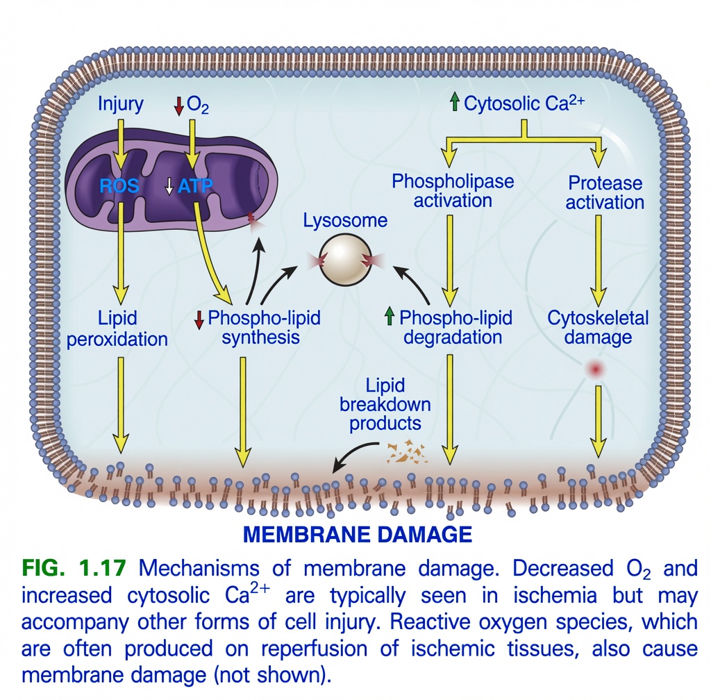
Mechanisms of Membrane Damage (Figure 1.17 )
This figure explains how different types of cell injury ultimately damage the cell membrane, which is a critical event leading to irreversible cell injury and necrosis.
Main Concept
Almost every severe cell injury eventually damages the cell membrane. Once the membrane is severely damaged, the cell leaks its contents, loses its normal function, and dies.
Think of the cell membrane as the wall of a water-filled balloon.
- A healthy balloon keeps water inside.
- If the balloon develops many holes, water leaks out.
- Eventually, the balloon bursts.
The same thing happens to a cell.
Overall Flow of the Figure
Everything in this figure leads to one final outcome:
Cell Injury
      ↓
Multiple damaging mechanisms
      ↓
MEMBRANE DAMAGE
      ↓
Cell contents leak out
      ↓
Cell death (Necrosis)
The Figure Shows 5 Main Pathways to Membrane Damage
1. ROS (Free radicals)
2. ATP depletion
3. Increased intracellular Ca²⁺
4. Lysosomal damage
5. Cytoskeletal damage
            ↓
      Membrane Damage
Let’s understand each pathway one by one.
PATHWAY 1 – Injury → ROS → Lipid Peroxidation
The figure starts with:
Injury
     ↓
ROS
Examples of injury:
- Ischemia
- Radiation
- Toxins
- Infection
These injuries increase Reactive Oxygen Species (ROS).
ROS attack membrane lipids
ROS attack the fatty phospholipids of the membrane.
This process is called:
Lipid Peroxidation
Easy Concept
Imagine the membrane is made of butter.
ROS are like fire.
The fire melts and damages the butter.
Eventually,
holes appear in the membrane.
Flow
Cell Injury
      ↓
ROS
      ↓
Lipid Peroxidation
      ↓
Membrane Damage
PATHWAY 2 – ↓ O₂ → ↓ ATP → ↓ Phospholipid Synthesis
The figure also shows
↓ O₂
     ↓
↓ ATP
Without oxygen,
mitochondria cannot make enough ATP.
Why is ATP important?
ATP is needed to manufacture new membrane phospholipids.
When ATP falls,
the cell cannot repair damaged membrane.
Result
↓ ATP
      ↓
↓ Phospholipid Synthesis
      ↓
Weak Membrane
      ↓
Membrane Damage
Easy Concept
Imagine the roof of your house develops holes.
Normally,
workers repair it.
But if you have no money (ATP),
the roof cannot be repaired.
Eventually,
the whole roof collapses.
PATHWAY 3 – Increased Cytosolic Ca²⁺
The figure shows
↑ Cytosolic Ca²⁺Normally,
intracellular calcium is very low.
During cell injury,
calcium enters the cell.
This activates harmful enzymes.
A. Calcium Activates Phospholipase
↑ Ca²⁺
      ↓
Phospholipase Activation
What do phospholipases do?
They digest membrane phospholipids.
Result
Phospholipid Degradation
        ↓
Lipid Breakdown Products
        ↓
Membrane Damage
Easy Concept
Phospholipase acts like scissors cutting the membrane into pieces.
B. Calcium Activates Proteases
↑ Ca²⁺
      ↓
Protease Activation
What do proteases do?
Proteases digest structural proteins.
These proteins normally support the membrane.
Result
Protease
      ↓
Cytoskeletal Damage
      ↓
Membrane Weakness
      ↓
Membrane Damage
Easy Concept
Imagine the membrane is a tent.
The cytoskeleton is the metal frame supporting it.
Proteases break the frame.
The tent collapses.
PATHWAY 4 – Lysosomal Damage
The figure shows
Lysosome
in the center.
Normally
Lysosomes safely store digestive enzymes.
During Injury
The lysosomal membrane becomes damaged.
Enzymes leak into the cytoplasm.
These enzymes digest:
- Membrane
- Proteins
- Organelles
Easy Concept
Imagine a bottle of acid breaks inside your room.
The acid begins dissolving everything.
The lysosome behaves the same way.
PATHWAY 5 – Cytoskeletal Damage
The figure shows
Protease
      ↓
Cytoskeletal Damage
The cytoskeleton normally:
- Supports the membrane
- Maintains cell shape
When damaged,
the membrane loses support.
It becomes fragile.
Eventually,
it ruptures.
Easy Concept
Think of a building.
If the steel pillars are removed,
the walls collapse.
Final Result
All pathways end at the bottom:
MEMBRANE DAMAGE
What happens after membrane damage?
Plasma membrane
- Cell contents leak out.
- Sodium enters.
- Water enters.
- Cell swells.
Mitochondrial membrane
- ATP production stops.
Lysosomal membrane
- Digestive enzymes leak out.
- Cell digests itself.
Final Outcome
Membrane Damage
      ↓
Leakage of Cell Contents
      ↓
Cell Death (Necrosis)
Complete Story of the Figure
Cell Injury
      ↓
────────────────────────────────────
1. ROS
      ↓
Lipid Peroxidation
      ↓
Membrane Damage
────────────────────────────────────
2. ↓ O₂
      ↓
↓ ATP
      ↓
↓ Phospholipid Synthesis
      ↓
Membrane Damage
────────────────────────────────────
3. ↑ Ca²⁺
      ↓
Phospholipase
      ↓
Phospholipid Degradation
      ↓
Membrane Damage
────────────────────────────────────
4. ↑ Ca²⁺
      ↓
Protease
      ↓
Cytoskeletal Damage
      ↓
Membrane Damage
────────────────────────────────────
5. Lysosomal Damage
      ↓
Digestive Enzymes Leak
      ↓
Membrane Damage
────────────────────────────────────
      ↓
CELL DEATH
Easy House Analogy
Imagine your house.
Different disasters can destroy it:
🔥 Fire → burns the walls (ROS)
💰 No money → cannot repair the walls (↓ ATP)
✂️ Workers cut the walls (Phospholipase)
🪚 Pillars are cut (Protease → Cytoskeleton)
🧪 Acid spills inside (Lysosomal enzymes)
Although the causes are different,
they all end with the same result:
🏚️ The house collapses.
Similarly,
all these pathways end in membrane damage and cell death.
Easy Memory Trick
Remember the 5 Causes of Membrane Damage
“RACPL”
- R = ROS → Lipid peroxidation
- A = ATP depletion → ↓ Phospholipid synthesis
- C = Calcium ↑
- P = Phospholipase & Protease activation
- L = Lysosomal enzyme leakage
All roads lead to:
➡️ Membrane Damage → Cell Death
Important Points from Figure 1.17
- Membrane damage is a hallmark of irreversible cell injury and an important cause of necrosis.
- Reactive oxygen species (ROS) cause lipid peroxidation, damaging membrane phospholipids.
- Hypoxia (↓ O₂) reduces ATP production, impairing phospholipid synthesis and membrane repair.
- Increased intracellular Ca²⁺ activates phospholipases, which degrade membrane phospholipids into lipid breakdown products.
- Elevated Ca²⁺ also activates proteases, which degrade cytoskeletal proteins, weakening membrane stability.
- Lysosomal membrane damage releases hydrolytic enzymes that digest cellular membranes and organelles.
- Damage to the plasma membrane causes leakage of intracellular contents and influx of sodium and water.
- Damage to the mitochondrial membrane further decreases ATP production, while lysosomal membrane damage promotes autodigestion.
- These mechanisms reinforce one another, leading to progressive membrane destruction and ultimately cell death (necrosis).
KEY CONCEPT (Figure 1.17)
Figure 1.17 demonstrates that multiple mechanisms converge to produce membrane damage, a critical step in irreversible cell injury. ROS induce lipid peroxidation, ATP depletion reduces phospholipid synthesis and membrane repair, and elevated cytosolic Ca²⁺ activates phospholipases and proteases, resulting in phospholipid degradation and cytoskeletal damage. Injury to lysosomal membranes releases digestive enzymes that further degrade cellular components. Together, these processes disrupt the integrity of the plasma membrane, mitochondrial membrane, and lysosomal membrane, leading to leakage of cell contents, loss of cellular homeostasis, and necrotic cell death.
Disturbance in Calcium Homeostasis
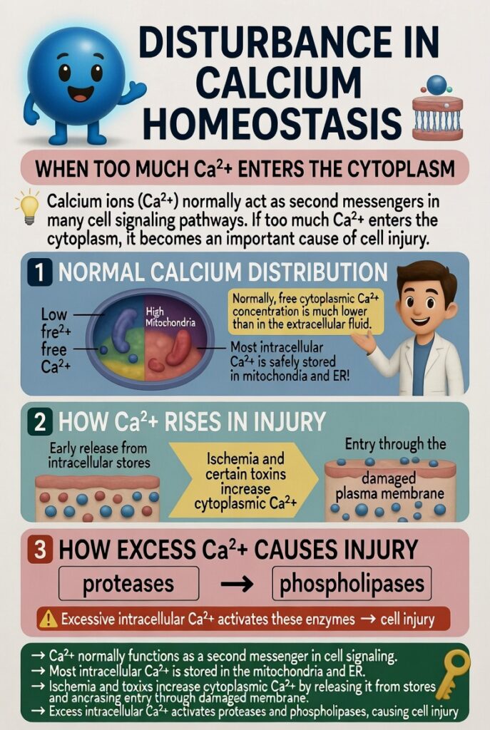
- Calcium ions (Ca²⁺) normally act as second messengers in many cell signaling pathways.
- If too much Ca²⁺ enters the cytoplasm, it becomes an important cause of cell injury.
- Normally, the concentration of free Ca²⁺ in the cytoplasm is much lower than in the extracellular fluid.
- Most intracellular Ca²⁺ is stored in:
- Mitochondria
- Endoplasmic reticulum (ER)
- Ischemia and certain toxins increase cytoplasmic Ca²⁺.
- At first, Ca²⁺ is released from intracellular stores.
- Later, more Ca²⁺ enters the cell through the damaged plasma membrane.
- Excessive intracellular Ca²⁺ causes cell injury by activating different enzymes.
- These enzymes include:
- Proteases, which damage cellular proteins.
- Phospholipases, which damage cell membranes.
KEY CONCEPT
- Ca²⁺ normally functions as a second messenger in cell signaling.
- Most intracellular Ca²⁺ is stored in the mitochondria and ER.
- Ischemia and toxins increase cytoplasmic Ca²⁺ by:
- Releasing Ca²⁺ from intracellular stores.
- Increasing Ca²⁺ entry through the damaged plasma membrane.
- Excess intracellular Ca²⁺ activates:
- Proteases
- Phospholipases
- Activation of these enzymes causes cell injury.
Endoplasmic Reticulum Stress
- The accumulation of misfolded proteins in a cell causes stress in the endoplasmic reticulum (ER).
- If this stress becomes severe, it can lead to cell death by apoptosis.
- During protein synthesis, chaperone proteins in the ER help newly made proteins fold into the correct shape.
- Protein folding is not always perfect.
- Some proteins become misfolded.
- Misfolded proteins are normally:
- Marked by ubiquitination.
- Broken down by proteolysis.
- When too many misfolded proteins accumulate in the ER, they trigger a protective response called the unfolded protein response (UPR) (Fig. 1.18).
- The unfolded protein response:
- Increases the production of chaperone proteins.
- Slows down protein synthesis.
- Reduces the amount of misfolded proteins in the cell.
- If the number of misfolded proteins becomes greater than the ER can handle, additional signals are produced.
- These signals activate proapoptotic sensors.
- This leads to apoptosis, mainly through the mitochondrial (intrinsic) pathway.
KEY CONCEPT
- Misfolded proteins cause endoplasmic reticulum (ER) stress.
- Chaperone proteins normally help proteins fold correctly.
- Misfolded proteins are removed by ubiquitination and proteolysis.
- Excess misfolded proteins activate the unfolded protein response (UPR) (Fig. 1.18).
- The UPR:
- Increases chaperone production.
- Decreases protein synthesis.
- If ER stress is too severe, proapoptotic signals are activated, causing apoptosis through the mitochondrial (intrinsic) pathway.
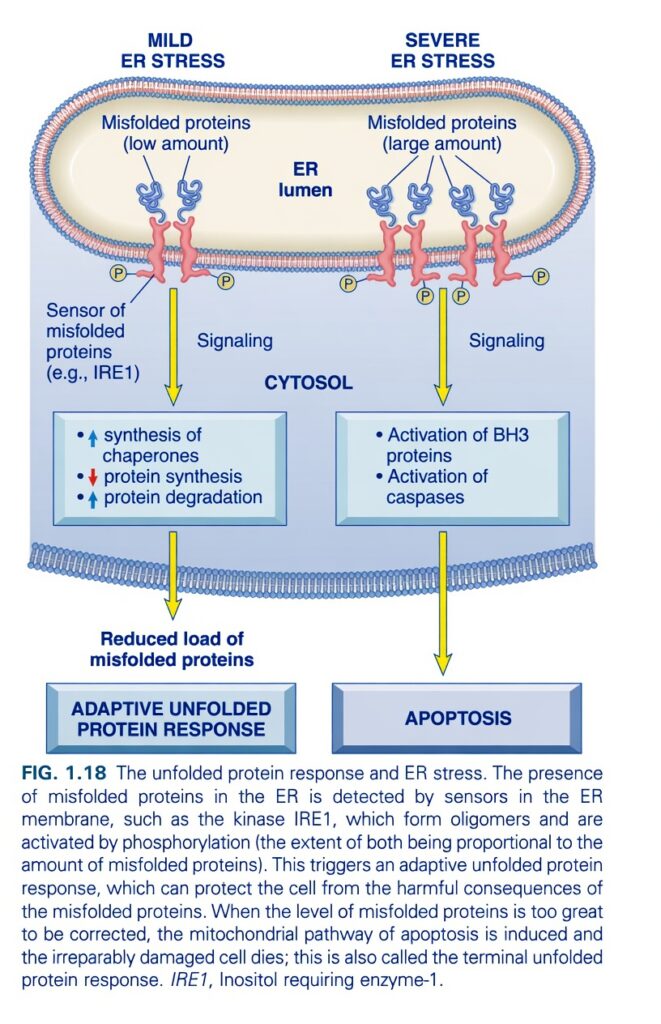
Unfolded Protein Response (UPR) & Endoplasmic Reticulum (ER) Stress (Figure 1.18)
his figure explains what happens when proteins are folded incorrectly inside the Endoplasmic Reticulum (ER).
The cell has two possible responses:
- Mild ER Stress → Repair the problem → Cell survives
- Severe ER Stress → Cannot repair → Cell undergoes apoptosis
Main Concept
The ER is the cell’s protein-folding factory. If only a few proteins are misfolded, the ER repairs them. If too many proteins are misfolded, the cell activates apoptosis to protect the body.
Think of the ER as a Shirt Factory
Imagine a factory making shirts.
Normally:
👕 Shirts are stitched correctly.
But sometimes,
some shirts are defective.
If only a few shirts are defective,
workers repair them.
If thousands of shirts are defective,
the factory cannot recover,
so it closes permanently.
The ER behaves in exactly the same way.
Overall Flow of the Figure
Misfolded Proteins
        ↓
ER Detects the Problem
        ↓
──────────────────────────────
Few Misfolded Proteins
        ↓
Repair System Activated
        ↓
Cell Survives
──────────────────────────────
Too Many Misfolded Proteins
        ↓
Apoptosis Activated
        ↓
Cell Dies
First Understand Normal ER Function
The Endoplasmic Reticulum (ER) is the place where proteins are:
- Made
- Folded
- Checked for quality
Every protein must have the correct shape.
Easy Concept
The ER is like a quality-control department in a factory.
What are Misfolded Proteins?
Normally,
proteins fold into a precise 3D shape.
Sometimes they fold incorrectly.
These are called:
Misfolded Proteins
Why do proteins misfold?
Examples:
- Starvation
- Hypoxia
- Viral infection
- Heat
- Mutations
- Toxins
Easy Concept
Imagine folding a paper airplane.
Correct folding →
It flies.
Wrong folding →
It crashes.
Proteins behave exactly the same way.
The Figure Has Two Parts
LEFT SIDE = Mild ER Stress
This is the good outcome.
Step 1 – Small Amount of Misfolded Proteins
The figure shows:
Misfolded proteins (Low amount)
Only a few defective proteins are present.
Easy Concept
Only a few defective shirts are coming off the production line.
Step 2 – ER Sensor Detects the Problem
The figure labels:
Sensor of Misfolded Proteins (e.g., IRE1)
This sensor continuously checks protein quality.
Easy Concept
IRE1 is the quality-control inspector.
It asks:
“Are these proteins folded correctly?”
Step 3 – Signalling Begins
Once IRE1 detects misfolded proteins,
it sends signals into the cytoplasm.
Easy Concept
The inspector calls the repair department.
Step 4 – Adaptive Unfolded Protein Response (UPR)
The figure shows three important responses.
A. Increase Chaperone Synthesis
The cell makes more
Chaperone proteins.
What do chaperones do?
They help proteins fold correctly.
Easy Concept
Chaperones are like experienced tailors who fix defective shirts.
B. Decrease Protein Synthesis
The figure shows:
↓ Protein synthesis
Why?
If the ER is overloaded,
making more proteins would worsen the problem.
So,
the cell temporarily slows protein production.
Easy Concept
Imagine a factory.
If many defective shirts are appearing,
the manager says:
“Stop making new shirts until we fix the problem.”
C. Increase Protein Degradation
The figure also shows:
↑ Protein degradation
Misfolded proteins are destroyed.
Easy Concept
Bad shirts are thrown into the recycling bin.
Step 5 – Misfolded Proteins Decrease
Because:
- Chaperones repair proteins.
- New protein production slows.
- Defective proteins are destroyed.
The amount of misfolded protein falls.
Result
The figure shows:
Reduced Load of Misfolded Proteins
The ER becomes normal again.
Final Result
The cell survives.
The figure labels this as:
Adaptive Unfolded Protein Response (UPR)
Easy Memory
Mild Stress → Repair → Survival
RIGHT SIDE = Severe ER Stress
Now look at the right side.
Step 1 – Large Amount of Misfolded Proteins
The figure shows:
Misfolded proteins (Large amount)
Now the ER is overwhelmed.
Easy Concept
Thousands of defective shirts are being produced.
The repair team cannot keep up.
Step 2 – ER Sensor Activates Strong Signals
The same sensors (such as IRE1) become highly activated.
They realize the damage cannot be repaired.
Easy Concept
The factory manager realizes:
“The factory cannot be saved.”
Step 3 – Activation of BH3 Proteins
The figure shows:
Activation of BH3 Proteins
What do BH3 proteins do?
They activate the mitochondrial apoptosis pathway.
Easy Concept
BH3 proteins are like workers who decide:
“Shut down the factory.”
Step 4 – Activation of Caspases
The figure also shows:
Activation of Caspases
What are caspases?
They are enzymes that carry out programmed cell death (apoptosis).
Easy Concept
Caspases are the demolition workers.
They carefully dismantle the cell.Final Result
The figure shows:
APOPTOSIS
The damaged cell dies in a controlled manner.
Easy Memory
Severe Stress → Apoptosis → Cell Death
Complete Story of the Figure
Misfolded Proteins
        ↓
ER Sensor (IRE1)
        ↓
──────────────────────────
Few Misfolded Proteins
        ↓
↑ Chaperones
↓ Protein Synthesis
↑ Protein Degradation
        ↓
Reduced Misfolded Proteins
        ↓
Cell Survives
──────────────────────────
Too Many Misfolded Proteins
        ↓
BH3 Activation
        ↓
Caspase Activation
        ↓
Apoptosis
Easy Factory Analogy
Imagine a shirt factory.
Situation 1
Only 10 shirts are defective.
Manager says:
- Hire more repair workers.
- Slow production.
- Throw away damaged shirts.
Factory survives.
Situation 2
100,000 shirts are defective.
Manager says:
“The factory cannot be repaired.”
Factory closes permanently.
The ER behaves exactly like this.
UPR vs Apoptosis
| Adaptive UPR | Apoptosis |
|---|---|
| Mild ER stress | Severe ER stress |
| Few misfolded proteins | Many misfolded proteins |
| Chaperones increase | BH3 proteins increase |
| Protein synthesis decreases | Caspases activated |
| Cell survives | Cell dies |
Easy Memory Trick
Think: “3 Rs of Mild Stress”
Repair Proteins
Reduce Protein Synthesis
Remove Bad Proteins
↓
Cell survives
Think: “2 Bs of Severe Stress”
BH3
        ↓
Bad Cell Dies
Important Points from Figure 1.18
- The endoplasmic reticulum (ER) is responsible for the proper folding and processing of newly synthesized proteins.
- Misfolded proteins accumulate in the ER during conditions such as hypoxia, nutrient deprivation, oxidative stress, viral infection, toxins, and genetic mutations.
- ER membrane sensors (e.g., IRE1) detect the accumulation of misfolded proteins and initiate intracellular signalling.
- During mild ER stress, the unfolded protein response (UPR) is activated:
- ↑ Chaperone synthesis improves protein folding.
- ↓ Protein synthesis reduces the workload on the ER.
- ↑ Protein degradation removes misfolded proteins.
- These adaptive responses reduce the burden of misfolded proteins and restore normal ER function.
- During severe or persistent ER stress, the adaptive response fails.
- Persistent stress activates BH3-only proteins and caspases, triggering the mitochondrial pathway of apoptosis.
- Thus, the UPR initially protects the cell, but if the damage cannot be corrected, it switches to programmed cell death (apoptosis).
KEY CONCEPT (Figure 1.18)
Figure 1.18 illustrates the unfolded protein response (UPR) during ER stress. When only a small number of misfolded proteins accumulate, ER sensors such as IRE1 activate an adaptive response that increases chaperone production, reduces new protein synthesis, and enhances degradation of misfolded proteins, allowing the cell to recover. However, if misfolded proteins accumulate excessively and the stress persists, the adaptive mechanisms become insufficient. The ER then activates BH3 proteins and caspases, initiating apoptosis to eliminate the irreversibly damaged cell. Thus, the ER first attempts repair, but if repair fails, it triggers programmed cell death.
Intracellular accumulation of misfolded proteins
- Abnormalities that make more misfolded proteins or reduce their removal can cause buildup inside cells.
- This can result from mutations that produce abnormal proteins.
- Aging lowers the cell’s ability to fix misfolded proteins.
- Viral infections produce large amounts of microbial proteins that overload the normal quality-control system for proper folding.
- Changes in cell pH or redox state can also lead to misfolding.
- Lack of glucose and oxygen (as in ischemia and hypoxia) increases the load of misfolded proteins.
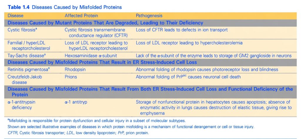
- Misfolded proteins inside cells can cause disease by creating a shortage of an essential protein or by triggering cell death (apoptosis) (Table 1.4).
- Misfolded proteins often lose their normal function and are quickly broken down, both of which can cause loss of function.
- If the lost function is essential, cell injury follows.
- One example is cystic fibrosis, caused by inherited mutations in a membrane transport protein that prevent its normal folding.
- Cell injury from protein misfolding occurs in several diseases (see Table 1.4).
KEY CONCEPT Misfolded proteins accumulate inside cells when production rises or clearance falls, leading to loss of essential protein function or apoptosis and resulting cell injury.
DNA Damage
- Exposure of cells to radiation or cancer drugs, production of ROS inside cells, and mutations can all cause DNA damage.
- If the damage is severe, it may start apoptotic cell death.
- Special sensor proteins inside the cell detect DNA damage and send signals that raise the level of p53 protein.
- p53 first stops the cell cycle at the G1 phase so the DNA can be repaired before it is copied (Chapter 6).
- If the damage is too severe to fix, p53 starts apoptosis, mainly through the mitochondrial pathway.
- When p53 is mutated or missing (as in some cancers), cells with damaged DNA that should die by apoptosis stay alive.
- In these cells, the DNA damage can produce genomic changes (such as chromosomal deletions) that lead to cancer development (Chapter 6).
KEY CONCEPT Severe DNA damage raises p53, which either pauses the cell cycle for repair or triggers apoptosis; loss of p53 allows damaged cells to survive and become cancerous.
Hypoxia and Ischemia
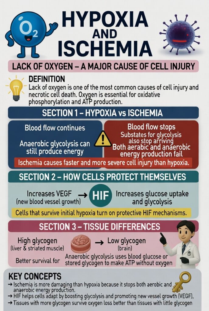
- Lack of oxygen is one of the most common causes of cell injury and necrotic cell death in clinical medicine.
- Oxygen is needed for oxidative phosphorylation and the production of ATP, the cell’s energy store.
- Therefore, cells without oxygen risk total failure of many essential functions.
- In hypoxia, blood flow continues and anaerobic glycolysis can still make energy.
- In ischemia, blood flow stops, so substrates for glycolysis also stop arriving.
- In ischemic tissues, aerobic metabolism ends and anaerobic energy production also fails once glycolytic substrates run out or glycolysis is blocked by buildup of metabolites that would normally be washed away by blood flow.
- For this reason, ischemia causes faster and more severe cell and tissue injury than hypoxia.
- Cells that face hypoxia but do not die at once turn on protective mechanisms controlled by hypoxia-inducible factor (HIF) transcription factors.
- HIF increases production of proteins that help the cell survive low oxygen.
- Some of these proteins, such as vascular endothelial growth factor (VEGF), promote growth of new blood vessels and thereby raise blood flow and oxygen supply.
- Other HIF-induced proteins change cell metabolism by increasing glucose uptake and glycolysis.
- Anaerobic glycolysis can make ATP without oxygen using glucose from the blood or from breakdown of stored glycogen inside the cell.
- Tissues that hold more glycogen (such as liver and striated muscle) survive oxygen loss and reduced oxidative phosphorylation better than tissues with little glycogen (such as the brain).
KEY CONCEPT Ischemia is more damaging than hypoxia because it stops both aerobic and anaerobic energy production; HIF helps cells adapt by boosting glycolysis and new vessel growth.
Persistent or Severe Hypoxia and Ischemia
- Persistent or severe hypoxia and ischemia cause depletion of ATP.
- Loss of this critical energy source leads to failure of the plasma membrane sodium pump.
- It also lowers intracellular pH, which changes the activities of many enzymes.
- Increased generation of ROS occurs.
- Defects in protein synthesis develop (Fig. 1.19).
- These alterations were described earlier in the discussion of mitochondrial damage.
KEY CONCEPT Severe or prolonged hypoxia and ischemia deplete ATP, causing membrane pump failure, lower pH, more ROS, and impaired protein synthesis.
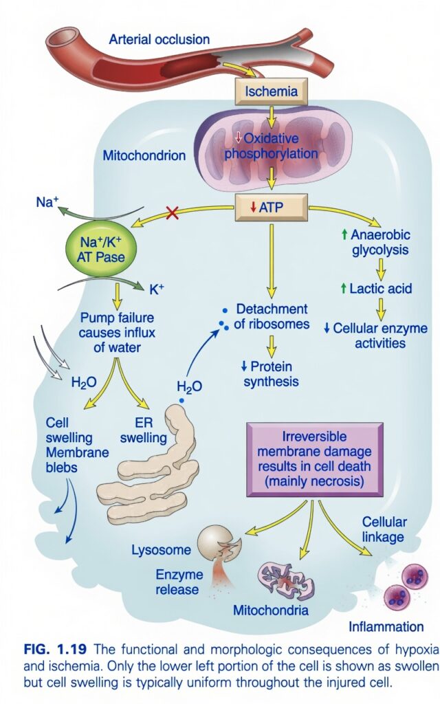
Hypoxia and Ischemia: Functional & Morphologic Consequences (Figure 1.19)
This figure explains what happens to a cell when its blood supply is blocked (ischemia).
The entire story can be remembered as:
No Blood → No Oxygen → No ATP → Cell Swelling → Membrane Damage → Cell Death (Necrosis)
Think of the cell as a factory.
- Blood = Electricity + Fuel delivery
- Mitochondria = Power station
- ATP = Electricity
- Cell membrane = Factory wall
If electricity stops, the whole factory gradually shuts down.
Main Concept
Ischemia reduces blood flow, causing oxygen deficiency (hypoxia). Without oxygen, ATP production falls, leading to pump failure, cell swelling, decreased protein synthesis, membrane damage, and finally necrosis.
The Whole Story in One Flow
Arterial Block
      ↓
Ischemia
      ↓
↓ Oxygen
      ↓
↓ ATP
      ↓
Cell Swelling
↓ Protein Synthesis
↑ Lactic Acid
      ↓
Membrane Damage
      ↓
Necrosis
STEP 1 – Arterial Occlusion
At the top of the figure, an artery is blocked.
This is called:
Arterial Occlusion
Examples
- Blood clot
- Atherosclerotic plaque
- Embolus
Easy Concept
Imagine a city water pipe becomes blocked.
No water reaches the houses.
Similarly,
blood cannot reach the cells.
STEP 2 – Ischemia
Because blood flow stops,
the tissue develops:
Ischemia
What is Ischemia?
Ischemia = Reduced blood supply to tissue.
Since blood carries oxygen and nutrients,
ischemia causes:
- ↓ Oxygen
- ↓ Glucose
- ↓ Nutrient delivery
Easy Concept
The cell is now starving and suffocating.
STEP 3 – Mitochondria Stop Making ATP
The figure shows:
Ischemia
      ↓
↓ Oxidative Phosphorylation
      ↓
↓ ATP
Normally,
mitochondria make ATP using oxygen.
Without oxygen,
ATP production falls.
Easy Concept
The mitochondria are the power station.
No oxygen = No electricity.
STEP 4 – ATP Falls
This is the most important event in the figure.
Everything that follows happens because ATP decreases.
PATHWAY 1 – Na⁺/K⁺ Pump Failure
Normally,
the Na⁺/K⁺ pump uses ATP.
When ATP decreases,
the pump stops.
Normal Pump
- 3 Na⁺ out
- 2 K⁺ in
During ATP Depletion
The pump cannot work.
Sodium stays inside the cell.
Water follows sodium.
Result
↓ ATP
      ↓
Na⁺/K⁺ Pump Failure
      ↓
Na⁺ Accumulates
      ↓
Water Enters
      ↓
Cell Swelling
The Figure Shows
- Cell swelling
- Membrane blebs
- ER swelling
Easy Concept
Imagine a balloon filled with more and more water.
It becomes swollen and stretched.
The injured cell behaves the same way.
What are Membrane Blebs?
Small balloon-like bulges form on the cell membrane.
They indicate:
The membrane is becoming weak.
ER Swelling
Water also enters the
Endoplasmic Reticulum (ER).
The ER enlarges and cannot function normally.
PATHWAY 2 – Ribosomes Detach
The figure shows:
↓ ATP
      ↓
Detachment of Ribosomes
      ↓
↓ Protein Synthesis
Why?
Ribosomes normally attach to the rough ER.
ATP depletion causes them to detach.
Without ribosomes,
proteins cannot be made efficiently.
Easy Concept
Imagine workers leaving the factory.
Production slows dramatically.
PATHWAY 3 – Anaerobic Glycolysis
Since mitochondria cannot make ATP,
the cell switches to:
Anaerobic Glycolysis
This produces only a small amount of ATP.
Problem
It also produces:
Lactic Acid
The figure shows:
↑ Anaerobic Glycolysis
        ↓
↑ Lactic Acid
What happens because of Lactic Acid?
The intracellular pH falls.
The acidic environment reduces
cellular enzyme activity.
The figure shows:
↑ Lactic Acid
      ↓
↓ Cellular Enzyme Activities
Easy Concept
Think of pouring acid into factory machines.
They stop working efficiently.
STEP 5 – Irreversible Membrane Damage
If ischemia continues,
the membrane becomes permanently damaged.
The figure highlights:
Irreversible Membrane Damage
At this point,
the injury can no longer be reversed.
Easy Concept
The balloon has burst.
Repair is impossible.
STEP 6 – Lysosomal Damage
The lysosome ruptures.
Digestive enzymes leak into the cytoplasm.
They digest the injured cell.
Easy Concept
Imagine a bottle of strong acid breaking inside a room.
Everything begins dissolving.
STEP 7 – Mitochondrial Damage
Mitochondria become severely damaged.
Now ATP production completely fails.
The injury becomes irreversible.
STEP 8 – Cellular Leakage
The damaged plasma membrane leaks.
Cell contents escape into the surrounding tissue and bloodstream.
Clinical Importance
Doctors detect this leakage by measuring enzymes such as:
- Troponin (heart)
- AST/ALT (liver)
- CK (muscle)
STEP 9 – Inflammation
Leaked cellular contents attract inflammatory cells.
Inflammation develops around the dead tissue.
STEP 10 – Cell Death (Necrosis)
The final outcome is:
Necrosis
The cell dies because it can no longer maintain its structure or function.
Complete Story of the Figure
Arterial Occlusion
        ↓
Ischemia
        ↓
↓ Oxygen
        ↓
↓ Oxidative Phosphorylation
        ↓
↓ ATP
        ↓
────────────────────────────
Na⁺/K⁺ Pump Failure
        ↓
Water Enters
        ↓
Cell Swelling
────────────────────────────
Ribosomes Detach
        ↓
↓ Protein Synthesis
────────────────────────────
↑ Anaerobic Glycolysis
        ↓
↑ Lactic Acid
        ↓
↓ Enzyme Activity
────────────────────────────
Persistent Injury
        ↓
Irreversible Membrane Damage
        ↓
Lysosomal Rupture
Mitochondrial Damage
Cell Leakage
Inflammation
        ↓
NECROSIS
Easy Factory Analogy
Imagine a factory.
Blood Supply Stops
🚫 No fuel arrives.
↓
Power Station Stops
⚡ No electricity.
↓
Machines Stop
- Water pipes burst
- Workers leave
- Production stops
- Waste accumulates
↓
Factory Walls Collapse
↓
Factory Closes Forever
That is necrosis.
Easy Memory Trick
Remember: “6 Ds”
↓ Blood Flow
        ↓
↓ Oxygen
        ↓
↓ ATP
        ↓
↓ Pumps
        ↓
Damage to Membrane
        ↓
Death (Necrosis)
Important Points from Figure 1.19
- Arterial occlusion causes ischemia, reducing blood flow and oxygen delivery.
- Hypoxia decreases oxidative phosphorylation in mitochondria, leading to ATP depletion.
- Reduced ATP causes Na⁺/K⁺ ATPase failure, allowing sodium and water to enter the cell, producing cell swelling, ER swelling, and membrane blebs.
- ATP depletion causes ribosomes to detach from the rough ER, reducing protein synthesis.
- The cell shifts to anaerobic glycolysis, increasing lactic acid and lowering intracellular pH, which decreases enzyme activity.
- Persistent ischemia results in irreversible membrane damage, lysosomal enzyme release, and mitochondrial dysfunction.
- Loss of membrane integrity causes cellular leakage, which can release intracellular enzymes into the bloodstream and trigger inflammation.
- The final outcome of prolonged ischemia is necrosis, the characteristic form of cell death caused by severe ischemic injury.
Figure 1.19 demonstrates the sequence of events during ischemic cell injury. Arterial occlusion reduces blood flow, causing hypoxia and decreased oxidative phosphorylation, which lowers ATP production. ATP depletion leads to Na⁺/K⁺ pump failure, resulting in cell swelling, ER swelling, and membrane blebbing. It also causes ribosome detachment with reduced protein synthesis and forces the cell to rely on anaerobic glycolysis, producing lactic acid and reducing enzyme activity. If ischemia persists, irreversible membrane damage develops, lysosomal enzymes are released, mitochondria fail, cellular contents leak out, inflammation occurs, and the cell ultimately dies by necrosis.
Ischemia-Reperfusion Injury
- In some cases, restoring blood flow to ischemic but still living tissues paradoxically increases cell injury and necrosis.
- This is the opposite of the expected result, which should be recovery of cells that were only reversibly injured.
- This process, called ischemia-reperfusion injury, is clinically important and can add significantly to tissue damage, especially after heart or brain ischemia.
- Several mechanisms may explain why reperfusion worsens injury in ischemic tissues:
- Increased ROS production can occur during reoxygenation and make the damage worse (described earlier).
- Some ROS may come from injured cells whose damaged mitochondria cannot fully reduce oxygen.
- Cellular antioxidant defenses may be weakened by ischemia, making the situation worse.
- ROS produced by incoming leukocytes may also harm vulnerable injured cells.
- Influx of calcium may cause injury by mechanisms described earlier.
- Inflammation triggered by ischemic injury increases with reperfusion because more leukocytes enter and become activated; their products cause further tissue injury (Chapter 2).
- Activation of the complement system may also contribute to ischemia-reperfusion injury.
KEY CONCEPT Restoring blood flow to ischemic tissue can worsen injury through excess ROS, calcium influx, increased inflammation, and complement activation.
Cell Injury Caused by Toxins
- Toxins, including environmental chemicals and substances made by infectious pathogens, cause cell injury that usually ends in necrosis.
- Different toxins injure cells by two general mechanisms:
- Direct-acting toxins combine directly with a critical molecular component or cellular organelle.
- For example, in mercuric chloride poisoning (from eating contaminated seafood, Chapter 7), mercury binds to sulfhydryl groups of cell membrane proteins, blocks ATP-dependent transport, and increases membrane permeability.
- Many cancer chemotherapy drugs damage cells, often DNA, by direct toxic effects.
- Toxins from infectious pathogens often harm host cell proteins needed for essential functions such as protein synthesis and ion transport.
- For instance, diphtheria toxin from Corynebacterium diphtheriae blocks protein synthesis.
- Different subunits of anthrax toxin from Bacillus anthracis cause water to enter cells and break down critical enzymes such as MAP kinases that control many cell functions.
- Latent toxins become active only after conversion to reactive metabolites that then act on target cells.
- This conversion is usually done by cytochrome P-450 in the smooth ER of the liver and other organs.
- Although the metabolites may damage membranes by direct binding to proteins and lipids, the main way they injure cells is by forming free radicals.
- Two classic examples are the solvent carbon tetrachloride and the drug acetaminophen.
- Carbon tetrachloride (CCl4) was once used in dry cleaning but is now banned.
- CCl4 is changed to a toxic free radical CCl3•, mainly in the liver; this free radical causes cell injury chiefly by peroxidation of membrane phospholipids.
- In less than 30 minutes after CCl4 exposure, enough damage to hepatocyte ER membranes occurs to lower synthesis of enzymes and plasma proteins.
- Within 2 hours, the smooth ER swells and ribosomes detach from the rough ER.
- Reduced synthesis of transport proteins also lowers triglyceride secretion, producing the fatty liver of CCl4 poisoning.
- Mitochondrial injury and lower ATP stores follow, causing defective ion transport and progressive cell swelling.
- Plasma membranes are further damaged by lipid peroxidation, and the final result can be cell death.
- Acetaminophen, a common pain and fever medicine, is the leading cause of acute liver failure in the United States (Chapter 14).
- At recommended doses, pathways that turn acetaminophen into nontoxic products dominate.
- At high doses these pathways become saturated and the drug is metabolized in the liver by the P-450 system into a highly toxic intermediate that can injure hepatocytes.
KEY CONCEPT Toxins cause cell injury (usually necrosis) either by acting directly on cell components or after conversion to reactive metabolites that generate free radicals.
MADE BY SELF LEARNING CEO AND FOUNDER DR SHEEN.
