


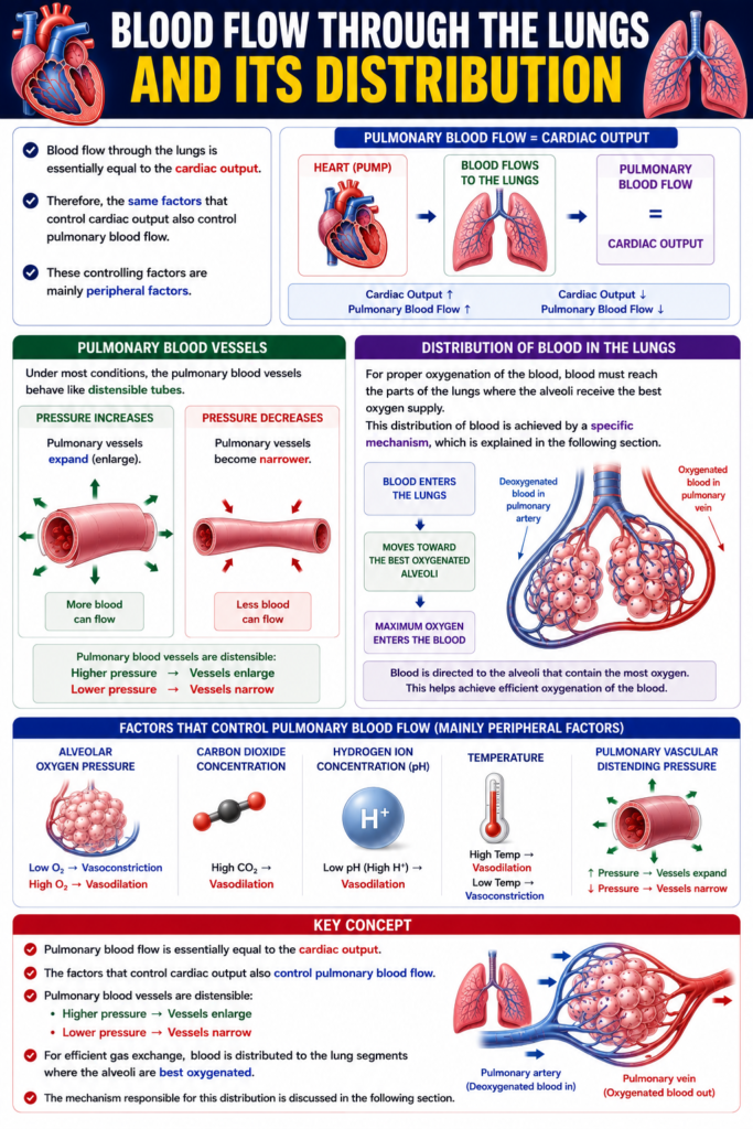
Blood flow through the lungs is essentially equal to the cardiac output.
- Therefore, the same factors that control cardiac output also control pulmonary blood flow.
- These controlling factors are mainly peripheral factors.
Pulmonary Blood Vessels
- Under most conditions, the pulmonary blood vessels behave like distensible tubes.
- When pressure increases, the pulmonary vessels expand (enlarge).
- When pressure decreases, the pulmonary vessels become narrower.
Distribution of Blood in the Lungs
- For proper oxygenation of the blood, blood must reach the parts of the lungs where the alveoli receive the best oxygen supply.
- This distribution of blood is achieved by a specific mechanism, which is explained in the following section.
Easy Concept
Think of the heart as a water pump and the lungs as a sponge with many tiny air sacs (alveoli).
Heart Pumps Blood
        │
        ▼
Blood Flows to the Lungs
        │
        ▼
Pulmonary Blood Flow
=
Cardiac Output
Concept:
- Whatever amount of blood the heart pumps, the same amount flows through the lungs.
- Therefore,
Cardiac Output ↑ → Pulmonary Blood Flow ↑
Cardiac Output ↓ → Pulmonary Blood Flow ↓
Easy Concept
Effect of Pressure on Pulmonary Blood Vessels
Pressure Increases
        │
        ▼
Pulmonary Vessels Expand
(Wider)
        │
        ▼
More Blood Can Flow
Pressure Decreases
│
▼
Pulmonary Vessels Narrow
│
▼
Less Blood Can FlowConcept:
- Pulmonary blood vessels are stretchable (distensible).
- They become wider when pressure increases.
- They become narrower when pressure decreases.
Easy Concept
Distribution of Blood
Blood Enters the Lungs
        │
        ▼
Moves Toward the
Best Oxygenated Alveoli
        │
        ▼
Maximum Oxygen
Enters the Blood
Concept:
- Blood is directed to the alveoli that contain the most oxygen.
- This helps achieve efficient oxygenation of the blood.
Key Concept
- Pulmonary blood flow is essentially equal to the cardiac output.
- Factors controlling cardiac output also control pulmonary blood flow.
- Pulmonary blood vessels are distensible:
- Higher pressure → Vessels enlarge
- Lower pressure → Vessels narrow
- For efficient gas exchange, blood is distributed to the lung segments where the alveoli are best oxygenated.
- The mechanism responsible for this distribution is discussed in the following section.
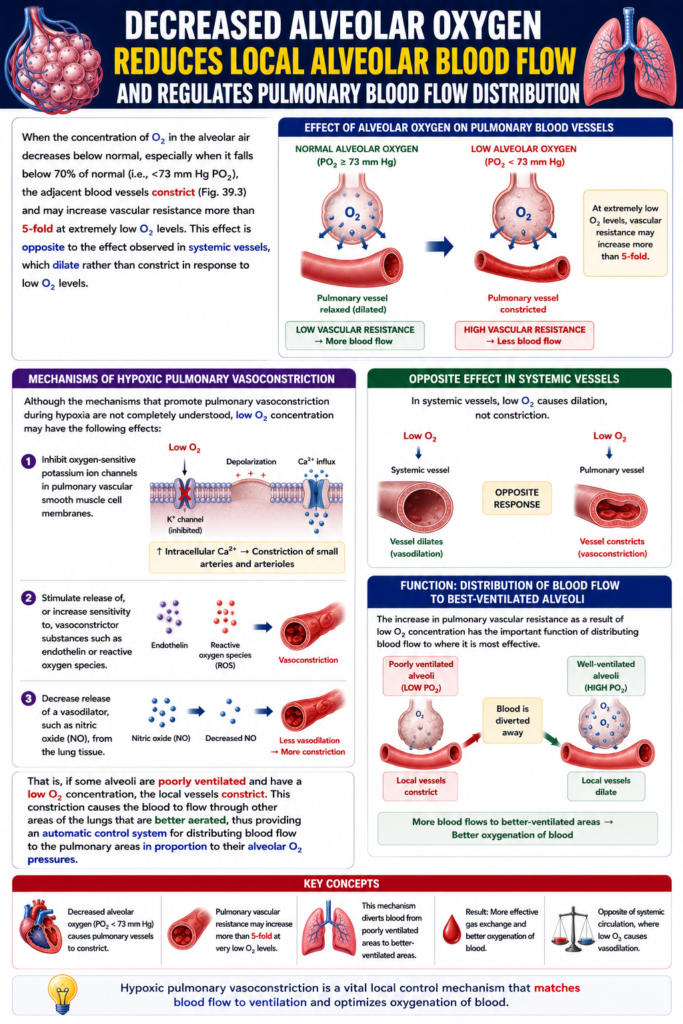
Decreased Alveolar Oxygen Reduces Local Alveolar Blood Flow and Regulates Pulmonary Blood Flow Distribution
Figure: Fig. 39.3
- When the oxygen (O₂) concentration in the alveoli decreases below normal, the nearby pulmonary blood vessels constrict.
- This effect becomes especially important when the alveolar PO₂ falls below about 70% of normal (less than 73 mm Hg PO₂).
- As shown in Fig. 39.3, the nearby blood vessels become narrower (vasoconstriction).
- At very low oxygen levels, the pulmonary vascular resistance may increase more than 5 times.
Comparison with Systemic Blood Vessels
- This response is opposite to the systemic circulation.
- In the pulmonary circulation, low O₂ causes vasoconstriction.
- In the systemic circulation, low O₂ causes vasodilation.
Possible Mechanisms of Pulmonary Vasoconstriction During Hypoxia
- The exact mechanism is not completely understood.
- Low oxygen may produce the following effects.
Inhibition of Oxygen-Sensitive Potassium Channels
- Low oxygen inhibits oxygen-sensitive potassium (K⁺) channels in the pulmonary vascular smooth muscle cells.
- This inhibition depolarizes the cell membrane.
- Depolarization opens voltage-gated calcium (Ca²⁺) channels.
- Calcium ions enter the smooth muscle cells.
- The increase in intracellular calcium causes constriction of the small pulmonary arteries and arterioles.
Easy Concept
Low Alveolar O₂
        │
        ▼
K⁺ Channels Close
        │
        ▼
Cell Membrane Depolarizes
        │
        ▼
Ca²⁺ Channels Open
        │
        ▼
Ca²⁺ Enters the Cell
        │
        ▼
Smooth Muscle Contracts
        │
        ▼
Pulmonary Vasoconstriction
Concept:
- Low oxygen starts a chain reaction.
- This chain reaction raises calcium inside the smooth muscle cells.
- More calcium causes the pulmonary blood vessels to constrict.
Release of Vasoconstrictor Substances
- Low oxygen may stimulate the release of vasoconstrictor substances.
- It may also increase the sensitivity to vasoconstrictors, such as:
- Endothelin
- Reactive oxygen species
Decreased Release of Vasodilators
- Low oxygen may reduce the release of nitric oxide from the lung tissue.
- Nitric oxide is a vasodilator.
- Reduced nitric oxide promotes vasoconstriction.
Importance of Pulmonary Vasoconstriction
- The increase in pulmonary vascular resistance helps distribute blood to the areas of the lungs where gas exchange is most effective.
- If some alveoli are poorly ventilated, they contain low oxygen.
- The blood vessels around these alveoli constrict.
- As a result, less blood flows to poorly ventilated alveoli.
- The blood is redirected to better-ventilated alveoli, where oxygen levels are higher.
- This provides an automatic mechanism for matching blood flow with alveolar oxygen levels.
Easy Concept
Poorly Ventilated Alveolus
(Low O₂)
        │
        ▼
Pulmonary Blood Vessels Constrict
        │
        ▼
Less Blood Flows Here
Well-Ventilated Alveolus
(High O₂)
│
▼
Pulmonary Blood Vessels Stay Open
│
▼
More Blood Flows Here
│
▼
Better Gas ExchangeConcept:
- Low O₂ alveoli receive less blood.
- High O₂ alveoli receive more blood.
- This ensures that blood flows to the alveoli that can oxygenate it best.
Key Concept
- Fig. 39.3 shows that low alveolar oxygen causes local pulmonary vasoconstriction.
- This response becomes important when alveolar PO₂ falls below about 73 mm Hg.
- Pulmonary vascular resistance can increase more than fivefold at very low oxygen levels.
- This response is opposite to the systemic circulation, where low oxygen causes vasodilation.
- Possible mechanisms include:
- Closure of oxygen-sensitive K⁺ channels → Depolarization → Opening of Ca²⁺ channels → Calcium influx → Vasoconstriction
- Increased release or sensitivity to endothelin and reactive oxygen species
- Decreased release of nitric oxide
- Pulmonary vasoconstriction diverts blood away from poorly ventilated alveoli toward well-ventilated alveoli, improving the efficiency of gas exchange.
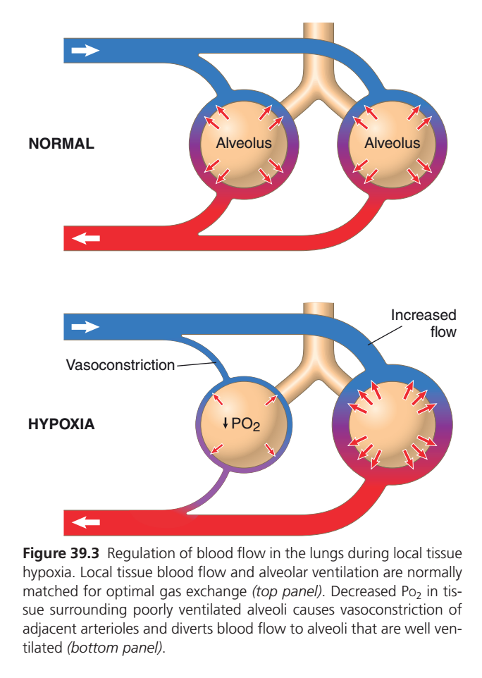
Figure 39.3: Regulation of Pulmonary Blood Flow During Local Alveolar Hypoxia (Guyton Physiology 15th Edition)
🎯 One-Line Core Concept
The lungs automatically send more blood to alveoli that have more oxygen and less blood to alveoli that have less oxygen. This perfectly matches blood flow (perfusion) with airflow (ventilation) for maximum gas exchange.
🧠 First Understand the Big Idea
Imagine there are two classrooms:
- Classroom A = Plenty of students (oxygen)
- Classroom B = Very few students (oxygen)
If a teacher (blood) wants to teach, where should they go?
✅ Obviously to Classroom A, where more students are present.
The lungs do exactly the same thing.
- Good oxygen → More blood flow
- Poor oxygen → Less blood flow
This is called:
Hypoxic Pulmonary Vasoconstriction (HPV)
TOP PICTURE: NORMAL LUNG
What is happening?
Both alveoli receive normal ventilation.
Both contain plenty of oxygen.
Therefore:
- Pulmonary arterioles remain relaxed (dilated).
- Blood flows equally to both alveoli.
- Gas exchange is excellent.
Blue Blood Vessel
🔵 Represents the Pulmonary Artery
- Carries deoxygenated blood from the right ventricle.
- Blood flows toward both alveoli.
White arrow →
Shows the direction of blood flow.
Red Blood Vessel
🔴 Represents the Pulmonary Vein
- Carries oxygenated blood back to the left atrium.
White arrow ←
Shows blood returning to the heart.
Two Alveoli
Each alveolus has:
- Normal oxygen
- Normal ventilation
- Normal gas exchange
Therefore both receive equal blood flow.
Red Small Arrows Around Each Alveolus
These arrows represent:
Oxygen diffusing from the alveolus into the surrounding blood.
Because oxygen inside the alveoli is normal,
blood becomes oxygenated efficiently.
Result in Normal Lung
Good ventilation
        ↓
High alveolar PO₂
        ↓
Pulmonary arterioles stay relaxed
        ↓
Normal blood flow
        ↓
Excellent gas exchange
BOTTOM PICTURE: HYPOXIA
Now look carefully.
One alveolus has become poorly ventilated.
Left Alveolus
Inside it is written
↓ PO₂
This means
Low oxygen inside this alveolus.
Why?
Possible reasons include:
- Airway obstruction
- Pneumonia
- Asthma
- COPD
- Atelectasis (collapsed alveolus)
This alveolus is receiving little fresh air.
What Happens Because Oxygen Falls?
This is the most important concept.
Most tissues of the body respond to low oxygen by vasodilation.
❌ The lungs behave differently.
Instead,
low alveolar oxygen causes
Pulmonary arterioles to constrict.
This is called
Hypoxic Pulmonary Vasoconstriction (HPV)
Vasoconstriction Label
The image points to the narrow blue blood vessel.
This vessel has become thinner.
Why?
Because low alveolar oxygen causes the nearby pulmonary arteriole to constrict.
Think of it like partially closing a water pipe.
Less blood can pass through.
Why Does the Lung Do This?
Suppose blood continues flowing to this poorly ventilated alveolus.
Can that blood pick up oxygen?
❌ No.
The alveolus contains very little oxygen.
So blood would leave still poorly oxygenated.
That would waste blood flow.
Instead,
the lungs divert blood elsewhere.
Increased Flow Label
Notice the right alveolus.
Its blood vessel has become larger.
The image labels this as
“Increased flow.”
Why?
Because blood that was blocked from the poorly ventilated alveolus is redirected here.
This alveolus has:
- Good ventilation
- Plenty of oxygen
So more blood arrives here and gets oxygenated efficiently.
Right Alveolus
This alveolus is healthy.
It still has:
- Normal oxygen
- Normal ventilation
Since blood is redirected here,
gas exchange becomes even more efficient.
Red Arrows Around the Right Alveolus
There are many arrows.
These indicate active oxygen transfer into the increased blood flow.
Because this alveolus receives:
- More oxygen
- More blood
Gas exchange remains highly efficient.
Why Is This Mechanism So Important?
Without this mechanism:
Poorly ventilated alveolus
        ↓
Blood still flows there
        ↓
Very little oxygen enters blood
        ↓
Low blood oxygen level (Hypoxemia)
With this mechanism:
Poorly ventilated alveolus
        ↓
Pulmonary arteriole constricts
        ↓
Blood is diverted
        ↓
Well-ventilated alveolus
        ↓
Better oxygenation
Normal vs Hypoxia
| Feature | Normal Lung | Hypoxic Lung |
|---|---|---|
| Alveolar PO₂ | Normal | Decreased |
| Pulmonary arteriole | Dilated | Constricted |
| Blood flow | Equal to both alveoli | Reduced to hypoxic alveolus |
| Blood redirected? | No | Yes |
| Gas exchange | Normal | Optimized by diverting blood |
Easy Flowchart
↓ Alveolar PO₂
        ↓
Pulmonary arteriole constricts
        ↓
↓ Blood flow to hypoxic alveolus
        ↓
Blood diverted
        ↓
Well-ventilated alveolus
        ↓
Better oxygen uptake
Memory Trick
“Air decides Blood.”
If an alveolus has:
✅ Good Air (High PO₂)
➡ Blood comes.
If an alveolus has:
❌ Poor Air (Low PO₂)
➡ Blood goes somewhere else.
Why Is This Different From the Rest of the Body?
| Most Body Tissues | Lungs |
|---|---|
| Low oxygen → Vasodilation | Low oxygen → Vasoconstriction |
| Increases blood supply to tissues | Decreases blood supply to poorly ventilated alveoli |
This unique response is exclusive to the pulmonary circulation.
📚 High-Yield MBBS Points
- Normal alveoli (high PO₂): Pulmonary arterioles remain dilated, allowing normal blood flow.
- Hypoxic alveoli (low PO₂): Nearby pulmonary arterioles undergo vasoconstriction (hypoxic pulmonary vasoconstriction).
- Purpose: Divert blood away from poorly ventilated alveoli toward well-ventilated alveoli.
- Benefit: Improves ventilation–perfusion (V/Q) matching, leading to more efficient oxygenation of blood.
- Clinical significance: This mechanism helps maintain arterial oxygen levels in localized lung disease. However, widespread alveolar hypoxia (e.g., high altitude or severe COPD) causes widespread pulmonary vasoconstriction, increasing pulmonary vascular resistance and potentially leading to pulmonary hypertension and right ventricular strain (cor pulmonale).
MADE BY SELF LEARNING CEO AND FOUNDER DR SHEEN
