


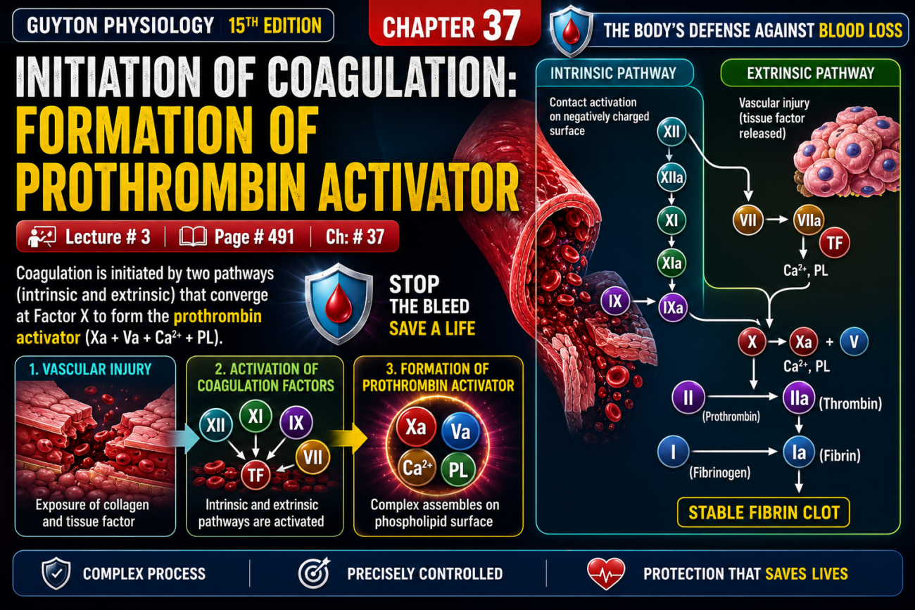
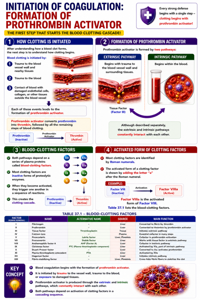
- After understanding how a blood clot forms, the next step is to understand how clotting begins.
- Blood clotting is initiated by:
- (1) Trauma to the blood vessel wall and nearby tissues
- (2) Trauma to the blood
- (3) Contact of blood with damaged endothelial cells, collagen, or other tissues outside the blood vessel
- Each of these events leads to the formation of prothrombin activator.
- Prothrombin activator converts prothrombin into thrombin.
- This is followed by all the remaining steps of blood clotting.
Formation of Prothrombin Activator
- Prothrombin activator is formed by two pathways:
- (1) Extrinsic pathway
- Begins with trauma to the blood vessel wall and surrounding tissues.
- (2) Intrinsic pathway
- Begins within the blood.
- (1) Extrinsic pathway
- Although described separately, the extrinsic and intrinsic pathways constantly interact with each other.
Blood-Clotting Factors
- Both pathways depend on a series of plasma proteins called blood-clotting factors.
- Most clotting factors are inactive forms of proteolytic enzymes.
- When they become activated, they trigger one another in a sequence of reactions.
- This creates the clotting cascade.
- Most clotting factors are identified by Roman numerals.
- The activated form of a clotting factor is shown by adding the letter “a” after the Roman numeral.
- Example: Factor VIIIa is the activated form of Factor VIII.
- Table 37.1 lists the blood-clotting factors.
Key Concept
- Blood coagulation begins with the formation of prothrombin activator. This is initiated by trauma to the vessel wall, blood, or exposure to damaged tissues. Prothrombin activator is produced through the extrinsic and intrinsic pathways, both of which depend on activation of clotting factors in a cascading sequence.
Extrinsic Pathway for Initiating Clotting
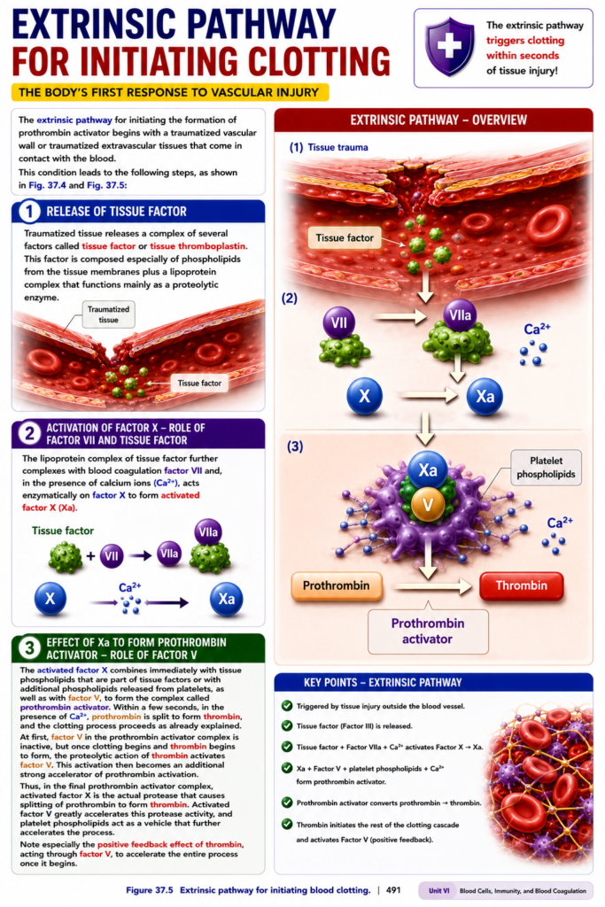
- The extrinsic pathway begins when a blood vessel wall or tissues outside the blood vessel are injured and come into contact with blood.
- This pathway proceeds through the following steps.
- Figure 37.4 and Figure 37.5 illustrate these steps.
1. Release of Tissue Factor
- Injured tissue releases tissue factor (tissue thromboplastin).
- Tissue factor is a complex of several factors.
- It contains:
- Phospholipids from tissue membranes
- A lipoprotein complex
- The lipoprotein complex mainly acts as a proteolytic enzyme.
2. Activation of Factor X — Role of Factor VII and Tissue Factor
- The lipoprotein complex of tissue factor combines with clotting factor VII.
- In the presence of calcium ions (Ca²⁺), this complex acts on factor X.
- Factor X is converted into activated factor X (Xa).
3. Formation of Prothrombin Activator — Role of Factor V
- Activated factor X (Xa) immediately combines with:
- Tissue phospholipids from tissue factor or
- Additional phospholipids released from platelets
- Factor V
- Together, they form the prothrombin activator complex.
- Within a few seconds, in the presence of Ca²⁺, prothrombin is converted into thrombin.
- The blood clotting process then continues.
- At first, factor V in the prothrombin activator complex is inactive.
- After thrombin begins to form, thrombin activates factor V.
- Activated factor V strongly accelerates prothrombin activation.
- In the final prothrombin activator complex:
- Activated factor X (Xa) is the enzyme (protease) that splits prothrombin into thrombin.
- Activated factor V greatly increases the activity of Xa.
- Platelet phospholipids act as a surface (vehicle) that further accelerates the reaction.
- Thrombin produces a positive feedback effect by activating factor V, which speeds up the entire clotting process after it has started.
Key Concept
- The extrinsic pathway is initiated by tissue injury. Tissue factor combines with factor VII and Ca²⁺ to activate factor X. Activated factor Xa, together with factor V, platelet phospholipids, and Ca²⁺, forms the prothrombin activator complex, which converts prothrombin into thrombin. Thrombin further activates factor V, creating a positive feedback loop that rapidly accelerates clot formation.
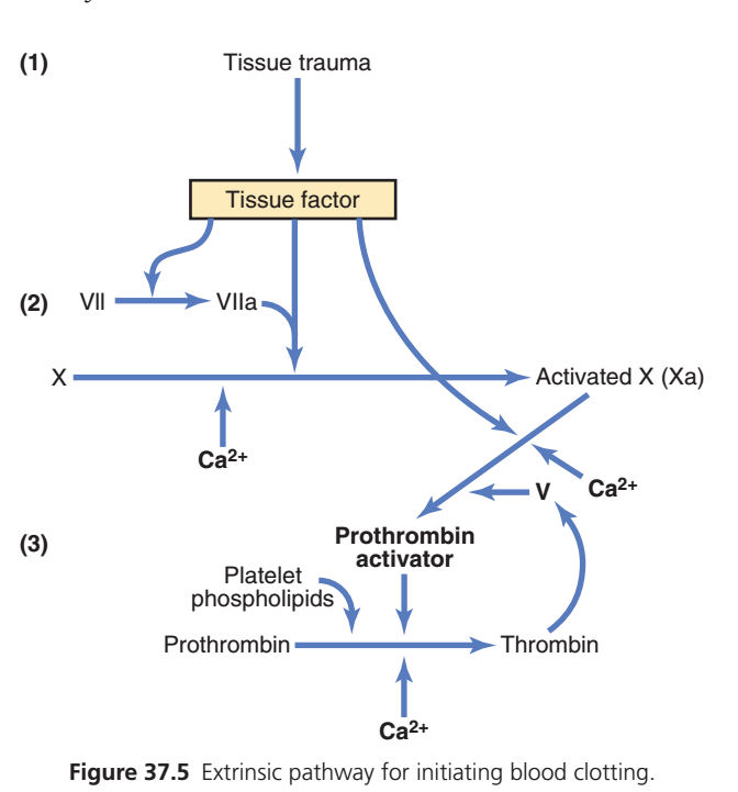
Extrinsic Pathway of Blood Clotting (Guyton Figure 37.5)
One-Line Concept
The extrinsic pathway is the fastest clotting pathway. It starts when damaged tissue releases Tissue Factor, which rapidly activates Factor X, leading to thrombin formation and finally a fibrin clot.
Easy Memory: Damage Outside Vessel → Tissue Factor → Factor X → Thrombin → Fibrin Clot
Complete Flow (Simple)
Tissue Injury
      ↓
Release of Tissue Factor (Factor III)
      ↓
Activates Factor VII → VIIa
      ↓
TF + VIIa + Ca²⁺
      ↓
Activates Factor X → Xa
      ↓
Xa + Factor V + Ca²⁺
      ↓
Prothrombin Activator
      ↓
Prothrombin → Thrombin
      ↓
Thrombin → Fibrin Formation
      ↓
Stable Blood Clot
Step 1: Tissue Trauma (Injury) 🩸
What happens?
A blood vessel and the surrounding tissues are injured.
Damaged tissue immediately releases:
Tissue Factor (TF)
Also called:
- Factor III
- Thromboplastin
Easy Concept
Think of Tissue Factor as an emergency alarm.
📢 “The tissue is injured—start clotting immediately!”
Step 2: Tissue Factor Activates Factor VII ⚡
What happens?
Tissue Factor combines with:
Factor VII
↓
Converts it into
↓
Factor VIIa (Active Form)
This reaction requires:
✅ Calcium (Ca²⁺)
Easy Concept
Imagine Tissue Factor switches ON Factor VII.
VII → VIIa (Activated)
Step 3: Activation of Factor X 🎯
Now,
Tissue Factor + Factor VIIa + Ca²⁺
work together to activate:
Factor X
↓
Factor Xa
Why is Factor Xa Important?
Factor Xa is the central enzyme that drives clot formation.
Easy Concept
Think of Factor Xa as the main machine that builds the clot.
Step 4: Formation of Prothrombin Activator 🏗️
Activated Factor X (Xa) combines with:
- Factor V
- Calcium (Ca²⁺)
- Platelet phospholipids
Together they form:
Prothrombin Activator
Easy Concept
Imagine several workers joining together to build a clotting machine.
Step 5: Prothrombin → Thrombin 🔥
Prothrombin Activator converts:
Prothrombin (Factor II)
↓
Thrombin
This step also requires:
✅ Calcium (Ca²⁺)
Easy Concept
Thrombin is the master clotting enzyme.
Once thrombin is formed, clotting accelerates rapidly.
Step 6: Positive Feedback by Thrombin 🔄
The figure also shows:
Thrombin activates:
➡️ Factor V
Activated Factor V helps produce even more Prothrombin Activator.
More Prothrombin Activator produces:
➡️ More Thrombin
Easy Concept
This is a positive feedback loop.
More thrombin → More Factor V activation → More thrombin
The clot grows very quickly.
Step 7: Formation of Fibrin 🕸️
Thrombin converts:
Fibrinogen
↓
Fibrin
Fibrin fibers weave around the platelet plug.
The clot becomes:
✔ Strong
✔ Stable
✔ Stops bleeding
Easy Story (Remember Forever)
Imagine a building catches fire.
🚨 1. Fire alarm rings → Tissue Factor released
👨🚒 2. Firefighter wakes up → Factor VII → VIIa
🚒 3. Fire truck starts → Factor X → Xa
⚙️ 4. Fire station is assembled → Prothrombin Activator
🔥 5. Water pump starts → Thrombin
🕸️ 6. Safety net is built → Fibrin
🛡️ 7. Fire is controlled → Stable blood clot
High-Yield Exam Table
| Step | Event | Main Factor | Function |
|---|---|---|---|
| 1 | Tissue injury | Tissue Factor (Factor III) | Starts extrinsic pathway |
| 2 | Factor VII activation | VII → VIIa | Forms TF–VIIa complex |
| 3 | Factor X activation | Xa | Key step toward clotting |
| 4 | Prothrombin Activator | Xa + V + Ca²⁺ + Platelet phospholipids | Converts prothrombin |
| 5 | Prothrombin → Thrombin | Thrombin | Master clotting enzyme |
| 6 | Positive feedback | Factor V | Increases thrombin production |
| 7 | Fibrin formation | Fibrin | Stable blood clot |
Role of Calcium (Ca²⁺)
Calcium is required in three important steps shown in this figure:
✅ Activation of Factor X
✅ Formation of Prothrombin Activator
✅ Conversion of Prothrombin → Thrombin
Easy Memory
Easy Mnemonic
“3-7-10-5-2”
This follows the order of the extrinsic pathway:
- 3 = Tissue Factor (Factor III)
- 7 = Factor VII → VIIa
- 10 = Factor X → Xa
- 5 = Factor V helps form Prothrombin Activator
- 2 = Prothrombin (Factor II) → Thrombin
Complete Flow Chart
Tissue Trauma
      ↓
Tissue Factor (III)
      ↓
Factor VII → VIIa
      ↓
TF + VIIa + Ca²⁺
      ↓
Factor X → Xa
      ↓
Xa + Factor V + Ca²⁺ + Platelet Phospholipids
      ↓
Prothrombin Activator
      ↓
Prothrombin (II)
      ↓
Thrombin
      ↓
Fibrinogen
      ↓
Fibrin
      ↓
Stable Blood Clot
Why Is the Extrinsic Pathway Called “Extrinsic”?
It is called the extrinsic pathway because it is triggered by Tissue Factor released from damaged tissues outside the blood vessel. Since Tissue Factor is immediately available after injury, this pathway is rapid and initiates clot formation within seconds.
Final Concept in One Sentence
In the extrinsic pathway, tissue injury releases Tissue Factor (Factor III), which activates Factor VII and then Factor X. Activated Factor X, together with Factor V, calcium, and platelet phospholipids, forms Prothrombin Activator, which converts Prothrombin into Thrombin. Thrombin then produces fibrin, resulting in a strong and stable blood clot.
Intrinsic Pathway for Initiating Clotting
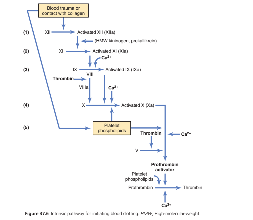
- The intrinsic pathway is the second mechanism for forming prothrombin activator and initiating blood clotting.
- It begins when:
- Blood is traumatized, or
- Blood comes into contact with collagen from a damaged blood vessel wall.
- The process continues through a series of cascading reactions.
- Figure 37.6 shows these reactions.
1. Blood Trauma Activates Factor XII and Releases Platelet Phospholipids
- Blood trauma or contact with vascular collagen changes:
- Factor XII
- Platelets
- When factor XII contacts collagen or a wettable surface (such as glass), it changes its molecular structure.
- It is converted into activated factor XII.
- Activated factor XII acts as a proteolytic enzyme.
- At the same time, blood trauma damages platelets.
- Platelets are damaged because they stick to collagen or another wettable surface, or by other types of injury.
- Damaged platelets release platelet phospholipids.
- These phospholipids contain platelet factor 3.
- Platelet factor 3 participates in the later clotting reactions.
2. Activation of Factor XI
- Activated factor XII activates factor XI.
- This step also requires high-molecular-weight kininogen.
- Prekallikrein accelerates this reaction.
3. Activation of Factor IX
- Activated factor XI activates factor IX.
4. Activation of Factor X — Role of Factor VIII
- Activated factor IX works together with:
- Activated factor VIII
- Platelet phospholipids
- Factor III from damaged platelets
- Together, they activate factor X.
- If factor VIII is deficient, this step becomes defective.
- If platelets are deficient, this step also becomes defective.
- Factor VIII is the antihemophilic factor.
- Classic hemophilia occurs because factor VIII is absent.
- Thrombocytopenia occurs because platelets are deficient.
5. Formation of Prothrombin Activator — Role of Factor V
- This step is the same as the final step of the extrinsic pathway.
- Activated factor X combines with:
- Factor V
- Platelet phospholipids or tissue phospholipids
- Together, they form the prothrombin activator complex.
- Prothrombin activator rapidly converts prothrombin into thrombin within a few seconds.
- Thrombin then starts the final steps of blood clot formation.
Key Concept
- The intrinsic pathway begins with blood trauma or exposure to collagen. Activated factor XII triggers a cascade (XII → XI → IX → VIII → X). Activated factor Xa combines with factor V and platelet phospholipids to form prothrombin activator, which rapidly converts prothrombin into thrombin and initiates the final clotting process.
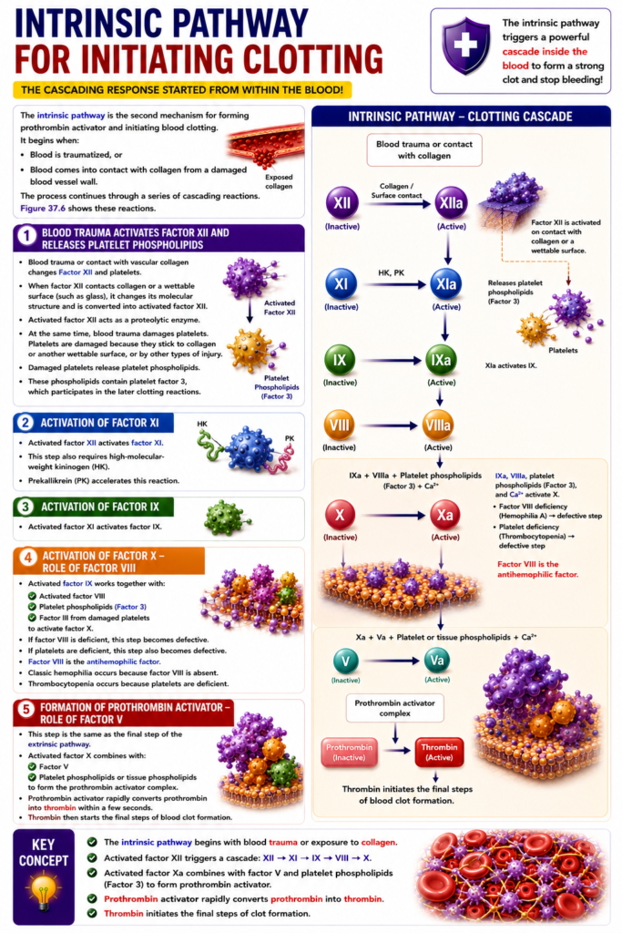
Intrinsic Pathway of Blood Clotting (Guyton Figure 37.6) — Easiest & Most Conceptual Summary
One-Line Concept
The intrinsic pathway starts when blood comes in contact with damaged collagen inside the blood vessel. It activates a chain of clotting factors (XII → XI → IX → VIII → X), finally producing thrombin and a stable fibrin clot.
Easy Memory: Inside Vessel Injury → 12 → 11 → 9 → 8 → 10 → 5 → 2 → Thrombin → Fibrin
Complete Flow (Simplified)
Blood contacts collagen
          ↓
Factor XII → XIIa
          ↓
Factor XI → XIa
          ↓
Factor IX → IXa
          ↓
Factor VIII → VIIIa
          ↓
Factor X → Xa
          ↓
Factor V + Platelet phospholipids + Ca²⁺
          ↓
Prothrombin Activator
          ↓
Prothrombin → Thrombin
          ↓
Fibrinogen → Fibrin
          ↓
Stable Blood Clot
Step 1: Blood Contacts Collagen 🩸
What happens?
The blood vessel is damaged.
Blood touches the exposed collagen beneath the endothelium.
This immediately activates:
Factor XII (Hageman Factor)
↓
Factor XIIa
Easy Concept
Imagine collagen is an alarm button inside the blood vessel.
When blood touches it,
➡️ Factor XII wakes up first.
Step 2: Factor XII Activates Factor XI ⚡
Activated Factor XII (XIIa) activates:
Factor XI
↓
Factor XIa
This step is helped by:
- High-Molecular-Weight (HMW) Kininogen
- Prekallikrein
Easy Concept
Think of Factor XII as the team leader.
It wakes up the next worker:
Factor XI
Step 3: Factor XI Activates Factor IX 🔄
Activated Factor XI (XIa), together with:
✅ Calcium (Ca²⁺)
activates:
Factor IX
↓
Factor IXa
Easy Concept
The clotting message continues down the chain.
12 → 11 → 9
Step 4: Factor VIII Helps Activate Factor X 🎯
Now,
Activated Factor IX (IXa) joins with:
- Factor VIIIa
- Calcium (Ca²⁺)
- Platelet phospholipids
Together they activate:
Factor X
↓
Factor Xa
Important Point
Factor VIII is first activated by Thrombin:
VIII → VIIIa
Easy Concept
Factor VIII acts like an assistant helping Factor IX activate Factor X.
Step 5: Platelet Phospholipids 🟣
Activated platelets provide:
Platelet phospholipids
These serve as a platform (working surface) where clotting factors assemble.
Easy Concept
Imagine building a house.
Before construction starts,
workers need a floor to stand on.
Platelet phospholipids are that floor.
Step 6: Formation of Prothrombin Activator 🏗️
Activated Factor X (Xa) combines with:
- Factor V
- Platelet phospholipids
- Calcium (Ca²⁺)
to form:
Prothrombin Activator
Easy Concept
This is the clot-making machine.
Step 7: Prothrombin → Thrombin 🔥
Prothrombin Activator converts:
Prothrombin (Factor II)
↓
Thrombin
Requires:
✅ Calcium (Ca²⁺)
Why is Thrombin Important?
Thrombin is the master enzyme of coagulation.
It produces fibrin and strengthens the clot.
Step 8: Positive Feedback 🔁
The figure also shows:
Thrombin activates:
- Factor VIII
- Factor V
These activated factors help generate even more thrombin.
Easy Concept
This is a snowball effect.
Small clot →
More thrombin →
Bigger clot →
Even more thrombin.
Step 9: Formation of Fibrin 🕸️
Thrombin converts:
Fibrinogen
↓
Fibrin
Fibrin fibers form a mesh around platelets.
Result:
✔ Strong clot
✔ Bleeding stops
Easy Story (Remember Forever)
Imagine a factory inside the blood vessel.
🚨 1. Alarm rings → Blood touches collagen
👷 2. Worker 12 wakes Worker 11
👷 3. Worker 11 wakes Worker 9
👷 4. Worker 9 and Worker 8 start Machine 10
🏭 5. Machine 10 builds the clotting machine (Prothrombin Activator)
🔥 6. Clotting machine produces Thrombin
🕸️ 7. Thrombin makes Fibrin
🛡️ 8. Fibrin forms a strong blood clot
High-Yield Exam Table
| Step | Activated Factor | Main Function |
|---|---|---|
| 1 | XII → XIIa | Starts intrinsic pathway |
| 2 | XI → XIa | Activates Factor IX |
| 3 | IX → IXa | Works with Factor VIII |
| 4 | VIII → VIIIa | Helps activate Factor X |
| 5 | X → Xa | Forms Prothrombin Activator |
| 6 | V | Helps Prothrombin Activator |
| 7 | II → Thrombin | Master clotting enzyme |
| 8 | Fibrin | Stable blood clot |
Role of Calcium (Ca²⁺)
Calcium is required for several major reactions shown in this figure:
- Factor XI → IX
- Factor IX + VIII → X
- Formation of Prothrombin Activator
- Prothrombin → Thrombin
Easy Memory
Calcium is the “helper ion” that keeps the clotting cascade moving.
Easy Mnemonic
“12 → 11 → 9 → 8 → 10 → 5 → 2”
- 12 = Starts the intrinsic pathway
- 11 = Activated by XII
- 9 = Activated by XI
- 8 = Cofactor for IX
- 10 = Activated by IXa + VIIIa
- 5 = Helps form Prothrombin Activator
- 2 = Prothrombin → Thrombin
Intrinsic vs Extrinsic Pathway
| Feature | Intrinsic Pathway | Extrinsic Pathway |
|---|---|---|
| Trigger | Blood contacts exposed collagen | Tissue injury releases Tissue Factor |
| First factor | Factor XII | Tissue Factor (Factor III) |
| Number of steps | More (slower) | Fewer (faster) |
| Speed | Slower | Very rapid |
| Both pathways meet at | Factor X (Xa) | Factor X (Xa) |
Complete Flow Chart
Blood contacts collagen
          ↓
Factor XII → XIIa
          ↓
Factor XI → XIa
          ↓
Factor IX → IXa
          ↓
Factor VIII → VIIIa (activated by thrombin)
          ↓
Factor X → Xa
          ↓
Xa + Factor V + Platelet phospholipids + Ca²⁺
          ↓
Prothrombin Activator
          ↓
Prothrombin
          ↓
Thrombin
          ↓
Fibrinogen
          ↓
Fibrin
          ↓
Stable Blood Clot
Quick Exam Pearls
- Intrinsic pathway is initiated by blood contacting exposed collagen within the damaged vessel.
- Factor XII (Hageman factor) is the first clotting factor activated.
- HMW kininogen and prekallikrein assist the activation of Factor XII.
- Factor VIII is activated by thrombin, then works with Factor IXa to activate Factor X.
- Both the intrinsic and extrinsic pathways converge at Factor X (Xa), after which they share the common pathway leading to thrombin and fibrin formation.
Final Concept in One Sentence
In the intrinsic pathway, blood contacting exposed collagen activates Factor XII, which sequentially activates Factors XI, IX, VIII, and X. Activated Factor X, together with Factor V, platelet phospholipids, and calcium, forms Prothrombin Activator, which converts prothrombin into thrombin, ultimately producing fibrin and a stable blood clot.
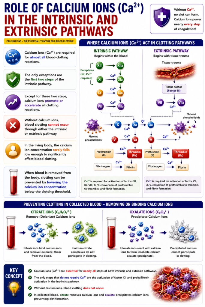
Role of Calcium Ions in the Intrinsic and Extrinsic Pathways
- Calcium ions (Ca²⁺) are required for almost all blood-clotting reactions.
- The only exceptions are the first two steps of the intrinsic pathway.
- Except for these two steps, calcium ions promote or accelerate all clotting reactions.
- Without calcium ions, blood clotting cannot occur through either the intrinsic or extrinsic pathway.
- In the living body, the calcium ion concentration rarely falls low enough to significantly affect blood clotting.
- When blood is removed from the body, clotting can be prevented by lowering the calcium ion concentration below the clotting threshold.
- Calcium ions can be removed (deionized) by reacting them with citrate ions.
- Calcium ions can also be precipitated by using oxalate ions.
Key Concept
- Calcium ions are essential for nearly all steps of both the intrinsic and extrinsic coagulation pathways. Without Ca²⁺, blood clotting does not occur. In collected blood, citrate or oxalate prevents clotting by removing or binding calcium ions.
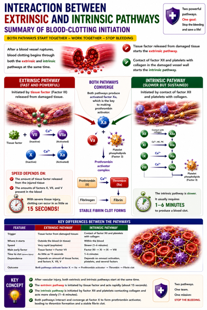
Interaction Between Extrinsic and Intrinsic Pathways—Summary of Blood-Clotting Initiation
- After a blood vessel ruptures, blood clotting begins through both the extrinsic and intrinsic pathways at the same time.
- Tissue factor starts the extrinsic pathway.
- Contact of factor XII and platelets with collagen in the damaged blood vessel wall starts the intrinsic pathway.
- An important difference between the two pathways is their speed.
- The extrinsic pathway is very rapid (explosive) once it starts.
- Its speed depends on:
- The amount of tissue factor released from the injured tissue
- The amounts of factors X, VII, and V present in the blood
- With severe tissue injury, blood clotting can occur in as little as 15 seconds.
- The intrinsic pathway is slower.
- It usually requires 1–6 minutes to produce blood clotting.
Key Concept
- After vascular injury, both coagulation pathways work simultaneously. The extrinsic pathway is initiated by tissue factor and acts rapidly (about 15 seconds), whereas the intrinsic pathway is initiated by factor XII and platelets contacting collagen and acts more slowly (1–6 minutes).
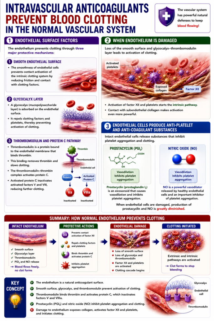
Intravascular Anticoagulants Prevent Blood Clotting in the Normal Vascular System
Endothelial Surface Factors
- Normal blood vessels contain natural anticoagulant mechanisms that prevent blood clotting.
- The most important endothelial surface factors are:
1. Smooth Endothelial Surface
- The smooth surface of endothelial cells prevents contact activation of the intrinsic clotting pathway.
2. Glycocalyx Layer
- The endothelium is covered by a glycocalyx layer.
- Glycocalyx is a mucopolysaccharide attached to the surface of endothelial cells.
- It repels clotting factors and platelets.
- This prevents activation of blood clotting.
3. Thrombomodulin
- Thrombomodulin is a protein attached to the endothelial membrane.
- It binds to thrombin.
- This binding slows blood clotting by removing free thrombin.
- The thrombomodulin–thrombin complex activates protein C.
- Protein C acts as an anticoagulant.
- It inactivates activated factor V and activated factor VIII.
Effect of Endothelial Damage
- When the endothelium is damaged, the following protective factors are lost:
- Smooth endothelial surface
- Glycocalyx layer
- Thrombomodulin
- This activates factor XII and platelets.
- As a result, the intrinsic clotting pathway begins.
- If factor XII and platelets come into contact with subendothelial collagen, the activation becomes even stronger.
Prostacyclin and Nitric Oxide (NO)
- Healthy endothelial cells also produce prostacyclin and nitric oxide (NO).
- Both substances inhibit platelet aggregation.
- They also prevent the initiation of blood clotting.
Prostacyclin (PGI₂)
- Prostacyclin (PGI₂) is a member of the eicosanoid family of lipids.
- It is a vasodilator.
- It also inhibits platelet aggregation.
Nitric Oxide (NO)
- Nitric oxide (NO) is a powerful vasodilator.
- It is released by healthy endothelial cells throughout the body.
- It is also an important inhibitor of platelet aggregation.
- When endothelial cells are damaged, the production of prostacyclin and NO decreases greatly.
Key Concept
- Normal endothelium prevents blood clotting through a smooth surface, glycocalyx, thrombomodulin, prostacyclin (PGI₂), and nitric oxide (NO). These factors inhibit platelet activation and the intrinsic pathway. Damage to the endothelium removes these protective mechanisms, allowing clot formation to begin.
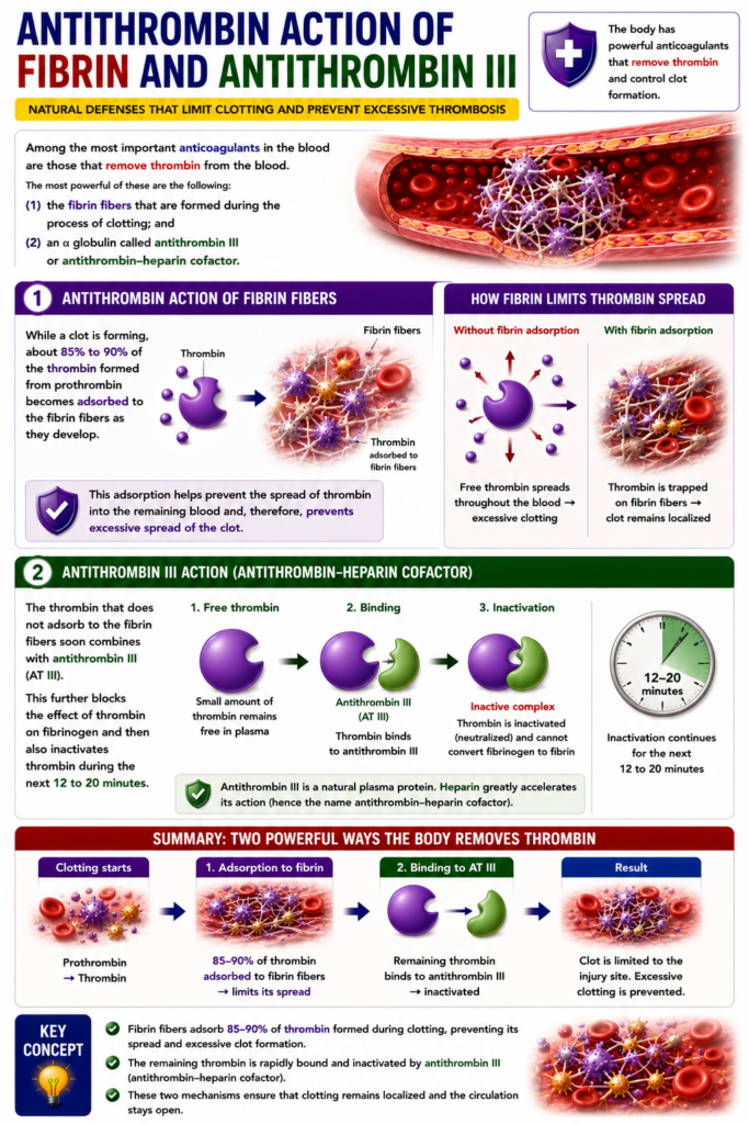
Antithrombin Action of Fibrin and Antithrombin III
- Some of the most important anticoagulants in the blood remove thrombin from the circulation.
- The two most powerful anticoagulants are:
- (1) Fibrin fibers formed during blood clotting.
- (2) Antithrombin III (antithrombin–heparin cofactor), an α-globulin.
- While a blood clot is forming, about 85%–90% of the thrombin produced from prothrombin becomes attached (adsorbed) to the developing fibrin fibers.
- This adsorption prevents thrombin from spreading into the remaining blood.
- As a result, it prevents excessive spread of the blood clot.
- The remaining thrombin that is not attached to fibrin fibers quickly binds with antithrombin III.
- Antithrombin III blocks the action of thrombin on fibrinogen.
- It also inactivates thrombin within the next 12–20 minutes.
Key Concept
- Fibrin fibers and antithrombin III are major natural anticoagulants. Fibrin fibers bind 85–90% of thrombin, limiting clot spread, while antithrombin III binds and inactivates the remaining thrombin within 12–20 minutes, preventing excessive coagulation.
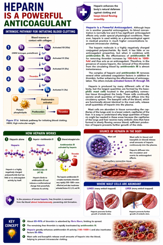
Heparin Is a Powerful Anticoagulant
- Heparin is a powerful anticoagulant.
- Normally, the amount of heparin in the blood is very low.
- Therefore, under normal conditions, its anticoagulant effect is small.
- Heparin is widely used as a medicine in much higher concentrations.
- It is used to prevent blood clotting inside blood vessels.
- Figure 37.6 shows the intrinsic pathway for initiating blood clotting.
Heparin and Antithrombin III
- Heparin is a highly negatively charged conjugated polysaccharide.
- By itself, heparin has little or no anticoagulant effect.
- When heparin combines with antithrombin III, the activity of antithrombin III increases by 100–1000 times.
- This greatly enhances its anticoagulant action.
- In the presence of large amounts of heparin, antithrombin III removes free thrombin from the blood almost immediately.
Effect on Other Clotting Factors
- The heparin–antithrombin III complex also inactivates several other activated clotting factors.
- These include activated factors IX, X, XI, and XII.
- This further increases the anticoagulant effect.
Production of Heparin
- Heparin is produced by many cells in the body.
- The largest amount is produced by basophilic mast cells.
- These mast cells are located in the connective tissue around capillaries throughout the body.
- Mast cells continuously release small amounts of heparin.
- This heparin diffuses into the bloodstream.
- Basophils in the blood are functionally similar to mast cells.
- Basophils also release small amounts of heparin into the plasma.
Heparin in the Lungs and Liver
- Mast cells are especially abundant around the capillaries of the lungs.
- They are also present around the capillaries of the liver, but in smaller numbers.
- The lungs and liver receive many embolic clots that develop in slowly flowing venous blood.
- Heparin produced in these areas prevents further growth of these clots.
Key Concept
- Heparin is a powerful natural and therapeutic anticoagulant. By binding to antithrombin III, it increases its activity by 100–1000 times, rapidly inactivating thrombin and activated factors IX–XII. Heparin is mainly produced by mast cells and basophils, especially around lung and liver capillaries, where it helps prevent the enlargement of embolic clots.
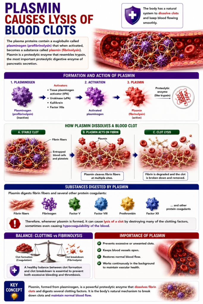
PLASMIN CAUSES LYSIS OF BLOOD CLOTS
- Plasma proteins contain a euglobulin called plasminogen (profibrinolysin).
- When plasminogen is activated, it becomes plasmin (fibrinolysin).
- Plasmin is a proteolytic enzyme.
- It resembles trypsin, the major proteolytic digestive enzyme of pancreatic secretion.
- Plasmin digests fibrin fibers.
- It also digests several other clotting proteins, including:
- Fibrinogen
- Factor V
- Factor VIII
- Prothrombin
- Factor XII
- Therefore, when plasmin is formed, it breaks down (lyses) the blood clot.
- It does this by destroying fibrin and many clotting factors.
- If too much plasmin is formed, it may cause hypocoagulability of the blood.
Key Concept
- Plasmin is the active form of plasminogen and is responsible for fibrinolysis. It dissolves blood clots by digesting fibrin fibers and several clotting factors (fibrinogen, factors V, VIII, XII, and prothrombin). Excess plasmin activity can reduce the blood’s ability to clot (hypocoagulability).
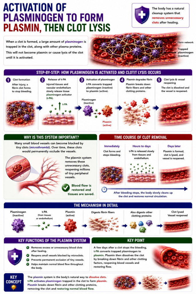
Activation of Plasminogen to Form Plasmin, Then Clot Lysis
- When a blood clot forms, a large amount of plasminogen becomes trapped inside the clot along with other plasma proteins.
- Plasminogen remains inactive and does not dissolve the clot until it is activated.
- Injured tissues and the vascular endothelium slowly release a powerful activator called tissue plasminogen activator (t-PA).
- A few days after the clot has stopped the bleeding, t-PA converts plasminogen into plasmin.
- Plasmin then breaks down and removes the remaining unnecessary blood clot.
- By this mechanism, many small blood vessels blocked by clots become open again.
- An important function of the plasmin system is to remove tiny blood clots from millions of small peripheral blood vessels.
- Without this clot-removal mechanism, these small blood vessels would eventually become blocked (occluded).
Key Concept
- After bleeding stops, tissue plasminogen activator (t-PA) slowly converts plasminogen trapped in the clot into plasmin. Plasmin dissolves the unnecessary clot, reopening blocked small blood vessels and preventing occlusion of the peripheral circulation.
MADE BY SELF LEARNING CEO AND FOUNDER DR SHEEN
