


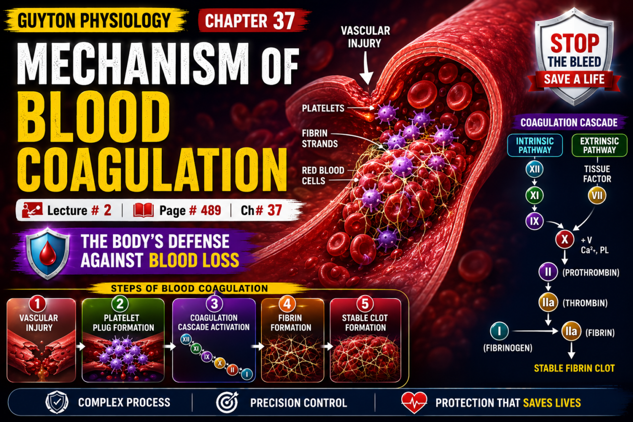
- More than 50 important substances in the blood and tissues affect blood coagulation.
- These substances are of two types:
- Procoagulants – promote blood clotting.
- Anticoagulants – prevent blood clotting.
- Whether blood clots or not depends on the balance between procoagulants and anticoagulants.
- In normal circulating blood, anticoagulants predominate.
- Therefore, blood does not clot while flowing inside blood vessels.
- When a blood vessel is ruptured, procoagulants from the damaged tissue become activated.
- These activated procoagulants overcome the anticoagulants.
- As a result, a blood clot forms.
GENERAL MECHANISM
- Blood clotting occurs in three essential steps:
Step 1
- When a blood vessel ruptures or blood is damaged, a complex cascade of chemical reactions begins.
- These reactions involve more than 12 blood coagulation factors.
- The final result is the formation of a group of activated substances called prothrombin activator.
Step 2
- Prothrombin activator converts prothrombin into thrombin.
Step 3
- Thrombin acts as an enzyme.
- It converts fibrinogen into fibrin fibers.
- Fibrin fibers trap platelets, blood cells, and plasma.
- This forms the blood clot.
- The blood clotting mechanism is first explained by conversion of prothrombin to thrombin.
- After that, the earlier steps that form prothrombin activator are explained.
Key Concept
- Blood coagulation depends on the balance between procoagulants and anticoagulants. Clot formation occurs in three main steps: formation of prothrombin activator, conversion of prothrombin to thrombin, and conversion of fibrinogen to fibrin to form the blood clot.
CONVERSION OF PROTHROMBIN TO THROMBIN
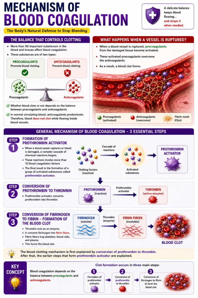
- Step 1:Prothrombin activator is formed after:
- Rupture of a blood vessel, or
- Damage to special substances in the blood.
- Step 2: In the presence of enough calcium ions (Ca²⁺), prothrombin activator converts prothrombin into thrombin.
- Figure 37.3 and Figure 37.4 illustrate this conversion.
- Step 3: Thrombin converts fibrinogen into fibrin fibers.
- This polymerization of fibrinogen occurs within 10–15 seconds.
- Formation of prothrombin activator is usually the rate-limiting step of blood coagulation.
- The later reactions occur rapidly.
- Therefore, once prothrombin activator is formed, the blood clot develops quickly.
- Platelets also help convert prothrombin into thrombin.
- Much of the prothrombin first binds to prothrombin receptors on platelets.
- These platelets are already attached to the damaged tissue.
Key Concept
- Blood coagulation depends mainly on the formation of prothrombin activator. In the presence of Ca²⁺, prothrombin activator converts prothrombin to thrombin, which rapidly converts fibrinogen into fibrin fibers. Platelets enhance this process by providing binding sites for prothrombin.
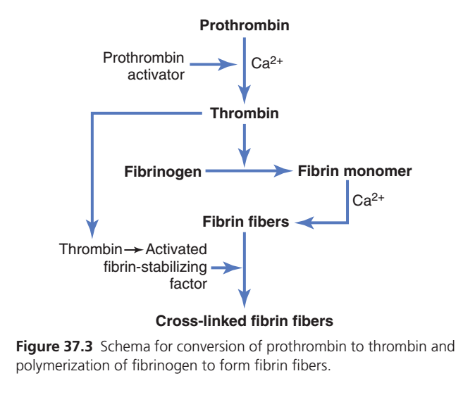
Conversion of Prothrombin to Thrombin & Formation of Fibrin (Guyton Figure 37.3) — Easiest Conceptual Summary
One-Line Concept
A weak platelet plug becomes a strong blood clot when prothrombin is converted into thrombin, which then converts fibrinogen into strong fibrin fibers.
Big Picture Flow
Blood vessel injury
        ↓
Prothrombin activator formed
        ↓
Prothrombin ──(Ca²⁺)──► Thrombin
        ↓
Fibrinogen ──► Fibrin monomers
        ↓
Fibrin fibers formed
        ↓
Factor XIII activated by thrombin
        ↓
Cross-linked fibrin fibers
        ↓
Strong, stable blood clot
Step 1: Prothrombin Activator is Formed 🚨
What happens?
After blood vessel injury, the body produces Prothrombin Activator.
Its job is to start the clotting process.
Easy Concept
Think of it as the “ON switch” of blood coagulation.
Without it, clotting cannot begin.
Step 2: Prothrombin → Thrombin (Requires Calcium) ⚡
What happens?
Inactive protein:
Prothrombin (Factor II)
↓
is converted into
↓
Thrombin (Active enzyme)
This reaction needs:
✅ Calcium ions (Ca²⁺)
Easy Concept
Imagine:
👨🎓 Prothrombin = Student
👨⚕️ Thrombin = Doctor
The student becomes a doctor only after training.
Similarly,
Prothrombin becomes active thrombin.
Step 3: Thrombin Converts Fibrinogen into Fibrin Monomers 🧵
What happens?
Thrombin acts on:
Fibrinogen
(a soluble protein floating in blood)
↓
converts it into
↓
Fibrin Monomers
Easy Concept
Imagine fibrinogen as a ball of soft yarn.
Thrombin unwinds the yarn into long strands.
Step 4: Fibrin Monomers Join to Form Fibrin Fibers 🕸️
What happens?
The fibrin monomers join together.
With the help of Ca²⁺, they form:
Fibrin fibers
These fibers create a mesh over the platelet plug.
Easy Concept
Like weaving many threads into a fishing net.
Step 5: Thrombin Activates Factor XIII 🔒
What happens?
Thrombin has another important job.
It activates:
Fibrin-Stabilizing Factor (Factor XIII)
Inactive Factor XIII
↓
Activated Factor XIII
Step 6: Cross-Linking of Fibrin Fibers 💪
What happens?
Activated Factor XIII links neighboring fibrin fibers together.
This process is called:
Cross-linking
Result
The fibrin mesh becomes:
✔ Thick
✔ Strong
✔ Stable
✔ Difficult to break
Final Result 🩸
The platelet plug is now reinforced by a cross-linked fibrin network, producing a strong and stable blood clot that effectively stops bleeding until the vessel heals.
Easy Story (Remember Forever)
Imagine repairing a broken bridge.
🚧 1. Accident occurs → Blood vessel injury
🔘 2. Start button pressed → Prothrombin activator
⚡ 3. Machine starts → Prothrombin becomes thrombin
🧶 4. Rope is made → Fibrinogen becomes fibrin
🕸️ 5. Rope forms a net → Fibrin fibers
🔩 6. Bolts tighten the net → Factor XIII cross-links fibrin
🌉 7. Bridge becomes strong → Stable blood clot
High-Yield Exam Table
| Step | Event | Important Molecule |
|---|---|---|
| 1 | Clotting starts | Prothrombin activator |
| 2 | Prothrombin → Thrombin | Ca²⁺ |
| 3 | Fibrinogen → Fibrin monomers | Thrombin |
| 4 | Fibrin fibers formed | Ca²⁺ |
| 5 | Factor XIII activated | Thrombin |
| 6 | Cross-linked fibrin fibers | Activated Factor XIII |
Key Functions of Thrombin
✅ Converts Prothrombin pathway into active clotting.
✅ Converts Fibrinogen → Fibrin.
✅ Activates Factor XIII.
✅ Produces a strong, stable clot.
Mnemonic
“PATFF”
- P = Prothrombin Activator
- A = Activation of Prothrombin
- T = Thrombin
- F = Fibrin
- F = Factor XIII fixes (cross-links) fibrin
Calcium’s Role (Ca²⁺) 🧪
Calcium is required at two major points in this figure:
- Prothrombin → Thrombin
- Formation and stabilization of Fibrin fibers
Easy Memory
“No Calcium → No Clot.”
Complete Flow Chart
Blood vessel injury
        ↓
Prothrombin activator
        ↓
Prothrombin + Ca²⁺
        ↓
Thrombin
      ↙        ↘
Fibrinogen      Factor XIII
      ↓              ↓
Fibrin monomers   Activated Factor XIII
      ↓
Fibrin fibers
      ↓
Cross-linked fibrin fibers
      ↓
Strong stable blood clot
Final Concept in One Sentence
Following blood vessel injury, prothrombin activator and calcium convert prothrombin into thrombin; thrombin then converts fibrinogen into fibrin, activates Factor XIII, and produces cross-linked fibrin fibers that strengthen the platelet plug into a stable blood clot.
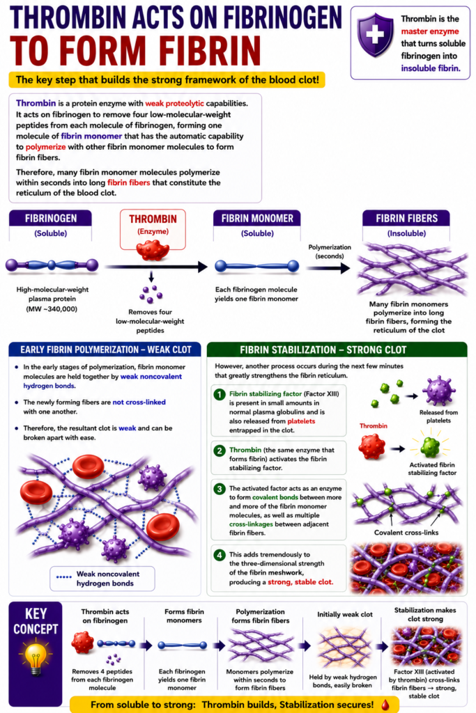
Prothrombin and Thrombin
- Prothrombin is a plasma protein.
- It is an α₂-globulin.
- It has a molecular weight of 68,700.
- The normal concentration of prothrombin in plasma is about 15 mg/dL.
- Prothrombin is an unstable protein.
- It can easily split into smaller compounds.
- One of these compounds is thrombin.
- Thrombin has a molecular weight of 33,700, which is almost half the molecular weight of prothrombin.
- Prothrombin is continuously produced by the liver.
- It is also continuously used in the body for blood clotting.
- If the liver cannot produce prothrombin, the plasma prothrombin level falls within about one day.
- When the prothrombin level becomes too low, normal blood coagulation cannot occur.
- Vitamin K is required by the liver for the normal activation of prothrombin.
- Vitamin K is also needed for the activation of some other clotting factors.
- Vitamin K deficiency can reduce prothrombin production.
- Liver disease can also prevent normal prothrombin formation.
- When prothrombin levels become very low, a bleeding tendency develops.
Key Concept
- Prothrombin is a liver-produced plasma protein that is converted into thrombin during blood coagulation. Normal production requires vitamin K and a healthy liver. Deficiency of vitamin K or liver disease lowers prothrombin levels, leading to impaired blood clotting and increased bleeding.
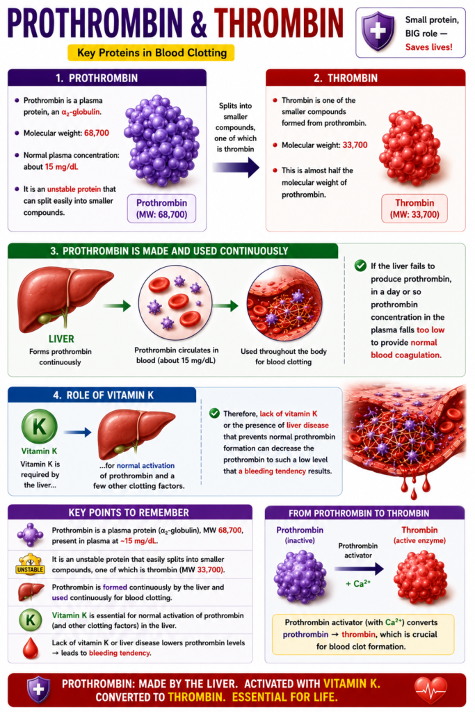
CONVERSION OF FIBRINOGEN TO FIBRIN—FORMATION OF THE CLOT
Fibrinogen Formed in the Liver Is Essential for Clot Formation
- Fibrinogen is a high-molecular-weight protein.
- It has a molecular weight of about 340,000.
- The normal concentration of fibrinogen in plasma is 100–700 mg/dL.
- Fibrinogen is produced by the liver.
- Liver disease can reduce the amount of circulating fibrinogen.
- Liver disease can also reduce prothrombin levels.
- Because fibrinogen is a very large molecule, very little normally leaks from blood vessels into the interstitial fluid.
- Fibrinogen is an essential factor for blood coagulation.
- Therefore, interstitial fluid normally does not clot.
- If capillary permeability becomes abnormally increased, fibrinogen leaks into the tissue fluid.
- When enough fibrinogen enters the tissue fluid, the tissue fluid can clot.
- This clotting occurs in much the same way as plasma and whole blood clot.
Key Concept
- Fibrinogen is a liver-produced plasma protein essential for clot formation. Because of its large size, it normally remains inside blood vessels, so interstitial fluid does not clot. Increased capillary permeability allows fibrinogen to enter tissues, where clotting can occur.
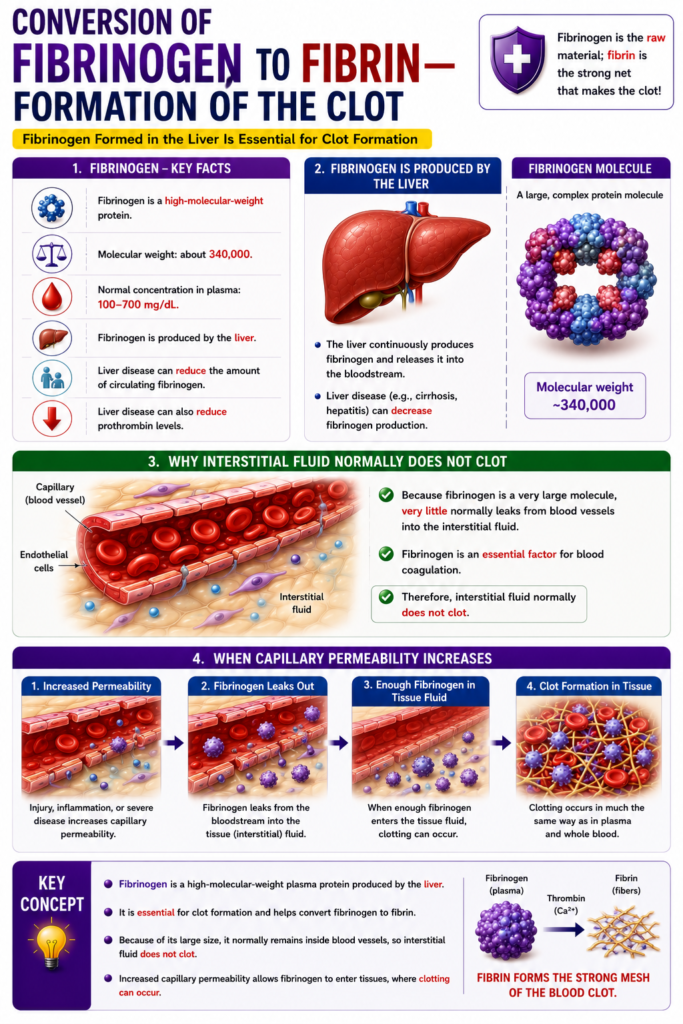
Thrombin Acts on Fibrinogen to Form Fibrin
- Thrombin is a protein enzyme.
- It has weak proteolytic (protein-splitting) activity.
- Thrombin acts on fibrinogen.
- It removes four low-molecular-weight peptides from each fibrinogen molecule.
- This forms one fibrin monomer molecule.
- Each fibrin monomer automatically joins with other fibrin monomers.
- This process is called polymerization.
- Polymerization forms fibrin fibers.
- Within seconds, many fibrin monomers polymerize into long fibrin fibers.
- These fibrin fibers form the meshwork (reticulum) of the blood clot.
- During the early stage of polymerization, fibrin monomers are held together by weak noncovalent hydrogen bonds.
- At this stage, the fibrin fibers are not cross-linked with each other.
- Therefore, the newly formed clot is weak.
- This weak clot can be broken easily.
- During the next few minutes, another process greatly strengthens the fibrin meshwork.
- This process involves fibrin-stabilizing factor.
- Fibrin-stabilizing factor is present:
- In small amounts in normal plasma globulins.
- It is also released from platelets trapped inside the clot.
- Before it can work, fibrin-stabilizing factor must be activated.
- The same thrombin that forms fibrin also activates fibrin-stabilizing factor.
- The activated fibrin-stabilizing factor acts as an enzyme.
- It forms covalent bonds between fibrin monomer molecules.
- It also creates multiple cross-links between adjacent fibrin fibers.
- These cross-links greatly increase the three-dimensional strength of the fibrin meshwork.
Key Concept
- Thrombin converts fibrinogen into fibrin monomers, which rapidly polymerize into fibrin fibers. Initially, the clot is weak because the fibers are joined by hydrogen bonds. Thrombin then activates fibrin-stabilizing factor, which forms covalent cross-links between fibrin fibers, producing a strong and stable blood clot.
Blood Clot
- A blood clot is made of a meshwork of fibrin fibers.
- The fibrin fibers run in all directions.
- These fibrin fibers trap blood cells, platelets, and plasma.
- Figure 37.4 shows the structure of a blood clot.
- The fibrin fibers also stick to the damaged surface of the blood vessel.
- Because of this, the blood clot firmly attaches to the opening in the damaged blood vessel.
- This prevents further blood loss.
Key Concept
- A blood clot is a fibrin mesh that traps blood cells, platelets, and plasma. The fibrin fibers adhere to the damaged vessel wall, sealing the vascular opening and stopping further bleeding.
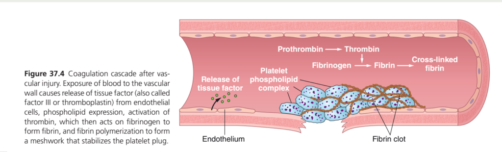
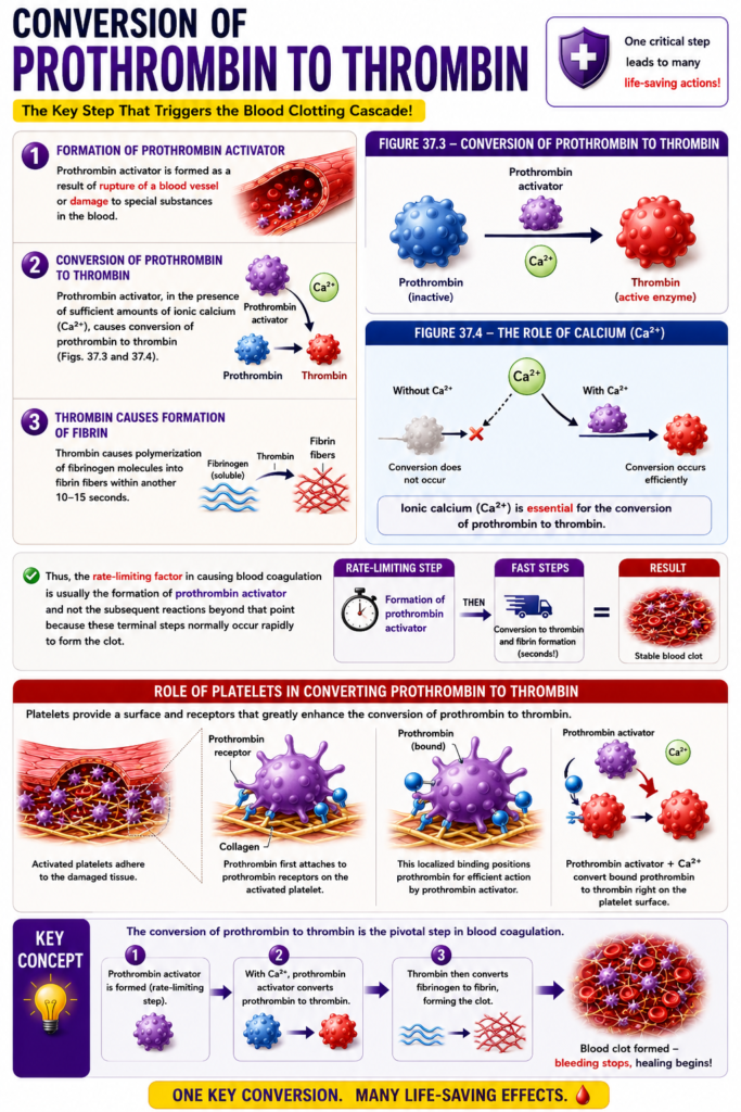
Coagulation Cascade After Blood Vessel Injury (Guyton Figure 37.4) — Easiest Conceptual Summary
One-Line Concept
After a blood vessel is injured, the body releases tissue factor, activates thrombin, produces fibrin, and forms a strong fibrin mesh that stabilizes the platelet plug and permanently stops bleeding.
Complete Process (Easy Flow)
Blood vessel injury
        ↓
Tissue Factor (Factor III) released
        ↓
Platelets become activated
        ↓
Platelet phospholipid complex forms
        ↓
Prothrombin → Thrombin
        ↓
Fibrinogen → Fibrin
        ↓
Cross-linked fibrin
        ↓
Stable fibrin clot
        ↓
Bleeding stops
Step 1: Blood Vessel Injury 🩸
What happens?
The blood vessel wall is damaged.
The endothelium (inner lining) breaks, exposing the underlying tissue.
Easy Concept
Imagine a pipe develops a crack and water begins leaking.
The body immediately starts its emergency repair process.
Step 2: Release of Tissue Factor (Factor III) 🚨
What happens?
The damaged cells release:
Tissue Factor (TF)
Also called:
- Factor III
- Thromboplastin
Function
Tissue factor is the signal that starts the coagulation cascade.
Easy Concept
Think of tissue factor as pressing the emergency alarm button.
📢 “The vessel is injured—start clotting!”
Step 3: Platelets Become Activated 🟣
What happens?
Platelets stick to the injured area.
Once activated, they expose platelet phospholipids on their surface.
These phospholipids provide a platform for clotting reactions.
Easy Concept
Think of platelets as construction workers.
Their phospholipid surface acts like a workbench where clotting proteins assemble.
Step 4: Platelet Phospholipid Complex ⚙️
What happens?
Clotting factors gather on the platelet surface.
Together they form the:
Platelet phospholipid complex
This greatly speeds up clotting.
Easy Concept
Imagine workers setting up a construction site before building begins.
Step 5: Prothrombin → Thrombin ⚡
What happens?
The clotting cascade converts:
Prothrombin (inactive)
↓
Thrombin (active enzyme)
Function of Thrombin
Thrombin is the main clotting enzyme.
It drives the next step.
Easy Concept
Thrombin is the chief engineer directing the repair.
Step 6: Fibrinogen → Fibrin 🧵
What happens?
Thrombin converts:
Fibrinogen (soluble protein)
↓
Fibrin (insoluble fibers)
The fibrin strands begin weaving around the platelet plug.
Easy Concept
Like weaving a strong fishing net over loose bricks.
Step 7: Cross-Linked Fibrin 🔗
What happens?
The fibrin strands become cross-linked (strengthened by Factor XIII, activated by thrombin).
This creates a strong mesh.
Easy Concept
Imagine tying many ropes together with strong knots.
Now the net cannot easily break.
Step 8: Stable Fibrin Clot 🛡️
What happens?
The fibrin mesh traps:
- Platelets
- Red blood cells (RBCs)
- White blood cells (WBCs)
This forms a stable fibrin clot that seals the damaged vessel.
Result
✔ Bleeding stops.
✔ The vessel is protected while healing occurs.
Easy Story (Remember Forever)
Imagine repairing a damaged bridge.
🌉 1. Bridge breaks → Blood vessel injury
🚨 2. Emergency alarm sounds → Tissue factor released
👷 3. Workers arrive → Platelets
🛠️ 4. Workers set up a workbench → Platelet phospholipid complex
👨🔧 5. Chief engineer arrives → Thrombin
🧶 6. Steel cables are produced → Fibrin
🔗 7. Cables are tied together → Cross-linked fibrin
🏗️ 8. Bridge becomes strong → Stable fibrin clot
High-Yield Exam Table
| Step | Event | Key Molecule | Function |
|---|---|---|---|
| 1 | Vessel injury | — | Starts clotting |
| 2 | Tissue factor release | Factor III (Thromboplastin) | Initiates coagulation cascade |
| 3 | Platelet activation | Platelets | Provide surface for clotting |
| 4 | Platelet phospholipid complex | Platelet phospholipids | Accelerates clotting reactions |
| 5 | Prothrombin → Thrombin | Thrombin | Main clotting enzyme |
| 6 | Fibrinogen → Fibrin | Fibrin | Forms clot fibers |
| 7 | Cross-linking | Factor XIII | Strengthens fibrin mesh |
| 8 | Stable clot | Fibrin + blood cells | Stops bleeding |
Easy Mnemonic
“TPTFC”
- T = Tissue factor released
- P = Platelet phospholipid complex
- T = Thrombin formed
- F = Fibrin produced
- C = Cross-linked stable clot
Difference Between the Platelet Plug and the Fibrin Clot
| Platelet Plug | Fibrin Clot |
|---|---|
| Forms first | Forms after the coagulation cascade |
| Made mainly of platelets | Made of fibrin mesh plus trapped platelets and blood cells |
| Temporary and relatively weak | Strong, stable, and long-lasting |
| Quickly reduces bleeding | Permanently seals the damaged vessel until healing |
Complete Flow Chart
Blood vessel injury
        ↓
Release of Tissue Factor (Factor III)
        ↓
Platelet activation
        ↓
Platelet phospholipid complex
        ↓
Prothrombin
        ↓
Thrombin
        ↓
Fibrinogen
        ↓
Fibrin
        ↓
Cross-linked fibrin
        ↓
Stable fibrin clot
        ↓
Bleeding stops
Final Concept in One Sentence
Blood vessel injury releases tissue factor, which initiates the coagulation cascade on the platelet phospholipid surface. This converts prothrombin into thrombin, thrombin converts fibrinogen into fibrin, and cross-linked fibrin forms a strong mesh that stabilizes the platelet plug into a firm blood clot, effectively stopping bleeding.
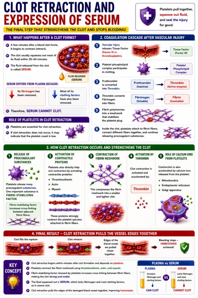
Clot Retraction and Expression of Serum
- A few minutes after a blood clot forms, it begins to contract (retract).
- The clot usually squeezes out most of its fluid within 20–60 minutes.
- The fluid released from the clot is called serum.
- Serum differs from plasma because:
- Its fibrinogen has been removed.
- Most of its clotting factors have also been removed.
- Therefore, serum cannot clot.
- Platelets are essential for clot retraction.
- If clot retraction does not occur, it may indicate that the platelet count is low.
- Figure 37.4 shows the coagulation cascade after vascular injury:
- Damage to the blood vessel releases tissue factor (Factor III or thromboplastin).
- Platelet phospholipid complex participates in clotting.
- Prothrombin is converted into thrombin.
- Thrombin converts fibrinogen into fibrin.
- Fibrin polymerizes into a meshwork that stabilizes the platelet plug.
- Inside the clot, platelets attach to fibrin fibers.
- They connect different fibrin fibers together.
- Platelets continue releasing procoagulant substances.
- One important substance is fibrin-stabilizing factor.
- Fibrin-stabilizing factor increases cross-linking between adjacent fibrin fibers.
- Platelets also directly help clot contraction.
- They activate the contractile proteins:
- Thrombosthenin
- Actin
- Myosin
- These proteins strongly contract the platelet spicules attached to fibrin fibers.
- This compresses the fibrin meshwork into a smaller and tighter clot.
- Clot contraction is activated and accelerated by thrombin.
- It is also accelerated by calcium ions released from the platelet:
- Mitochondria
- Endoplasmic reticulum
- Golgi apparatus
- As the clot retracts, the edges of the damaged blood vessel are pulled closer together.
- This further helps stop bleeding (hemostasis).
Key Concept
- Clot retraction begins within minutes after clot formation and depends on platelets. Platelets contract the fibrin meshwork using thrombosthenin, actin, and myosin, while fibrin-stabilizing factor strengthens the clot. The released fluid is serum, which cannot clot because it lacks fibrinogen and most clotting factors. Clot retraction pulls the vessel edges together, improving hemostasis.
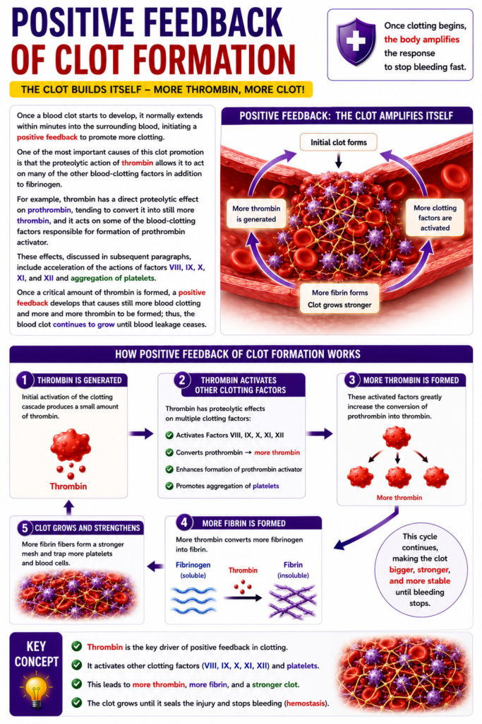
POSITIVE FEEDBACK OF CLOT FORMATION
- Once a blood clot starts to form, it usually spreads into the surrounding blood within a few minutes.
- This creates a positive feedback mechanism that promotes more blood clotting.
- One major reason for this positive feedback is the proteolytic action of thrombin.
- Thrombin acts on many blood-clotting factors, not just fibrinogen.
- Thrombin directly acts on prothrombin.
- This converts more prothrombin into thrombin.
- Thrombin also acts on clotting factors involved in forming prothrombin activator.
- Thrombin accelerates the activity of the following clotting factors:
- Factor VIII
- Factor IX
- Factor X
- Factor XI
- Factor XII
- Thrombin also promotes platelet aggregation.
- Once a critical amount of thrombin is produced, a positive feedback cycle begins.
- This cycle produces even more thrombin.
- As a result, more blood clotting occurs.
- The blood clot continues to grow until blood leakage stops.
Key Concept
- Thrombin creates a positive feedback loop by activating more prothrombin, multiple clotting factors (VIII, IX, X, XI, XII), and platelet aggregation. This rapidly increases clot formation until bleeding is completely stopped.
MADE BY SELF LEARNING CEO AND FOUNDER DR SHEEN
