


- Antibodies are gamma globulins called immunoglobulins (Igs).
- Their molecular weight ranges from 160,000 to 970,000.
- Immunoglobulins make up about 20% of all plasma proteins.
- All immunoglobulins are made of light polypeptide chains and heavy polypeptide chains.
- Most immunoglobulins contain:
- Two light chains
- Two heavy chains
- Some immunoglobulins contain up to 10 heavy chains and 10 light chains.
- These larger molecules form high-molecular-weight immunoglobulins.
- In every immunoglobulin:
- Each heavy chain is paired with one light chain.
- This forms a heavy-light pair.
- Every immunoglobulin contains at least 2 and up to 10 heavy-light pairs.
- Each light chain and heavy chain has a variable region.
- The remaining part of each chain is called the constant region.
- The variable region is different for each antibody.
- The variable region binds specifically to a particular antigen.
- The constant region determines the other properties of the antibody.
- The constant region determines:
- Diffusion of antibodies through tissues.
- Attachment to specific tissue structures.
- Binding to the complement complex.
- Ability to pass through membranes.
- Other biological properties of the antibody.
- The light and heavy chains are held together by:
- Noncovalent bonds
- Covalent (disulfide) bonds
Figure Number
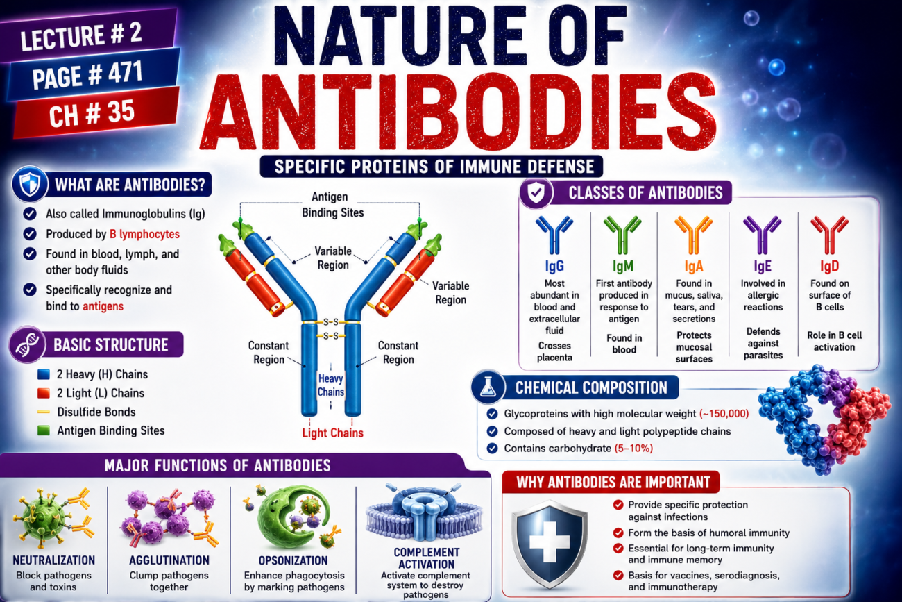
- Figure 35.4: Shows the basic structure of an immunoglobulin (antibody), including:
- Heavy chains
- Light chains
- Variable regions
- Constant regions
- Heavy-light chain pairing
- Figure 35.3: Shows the time course of:
- Primary antibody response after the first antigen injection.
- Secondary antibody response after a second antigen injection several weeks later.
Key Concept
Antibodies (immunoglobulins) are gamma globulin proteins composed of heavy and light polypeptide chains. Their variable regions specifically recognize and bind antigens, while their constant regions determine important biological functions such as tissue diffusion, complement activation, membrane transport, and interactions with body tissues. Heavy and light chains are linked by noncovalent and disulfide bonds.
Guyton Physiology Figure 35.4 Explained in Detail
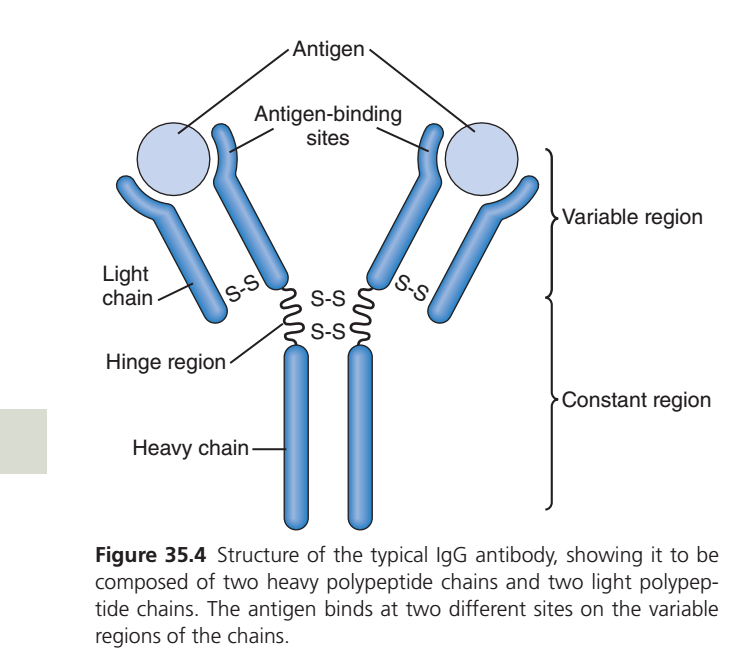
Structure of a Typical IgG Antibody (Immunoglobulin G)
This figure explains the molecular structure of an antibody (Immunoglobulin G, IgG), which is the most abundant antibody in human blood and the major antibody involved in the secondary immune response.
Understanding this figure is essential because it explains:
- How antibodies recognize antigens
- Why antibodies are highly specific
- Why one antibody can bind two antigens
- Why antibodies have different classes (IgG, IgA, IgM, IgE, IgD)
- How antibodies activate immune mechanisms
Overall Concept
Imagine an antibody as a special military robot.
It has:
- Two hands → to catch enemies (antigens)
- Flexible elbows → to move freely
- Strong body → determines its function
- Unique fingerprints → recognize only one specific enemy
This is exactly how an antibody is built.
First Look at the Figure
The antibody looks like the letter
“Y”
This Y-shape is formed by
- Two Heavy Chains
- Two Light Chains
Together,
they form one complete antibody molecule.
Components of the Figure
The figure labels:
- Heavy Chains
- Light Chains
- Variable Region
- Constant Region
- Hinge Region
- Disulfide Bonds (S-S)
- Antigen Binding Sites
- Antigen
We’ll understand each one individually.
PART 1
Overall Y-Shaped Structure
Look carefully.
The antibody has
- Two identical arms
- One stem
The two arms recognize antigen.
The stem performs immune functions.
Easy Analogy
Imagine
A slingshot
       \     /
        \   /
         \ /
          |
          |
The upper two arms catch the antigen.
The lower handle performs the immune functions.
PART 2
Heavy Chains
Look at the long blue chains.
These are
Heavy Chains
There are
Two Heavy Chains
Each heavy chain is identical.
Why are they called Heavy?
Because
they are larger proteins.
Approximately
50 kDa each.
What Do Heavy Chains Do?
Heavy chains determine
the class of antibody.
Depending upon the heavy chain,
an antibody becomes
- IgG
- IgA
- IgM
- IgE
- IgD
Easy Memory
Heavy Chain
↓
Determines
Identity of antibody
Clinical Example
If the heavy chain is Gamma (γ)
↓
The antibody is
IgG
If the heavy chain is Mu (μ)
↓
It becomes
IgM
PART 3
Light Chains
Look at the smaller chains attached to each arm.
These are
Light Chains
There are
Two Light Chains.
Why are they called Light?
Because
they are much smaller
Approximately
25 kDa each.
Function
They assist heavy chains
in binding antigen.
They do not determine antibody class.
Easy Analogy
Think of
Heavy chains = Main arm
Light chains = Supporting arm
Both are needed to catch the antigen firmly.
PART 4
Disulfide Bonds (S-S)
Notice the
S-S
written in the center.
These are
Disulfide Bonds
They join
Heavy chain ↔ Heavy chain
Heavy chain ↔ Light chain
Why are They Important?
Without these bonds
the antibody would fall apart.
They provide
- strength
- stability
- proper Y shape
Analogy
Imagine screws holding furniture together.
Remove the screws
↓
Furniture collapses.
Disulfide bonds are those screws.
PART 5
Hinge Region
Look at the center
where the two arms meet.
This is
Hinge Region
Function
Allows the two arms
to move freely.
Why is this Necessary?
Antigens are
not always the same distance apart.
Sometimes
they are close.
Sometimes
far apart.
The hinge lets the arms
open
close
bend
rotate
so both antigen-binding sites can engage efficiently.
Easy Analogy
Imagine your shoulder joint.
Your arm moves
- upward
- downward
- sideways
Similarly
the hinge region
makes antibody arms flexible.
PART 6
Variable Region
Look at the tips of the Y.
These are
Variable Regions
This is the
most important part of the antibody.
Why is it called Variable?
Because
this part changes
from one antibody
to another.
Every antibody has
a different variable region.Function
Recognizes
one specific antigen.
Example
Antibody against COVID virus
↓
Different variable region
Antibody against tetanus
↓
Different variable region
Antibody against measles
↓
Different variable region
Easy Analogy
Think of
Variable Region
=
Fingerprint
Every antibody has
its own fingerprint.
PART 7
Antigen-Binding Sites
Look carefully.
Each arm ends in
one
Antigen Binding Site.
Total
Two binding sites
per antibody.
Why Two?
Because
one antibody
can bind
two identical antigens simultaneously.
Advantage
This allows antibodies to
- cross-link antigens
- form immune complexes
- agglutinate bacteria
- improve pathogen removal
Analogy
Imagine
two hands
holding
two balloons.
One hand
↓
One balloon
Two hands
↓
Two balloons.
PART 8
Antigen
The grey circles represent
Antigens
These may be
- bacteria
- viruses
- toxins
- pollen
- proteins
Notice Something Important
Only
the variable region
touches
the antigen.
The constant region
never binds antigen directly.
Lock and Key Concept
Variable Region
↓
Lock
Antigen
↓
Key
Only
matching key
opens
matching lock.
PART 9
Constant Region
The lower stem
is called
Constant Region
Why Constant?
Because
it remains almost
the same
within each antibody class.
Function
After antigen binding,
the constant region performs the “effector functions” of the antibody.
These include:
- Activating the complement system (especially IgG and IgM)
- Binding to Fc receptors on macrophages and neutrophils
- Promoting phagocytosis (opsonization)
- Helping natural killer (NK) cells perform antibody-dependent cellular cytotoxicity (ADCC)
- Crossing the placenta (IgG)
Easy Analogy
Think of the antibody as a crane.
The hook (variable region)
grabs the load.
The engine (constant region)
does the heavy work.
Complete Structure
One antibody consists of
| Component | Number |
|---|---|
| Heavy Chains | 2 |
| Light Chains | 2 |
| Variable Regions | 2 |
| Constant Regions | 2 heavy-chain constant regions (plus constant portions of light chains) |
| Antigen Binding Sites | 2 |
| Hinge Region | 1 |
| Disulfide Bonds | Multiple |
How an Antibody Works
Let’s follow the entire process.
Step 1
Bacteria enter the body.
↓
Step 2
Plasma cells produce antibodies.
↓
Step 3
Variable regions recognize
the bacteria.
↓
Step 4
Both binding sites attach.
↓
Step 5
Constant region activates
complement,
phagocytes,
and other immune mechanisms.
↓
Step 6
Bacteria are destroyed.
Clinical Correlations
1. Vaccination
Vaccines stimulate plasma cells to produce antigen-specific antibodies. During later exposure to the real pathogen, these antibodies rapidly bind and neutralize the antigen through their variable regions, while the constant region recruits immune effector mechanisms.
2. Monoclonal Antibody Therapy
Therapeutic antibodies such as rituximab or trastuzumab have highly specific variable regions that bind target molecules, whereas their constant regions recruit immune cells or activate complement to eliminate the target.
3. Multiple Myeloma
In multiple myeloma, one plasma-cell clone produces excessive amounts of a single monoclonal immunoglobulin, all with identical variable and constant regions.
High-Yield MBBS Points
- IgG is a Y-shaped immunoglobulin composed of 2 identical heavy chains and 2 identical light chains.
- Heavy chains determine the antibody class (IgG, IgA, IgM, IgE, IgD).
- Variable regions are located at the tips of the Y and are responsible for specific antigen recognition.
- Each IgG molecule has two identical antigen-binding sites, allowing it to bind two identical epitopes.
- Constant (Fc) region mediates effector functions such as complement activation, Fc receptor binding, opsonization, ADCC, and placental transfer (IgG).
- Disulfide bonds stabilize the antibody by linking heavy-heavy and heavy-light chains.
- The hinge region provides flexibility, allowing the antibody arms to adjust to the spacing of antigenic epitopes.
Complete Flow Chart
Plasma Cell
      ↓
Produces IgG Antibody
      ↓
IgG Structure
      ↓
2 Heavy Chains + 2 Light Chains
      ↓
Variable Regions Recognize Specific Antigen
      ↓
Antigen Binds to Two Binding Sites
      ↓
Constant (Fc) Region Activates Immune Effector Mechanisms
      ↓
Complement Activation + Opsonization + Phagocytosis + ADCC
      ↓
Elimination of the Pathogen
Connecting Figures 35.1–35.4
| Figure | Main Concept |
|---|---|
| Figure 35.1 | Development of T and B lymphocytes and the origin of cell-mediated and humoral immunity. |
| Figure 35.2 | Clonal selection: only the B cell with the correct receptor is activated and undergoes clonal expansion. |
| Figure 35.3 | Primary and secondary antibody responses, highlighting the role of memory B cells. |
| Figure 35.4 | Molecular structure of the antibody produced by plasma cells, explaining how antibodies specifically recognize antigens and recruit immune effector functions. |
Together, these figures describe the complete pathway of humoral immunity:
B-cell development → Antigen recognition → Clonal expansion → Plasma-cell formation → Antibody production → Antigen binding and pathogen elimination.
Key Concept
Figure 35.4 illustrates the structure-function relationship of the IgG antibody. The two variable regions at the tips of the Y-shaped molecule provide highly specific recognition of antigens, while the constant (Fc) region recruits immune mechanisms such as complement activation and phagocytosis. The hinge region gives flexibility, and disulfide bonds maintain structural stability. This elegant design allows antibodies to recognize foreign molecules with remarkable specificity while simultaneously activating powerful effector mechanisms that eliminate pathogens.
Each Antibody Is Specific for a Particular Antigen
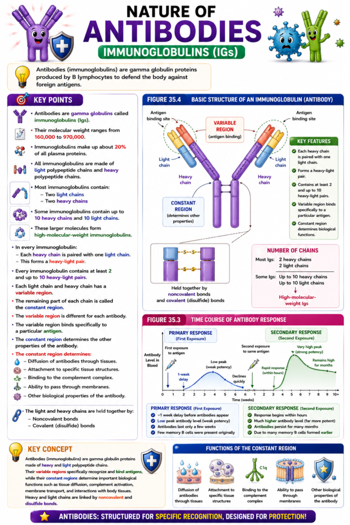
- Each antibody is specific for one particular antigen.
- This specificity is due to the unique arrangement of amino acids in the variable regions of the light and heavy chains.
- The amino acid arrangement gives each antibody a unique three-dimensional (steric) shape.
- When the correct antigen meets the antibody, the antigen fits the antibody like a mirror image.
- Multiple prosthetic groups of the antigen match the corresponding sites on the antibody.
- This close fit allows rapid and strong binding between the antibody and the antigen.
- When an antibody is highly specific, many binding sites interact at the same time.
- This makes the antibody-antigen complex very stable.
- The antibody and antigen are held together by:
- Hydrophobic bonds
- Hydrogen bonds
- Ionic attractions
- Van der Waals forces
- The binding between an antibody and an antigen follows the thermodynamic mass action law.
Mathematical Equation
Ka=Concentration of antibody×Concentration of antigenConcentration of bound antibody-antigen complex
Where:
- KaK_aKa = Affinity constant
- The affinity constant measures how tightly an antibody binds to its antigen.
- A higher KaK_aKa means stronger antibody-antigen binding.
- Most antibodies have two antigen-binding sites.
- Therefore, these antibodies are called bivalent antibodies.
- Some antibodies contain up to 10 light chains and 10 heavy chains.
- These larger antibodies can have as many as 10 antigen-binding sites.
Figure Number
- Figure 35.4: Shows:
- Heavy chains
- Light chains
- Variable regions
- Constant regions
- Two antigen-binding (variable) sites of a bivalent antibody
Key Concept
Each antibody recognizes only one specific antigen because of the unique amino acid arrangement in its variable regions. The antigen fits the antibody with high structural complementarity, producing strong binding through hydrophobic bonds, hydrogen bonds, ionic attractions, and van der Waals forces. The strength of this interaction is measured by the affinity constant (KaK_aKa). Most antibodies are bivalent, while some larger immunoglobulins possess up to 10 antigen-binding sites.
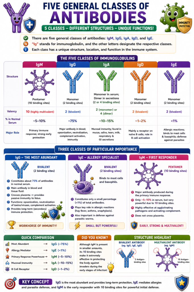
Five General Classes of Antibodies
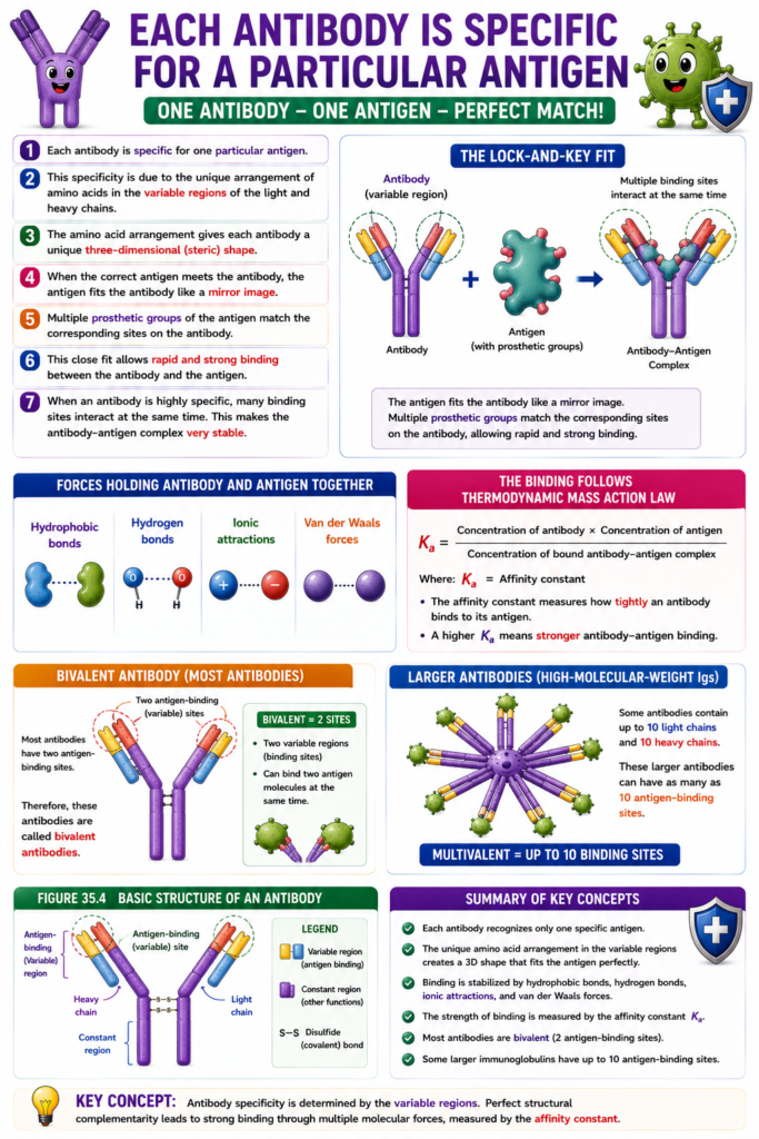
- There are five general classes of antibodies.
- These classes are:
- IgM
- IgG
- IgA
- IgD
- IgE
- Ig stands for immunoglobulin.
- The letters M, G, A, D, and E identify the different antibody classes.
- Among these classes, IgG is especially important.
- IgG is a bivalent antibody.
- It makes up about 75% of the antibodies in a normal person.
- Another important class is IgE.
- IgE is present in only a small percentage of the body’s antibodies.
- IgE is mainly involved in allergic reactions.
- IgM is also an important antibody class.
- A large proportion of the antibodies produced during the primary immune response are IgM antibodies.
- IgM antibodies have 10 antigen-binding sites.
- These multiple binding sites make IgM very effective in protecting the body against invading organisms.
- IgM provides strong protection even though its overall amount is relatively small.
Key Concept
The five classes of immunoglobulins are IgM, IgG, IgA, IgD, and IgE. IgG is the most abundant antibody (about 75%) and is bivalent. IgE is primarily involved in allergic reactions. IgM is the major antibody produced during the primary immune response and has 10 antigen-binding sites, making it highly effective against invading pathogens.
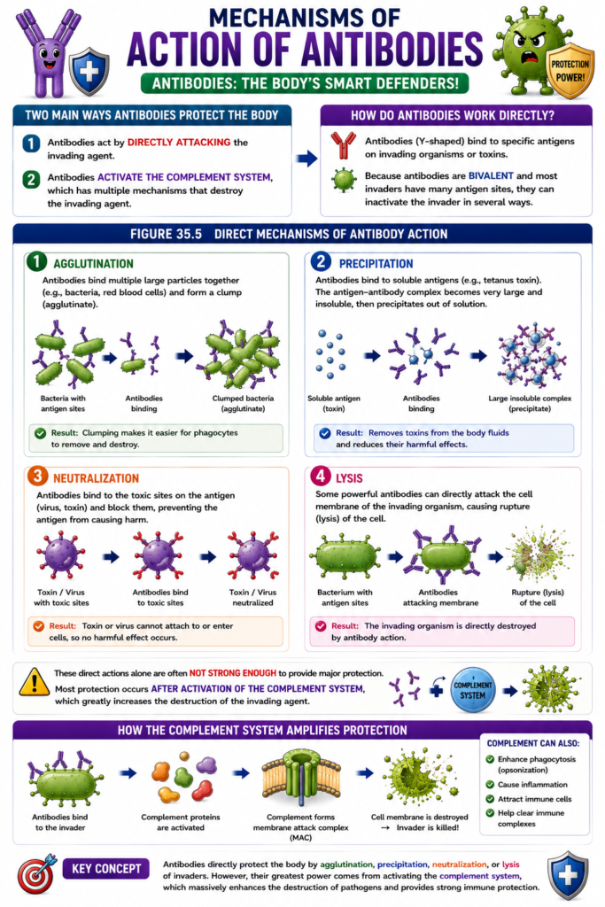
Mechanisms of Action of Antibodies
- Antibodies protect the body in two main ways.
- First, antibodies act by directly attacking the invading agent.
- Second, antibodies activate the complement system.
- The complement system has multiple mechanisms that help destroy the invading agent.
- During direct action, antibodies bind to specific antigens on the invading organism or toxin.
- Because antibodies are bivalent and most invading agents have many antigen sites, antibodies can inactivate the invader in several ways.
- Agglutination
- Antibodies bind multiple large particles together.
- Examples include bacteria and red blood cells.
- This forms a clump (agglutinate).
- Precipitation
- Antibodies bind to soluble antigens, such as tetanus toxin.
- The antigen-antibody complex becomes very large.
- The complex becomes insoluble.
- It then precipitates out of solution.
- Neutralization
- Antibodies cover the toxic sites on the antigen.
- This prevents the antigen from producing its harmful effects.
- Lysis
- Some powerful antibodies can directly attack the cell membrane of the invading organism.
- This causes rupture (lysis) of the cell.
- These direct antibody actions alone are often not strong enough to provide major protection.
- Most protection occurs after activation of the complement system, which greatly increases the destruction of the invading agent.
Figure Number
- Figure 35.5: Shows:
- Y-shaped antibodies binding to specific antigens.
- Direct mechanisms of antibody action, including:
- Agglutination
- Precipitation
- Neutralization
- Lysis
Key Concept
Antibodies defend the body by directly binding to antigens and causing agglutination, precipitation, neutralization, or lysis. However, their greatest protective effect occurs by activating the complement system, which greatly amplifies the destruction of invading organisms.
Guyton Physiology Figure 35.5 Explained in Detail
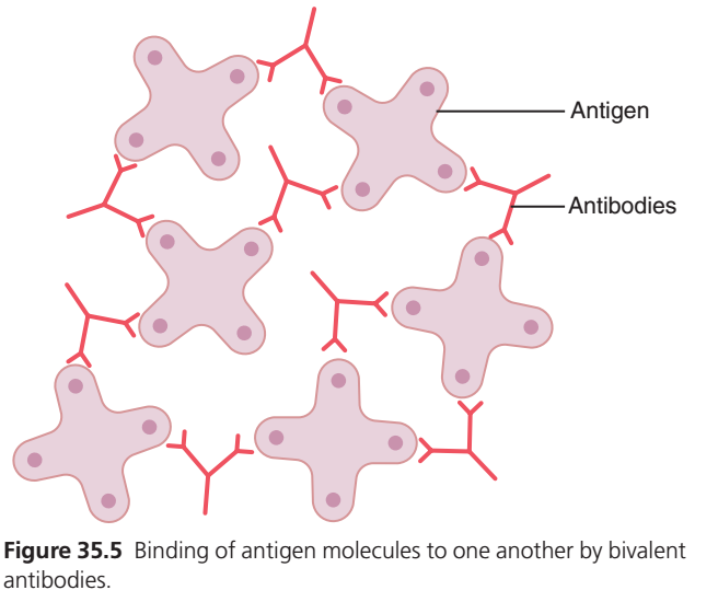
Agglutination: Binding of Antigen Molecules by Bivalent Antibodies
This figure explains one of the most important mechanisms of antibody action:
How antibodies bind multiple antigens together and help the immune system remove them.
This process is called:
Agglutination (Cross-Linking of Antigens)
It is one of the direct mechanisms by which antibodies protect the body, even before complement activation or phagocytosis occurs.
Overall Concept
Imagine children playing with Lego blocks.
Each Lego block has holes.
Now imagine you have a connector piece with two ends.
One end attaches to one Lego block.
The other end attaches to another Lego block.
Very quickly,
many Lego blocks become connected into one large structure.
Exactly the same thing happens in this figure.
The antibody acts like the connector.
The antigens act like the Lego blocks.
First Look at the Figure
The figure contains only two structures.
Pink Objects
These represent
Antigens
Red Y-shaped Structures
These represent
Antibodies
Notice carefully
every antibody is connected to
two different antigens.
This is the most important concept.
Why Can One Antibody Bind Two Antigens?
Recall Figure 35.4.
IgG antibody has
Two Antigen Binding Sites
        Antigen
           ●
          /\
         /  \
        |    |
        |    |
         \  /
          ||
One arm binds
Antigen A
The other arm binds
Antigen B
Thus,
one antibody connects
two antigens together.
This property is called
Bivalency
What Does “Bivalent” Mean?
“Bivalent” means
Having two antigen-binding sites.
Since IgG possesses
two Fab arms,
it can bind
two identical antigenic epitopes simultaneously.
Easy Analogy
Think of an antibody as a
Double-sided hook.
One hook catches one object.
The second hook catches another object.
Now both objects become connected.
PART 1
Antigen
The pink objects are
Antigens.
These may be
- bacteria
- viruses
- toxins
- pollen
- foreign proteins
Notice
each antigen has
many antigenic sites (epitopes).
Therefore,
many antibodies can attach simultaneously.
Important Concept
One bacterium
has
hundreds to thousands
of antigenic molecules.
Therefore,
hundreds of antibodies
may bind
the same bacterium.
PART 2
Antibody
The red structures are
IgG antibodies.
Each antibody has
Two Fab arms.
Each arm binds
one epitope.
Remember Figure 35.4
Upper arm
↓
Variable region
↓
Antigen recognition
Lower stem
↓
Constant region
↓
Immune functions
PART 3
Cross-Linking
Now look carefully.
Each antibody binds
one antigen
↓
then
another antigen.
This creates
bridges.
One antibody
↓
connects
two antigens.
Another antibody
↓
connects
two more antigens.
Soon
a large network develops.
This is called
Cross-Linking
because antibodies
cross-connect
different antigen molecules.
Easy Analogy
Imagine
people holding hands.
One person
holds
two people.
Those people
hold
others.
Soon
everyone becomes connected.
Exactly the same process occurs here.
PART 4
Agglutination
As more antibodies bind,
individual antigens begin
forming
large clumps.
This process is called
Agglutination
Definition
Agglutination is
The clumping together of particulate antigens (such as bacteria or cells) by antibodies.
Why Does This Happen?
Because every antibody
has
two antigen-binding sites.
Each antigen
has
many epitopes.
Therefore
large networks form.
Easy Analogy
Imagine
small magnets.
When connectors join them,
they become
one giant cluster.
Why Is Agglutination Useful?
Agglutination provides several advantages.
1. Immobilizes Pathogens
Normally
bacteria move freely.
After agglutination,
they become
one large clump.
Movement becomes difficult.
Clinical Example
Motile bacteria are easier to contain once they are agglutinated.
2. Prevents Spread
Large clumps
cannot spread
through tissues
as easily
as single bacteria.
Example
Instead of
100 separate bacteria,
the immune system now deals with
one large bacterial cluster.
3. Easier Phagocytosis
Macrophages
and
neutrophils
can easily recognize
large antibody-coated clumps.
They engulf them more efficiently.
This process is enhanced further because the antibody Fc (constant) region can bind Fc receptors on phagocytes.
Easy Analogy
Cleaning
100 scattered marbles
is difficult.
Cleaning
one bag containing all marbles
is much easier.
4. Activates Complement
When several IgG molecules—or a single IgM molecule—bind close together on a microbial surface,
the classical complement pathway can be activated.
Complement helps
- lyse bacteria
- increase inflammation
- promote opsonization
5. Neutralizes Antigens
Some antibodies bind
toxins
or
viruses
preventing them
from attaching
to human cells.
Difference Between Agglutination and Neutralization
Many students confuse these.
Neutralization
One antibody binds
one toxin
or
virus
and blocks its activity.
Example:
Tetanus toxin
↓
Antibody blocks toxin
↓
Toxin cannot enter nerve cells.
Agglutination
One antibody binds
multiple bacteria
↓
Large bacterial clumps form.
Easy Trick
Neutralization
↓
Stops function.
Agglutination
↓
Creates clumps.
Why Does IgM Agglutinate Better Than IgG?
Although this figure shows a typical bivalent antibody,
in the body
IgM
is the most efficient agglutinating antibody.
Why?
Because
IgM has
10 antigen-binding sites (a pentamer).
One IgM molecule can connect many antigen molecules simultaneously.
IgG has only
2 binding sites.
Therefore,
IgM is much better
at agglutination.
Clinical Correlations
1. Blood Group Testing
Agglutination is the principle behind ABO blood grouping.
Example:
A person’s red blood cells are mixed with anti-A serum.
- If agglutination occurs → A antigen is present.
- If no agglutination occurs → A antigen is absent.
2. Widal Test
The Widal test detects antibodies against Salmonella by observing agglutination between patient serum and bacterial antigens.
3. Latex Agglutination Tests
Many rapid diagnostic tests use latex particles coated with antigens or antibodies.
If the corresponding antibody or antigen is present in the patient’s sample,
visible clumping occurs.
Examples include tests for:
- Rheumatoid factor
- C-reactive protein (CRP)
- Certain bacterial antigens
Relationship with Previous Figures
Let’s connect all the figures.
Figure 35.1
B cells develop.
↓
Figure 35.2
One B cell selected.
↓
Figure 35.3
Memory B cells formed.
↓
Figure 35.4
Antibody structure explained.
↓
Figure 35.5
Shows how that antibody actually protects the body by physically linking antigens together into clumps that are easier for the immune system to eliminate.
High-Yield MBBS Points
- Agglutination is the clumping of particulate antigens by antibodies.
- It occurs because antibodies (such as IgG) are bivalent, while antigens have multiple epitopes.
- Cross-linking forms large antigen-antibody complexes.
- Agglutination:
- Immobilizes microorganisms
- Limits their spread
- Facilitates phagocytosis
- Can promote complement activation
- IgM is the most effective agglutinating antibody because it is a pentamer with 10 antigen-binding sites.
- Agglutination forms the basis of important laboratory tests such as ABO blood typing and the Widal test.
Complete Flow Chart
Foreign bacteria enter the body
          ↓
Plasma cells produce antibodies
          ↓
Antibodies bind specific antigens
          ↓
Each antibody binds two antigen molecules
          ↓
Cross-linking between multiple antigens
          ↓
Agglutination (Large antigen-antibody clumps)
          ↓
Immobilization of pathogens
          ↓
Enhanced phagocytosis + Complement activation
          ↓
Removal of pathogens from the body
Connecting Figures 35.1–35.5
| Figure | Main Concept |
|---|---|
| Figure 35.1 | Origin and maturation of T and B lymphocytes. |
| Figure 35.2 | Clonal selection and activation of the specific B-cell clone. |
| Figure 35.3 | Primary and secondary antibody responses and immunological memory. |
| Figure 35.4 | Molecular structure of the IgG antibody, including variable and constant regions. |
| Figure 35.5 | Functional action of antibodies: cross-linking antigens to produce agglutination, facilitating pathogen elimination. |
These figures together describe the entire pathway of humoral immunity:
B-cell development → Antigen recognition → Clonal expansion → Antibody production → Antigen binding → Agglutination → Efficient clearance by the immune system.
Key Concept
Figure 35.5 demonstrates that antibodies are not passive molecules—they actively organize pathogens into large antigen-antibody complexes. Because each antibody has at least two antigen-binding sites, it can bridge multiple antigen molecules, producing agglutination (cross-linking). This immobilizes microorganisms, prevents their spread, enhances phagocytosis, and promotes complement activation, making pathogen elimination faster and more efficient.
MADE BY SELF LEARNING CEO AND FOUNDER DR SHEEN
