


- Regulation of hydrogen ion (H⁺) balance is similar to the regulation of other body ions.
- There must be a balance between H⁺ intake or production and H⁺ removal.
- This balance is necessary to maintain homeostasis.
- The kidneys play a major role in removing H⁺ from the body.
- Regulation of extracellular fluid H⁺ concentration requires more than kidney excretion alone.
- Acid–base buffering mechanisms are also essential.
- The blood contains important buffering mechanisms.
- Body cells contain important buffering mechanisms.
- The lungs also help regulate H⁺ concentration.
- These mechanisms maintain normal H⁺ concentration in extracellular fluid.
- These mechanisms also maintain normal H⁺ concentration in intracellular fluid.
- This chapter explains the mechanisms that regulate H⁺ concentration.
- Special emphasis is given to renal H⁺ secretion.
- Special emphasis is given to renal H⁺ reabsorption.
- Special emphasis is given to renal production of HCO₃⁻.
- Special emphasis is given to renal excretion of HCO₃⁻.
- HCO₃⁻ is a key component of the body’s acid–base control system.
HYDROGEN ION CONCENTRATION IS PRECISELY REGULATED
- Precise regulation of H⁺ concentration is essential.
- Almost all enzyme systems are affected by H⁺ concentration.
- Changes in H⁺ concentration alter enzyme activity.
- Changes in H⁺ concentration affect almost all cell functions.
- Changes in H⁺ concentration affect almost all body functions.
- H⁺ concentration in body fluids is normally very low.
- Extracellular fluid sodium concentration is 142 mEq/L.
- Normal H⁺ concentration is only 0.00004 mEq/L.
- Sodium concentration is about 3.5 million times greater than H⁺ concentration.
- Normal variation in H⁺ concentration is extremely small.
- The normal variation in H⁺ concentration is only about one-millionth of the normal variation in sodium (Na⁺) concentration.
- This precise regulation shows the importance of H⁺ concentration for normal cellular function.
KEY CONCEPT
- H⁺ balance depends on a balance between H⁺ production/intake and H⁺ removal.
- The kidneys, blood, cells, and lungs work together to regulate H⁺ concentration.
- The kidneys regulate H⁺ secretion and HCO₃⁻ reabsorption, production, and excretion.
- H⁺ concentration strongly affects enzyme activity and cell function.
- Normal Na⁺ concentration = 142 mEq/L.
- Normal H⁺ concentration = 0.00004 mEq/L.
- Na⁺ concentration is about 3.5 million times greater than H⁺ concentration.
ACIDS AND BASES—DEFINITIONS AND MEANINGS
- A hydrogen ion (H⁺) is a single free proton released from a hydrogen atom.
- Molecules that release H⁺ in solution are called acids.
- Hydrochloric acid (HCl) is an example of an acid.
- HCl ionizes in water.
- HCl forms H⁺ and Cl⁻ ions.
- Carbonic acid (H₂CO₃) is also an acid.
- H₂CO₃ ionizes in water.
- H₂CO₃ forms H⁺ and HCO₃⁻.
- A base is an ion or molecule that accepts H⁺.
- HCO₃⁻ is a base.
- HCO₃⁻ combines with H⁺ to form H₂CO₃.
- HPO₄²⁻ is also a base.
- HPO₄²⁻ accepts H⁺ to form H₂PO₄⁻.
- Body proteins also act as bases.
- Some amino acids in proteins carry negative charges.
- These negative charges readily accept H⁺.
- Hemoglobin in red blood cells is an important body base.
- Proteins in other body cells are also important bases.
- The terms base and alkali are often used interchangeably.
- An alkali is formed by combining an alkaline metal with a highly basic ion.
- Examples of alkaline metals are sodium, potassium, and lithium.
- A common highly basic ion is OH⁻.
- The base portion quickly reacts with H⁺.
- This reaction removes H⁺ from the solution.
- Therefore, alkalis are typical bases.
- Alkalosis means excessive removal of H⁺ from body fluids.
- Acidosis means excessive addition of H⁺ to body fluids.
Strong and Weak Acids and Bases
- Strong acids dissociate rapidly.
- Strong acids release large amounts of H⁺.
- HCl is a strong acid.
- Weak acids dissociate less readily.
- Weak acids release H⁺ more slowly.
- H₂CO₃ is a weak acid.
- Strong bases react rapidly with H⁺.
- Strong bases quickly remove H⁺ from solution.
- OH⁻ is a strong base.
- OH⁻ combines with H⁺ to form H₂O.
- HCO₃⁻ is a weak base.
- HCO₃⁻ binds H⁺ less strongly than OH⁻.
- Most acids and bases in extracellular fluid are weak.
- These weak acids and bases regulate normal acid–base balance.
- The most important buffer pair is H₂CO₃ / HCO₃⁻.
Normal H⁺ Concentration and pH of Body Fluids and Changes That Occur in Acidosis and Alkalosis
- Normal blood H⁺ concentration is about 0.00004 mEq/L.
- This is equal to 40 nEq/L.
- Normal variation is only 3–5 nEq/L.
- Under extreme conditions, H⁺ concentration may fall to 10 nEq/L.
- Under extreme conditions, H⁺ concentration may rise to 160 nEq/L.
- These extreme values may still be compatible with life.
- H⁺ balance depends on a balance between H⁺ production or intake and H⁺ removal.
- The kidneys play a major role in removing H⁺.
- Kidney excretion alone is not enough to regulate H⁺.
- Blood buffering systems also regulate H⁺.
- Cells also regulate H⁺.
- The lungs also regulate H⁺.
- These systems maintain normal extracellular H⁺ concentration.
- These systems also maintain normal intracellular H⁺ concentration.
- This chapter focuses on H⁺ regulation.
- It emphasizes renal H⁺ secretion.
- It emphasizes renal H⁺ reabsorption.
- It emphasizes renal production of HCO₃⁻.
- It emphasizes renal excretion of HCO₃⁻.
- HCO₃⁻ is a major component of acid–base regulation.
HYDROGEN ION CONCENTRATION IS PRECISELY REGULATED
- Precise regulation of H⁺ is essential.
- Almost all enzyme systems are affected by H⁺ concentration.
- Changes in H⁺ concentration alter enzyme activity.
- Changes in H⁺ concentration affect almost all cell functions.
- Changes in H⁺ concentration affect almost all body functions.
- H⁺ concentration in body fluids is normally very low.
- Normal extracellular sodium concentration is 142 mEq/L.
- Normal H⁺ concentration is 0.00004 mEq/L.
- Sodium concentration is about 3.5 million times greater than H⁺ concentration.
- Normal variation in H⁺ concentration is only about one-millionth of the normal variation in Na⁺ concentration.
- This precise regulation highlights the importance of H⁺ in cell function.
ACIDS AND BASES—DEFINITIONS AND MEANINGS
- Because H⁺ concentration is very low, pH is used to express it.
- pH uses a logarithmic scale.
Formula
pH=log([H+]1)=−log[H+]
- H⁺ concentration is expressed in equivalents per liter (Eq/L).
Mathematical Calculation
Given:
- H⁺ = 40 nEq/L
- 40 nEq/L = 0.00000004 Eq/L = 4 × 10⁻⁸ Eq/L
Calculation:pH=−log(4×10⁻⁸) =−[log4+log10−8] =−(0.6−8) =7.4
Final Answer
- Normal pH = 7.4
- pH is inversely related to H⁺ concentration.
- Low pH means high H⁺ concentration.
- High pH means low H⁺ concentration.
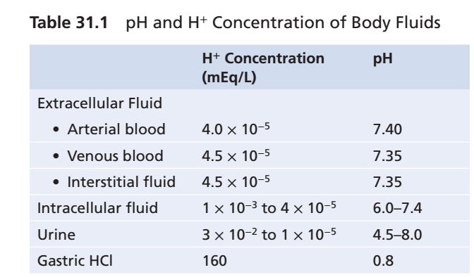
- Normal arterial blood pH is 7.4.
- Venous blood pH is about 7.35.
- Interstitial fluid pH is about 7.35.
- Venous blood contains more CO₂ than arterial blood.
- Extra CO₂ forms more H₂CO₃.
- More H₂CO₃ slightly lowers pH.
- These values are summarized in Table 31.1.
- Acidemia occurs when arterial blood pH falls significantly below 7.4.
- Alkalemia occurs when arterial blood pH rises above 7.4.
- The lower limit of survival is about pH 6.8.
- The upper limit of survival is about pH 8.0.
- Intracellular pH is usually lower than plasma pH.
- Cell metabolism produces acids.
- H₂CO₃ is an important intracellular acid.
- Intracellular pH ranges from 6.0 to 7.4.
- Hypoxia causes acid accumulation.
- Poor tissue blood flow causes acid accumulation.
- These conditions decrease intracellular pH.
- Acidosis is the process that leads to acidemia.
- Alkalosis is the process that leads to alkalemia.
- Urine pH ranges from 4.5 to 8.0.
- Urine pH depends on extracellular acid–base status.
- The kidneys correct abnormal H⁺ concentration.
- The kidneys excrete acids or bases as needed.
- Gastric HCl is an example of an extremely acidic body fluid.
- HCl is secreted by oxyntic (parietal) cells of the stomach.
- This process is discussed in Chapter 65.
- H⁺ concentration in these cells is about 4 million times greater than in blood.
- Gastric HCl has a pH of 0.8.
- The remainder of the chapter discusses regulation of extracellular H⁺ concentration.
KEY CONCEPT
- Acids donate H⁺; bases accept H⁺.
- Strong acids/bases dissociate rapidly; weak acids/bases dissociate slowly.
- The most important extracellular buffer system is H₂CO₃ / HCO₃⁻.
- Normal H⁺ = 40 nEq/L (0.00004 mEq/L).
- Normal arterial pH = 7.4.
- Venous/interstitial pH = 7.35.
- Intracellular pH = 6.0–7.4.
- Urine pH = 4.5–8.0.
- Survival pH range = 6.8–8.0.
- pH = −log[H⁺].
- Calculated pH = 7.4.
- Table Mentioned: Table 31.1.
DEFENDING AGAINST CHANGES IN H⁺ CONCENTRATION: BUFFERS, LUNGS, AND KIDNEYS
- Three primary systems regulate H⁺ concentration in body fluids.
- The first system is the chemical acid–base buffer system.
- Buffer systems immediately combine with acids or bases.
- Buffer systems prevent excessive changes in H⁺ concentration.
- The second system is the respiratory center.
- The respiratory center regulates the removal of CO₂.
- Removal of CO₂ also removes H₂CO₃ from the extracellular fluid.
- The third system is the kidneys.
- The kidneys can excrete acidic urine.
- The kidneys can excrete alkaline urine.
- The kidneys return extracellular H⁺ concentration toward normal during acidosis.
- The kidneys return extracellular H⁺ concentration toward normal during alkalosis.
- When H⁺ concentration changes, buffer systems respond within seconds.
- Buffer systems minimize changes in H⁺ concentration.
- Buffer systems do not remove H⁺ from the body.
- Buffer systems do not add H⁺ to the body.
- Buffer systems temporarily bind H⁺ until balance is restored.
- The respiratory system is the second line of defense.
- The respiratory system responds within a few minutes.
- It removes CO₂ from the body.
- Removal of CO₂ also removes H₂CO₃.
- The first two defense systems limit changes in H⁺ concentration.
- They protect the body until the kidneys respond.
- The kidneys are the third line of defense.
- The kidneys remove excess acid from the body.
- The kidneys remove excess base from the body.
- The kidneys respond more slowly than buffers and lungs.
- Kidney responses take several hours to several days.
- The kidneys are the most powerful acid–base regulatory system.
BUFFERING OF H⁺ IN THE BODY FLUIDS
- A buffer is any substance that can reversibly bind H⁺.
Buffer Reaction
Buffer+H+⇌H-Buffer
- A free H⁺ combines with the buffer.
- This forms a weak acid called H-Buffer.
- H-Buffer may remain as an undissociated molecule.
- H-Buffer may dissociate back into Buffer and H⁺.
- When H⁺ concentration increases, the reaction moves to the right.
- More H⁺ binds to the buffer.
- This continues as long as buffer is available.
- When H⁺ concentration decreases, the reaction moves to the left.
- H⁺ is released from the buffer.
- These reactions minimize changes in H⁺ concentration.
- Body fluid buffers are very important.
- H⁺ concentration in body fluids is normally very low.
- The body produces relatively large amounts of acid every day.
- About 80 mEq of H⁺ is ingested or produced daily by metabolism.
- Normal H⁺ concentration in body fluids is only about 0.00004 mEq/L.
- Without buffers, daily acid production would rapidly cause life-threatening changes in H⁺ concentration.
- The bicarbonate buffer system is the most important extracellular buffer.
- The bicarbonate buffer system is explained next.
KEY CONCEPT
- The body has three lines of defense against changes in H⁺ concentration:
- 1st: Chemical buffer systems (seconds).
- 2nd: Respiratory system (minutes).
- 3rd: Kidneys (hours to days, most powerful).
- Buffers temporarily bind or release H⁺ to minimize changes in H⁺ concentration.
- Buffer Reaction: Buffer + H⁺ ⇌ H-Buffer
- When H⁺ increases → Reaction shifts right → More H⁺ binds to buffer.
- When H⁺ decreases → Reaction shifts left → Buffer releases H⁺.
- The body produces about 80 mEq of H⁺ per day, while normal H⁺ concentration is only 0.00004 mEq/L.
- The bicarbonate buffer system is the most important extracellular buffer.
- Figure Mentioned: None in the provided text.
- Table Mentioned: Table 31.1
- Mathematical Equations:
- Buffer + H⁺ ⇌ H-Buffer (Buffering reaction)
BICARBONATE BUFFER SYSTEM
- The bicarbonate buffer system contains two components.
- Component 1: A weak acid (H₂CO₃).
- Component 2: A bicarbonate salt, usually NaHCO₃.
Formation of Carbonic Acid (H₂CO₃)
Biochemical EquationCO2+H2OCarbonic AnhydraseH2CO3
Easy Concept
- CO₂ combines with H₂O.
- Carbonic anhydrase speeds up this reaction.
- H₂CO₃ (carbonic acid) is formed.
- Without carbonic anhydrase, this reaction is very slow.
- Only a very small amount of H₂CO₃ is formed without the enzyme.
- Carbonic anhydrase is abundant in the walls of the lung alveoli.
- In the lungs, CO₂ is released.
- Carbonic anhydrase is also present in renal tubular epithelial cells.
- In the kidneys, CO₂ reacts with H₂O to form H₂CO₃.
Ionization of Carbonic Acid
Biochemical EquationH2CO3H++HCO3−
Easy Concept
- Carbonic acid breaks into:
- H⁺
- HCO₃⁻
- This ionization is weak.
- Therefore, only a small amount of H⁺ is produced.
- H₂CO₃ ionizes weakly.
- Small amounts of H⁺ are formed.
- Small amounts of HCO₃⁻ are formed.
Ionization of Sodium Bicarbonate
Biochemical EquationNaHCO3Na++HCO3−
Easy Concept
- NaHCO₃ separates into:
- Na⁺
- HCO₃⁻
- This ionization is almost complete.
- NaHCO₃ is the main bicarbonate salt in extracellular fluid.
- It dissociates almost completely.
- It produces Na⁺.
- It produces HCO₃⁻.
Complete Bicarbonate Buffer System
Biochemical EquationCO2+H2OH2CO3H++HCO3−
Easy Concept
- Step 1: CO₂ + H₂O → H₂CO₃
- Step 2: H₂CO₃ → H⁺ + HCO₃⁻
- H₂CO₃ dissociates only slightly.
- Therefore, H⁺ concentration remains very low.
- Because H₂CO₃ dissociates weakly, H⁺ concentration stays extremely low.
Addition of a Strong Acid (HCl)
Biochemical EquationHCl→H++Cl−
Easy Concept
- HCl releases a large amount of H⁺.
Buffer ReactionH++HCO3−→H2CO3→CO2+H2O
Easy Concept
- Step 1: HCl releases H⁺.
- Step 2: HCO₃⁻ immediately binds H⁺.
- Step 3: H₂CO₃ is formed.
- Step 4: H₂CO₃ breaks into CO₂ and H₂O.
- Step 5: CO₂ is removed by the lungs.
- H⁺ released from HCl is buffered by HCO₃⁻.
- More H₂CO₃ is formed.
- More CO₂ is produced.
- More H₂O is produced.
- The extra CO₂ strongly stimulates respiration.
- Increased respiration removes CO₂ from the extracellular fluid.
Addition of a Strong Base (NaOH)
Biochemical EquationNaOH+H2CO3→NaHCO3+H2O
Easy Concept
- Step 1: NaOH provides OH⁻.
- Step 2: OH⁻ combines with H₂CO₃.
- Step 3: NaHCO₃ and H₂O are formed.
- Step 4: The strong base becomes a weak base.
- OH⁻ combines with H₂CO₃.
- More HCO₃⁻ is formed.
- NaHCO₃ replaces the strong base NaOH.
- H₂CO₃ concentration decreases.
Replacement of Carbonic Acid
Biochemical EquationCO2+H2O→H2CO3
Easy Concept
- As H₂CO₃ decreases,
- More CO₂ combines with H₂O.
- New H₂CO₃ is formed.
- More CO₂ reacts with H₂O to replace H₂CO₃.
- Blood CO₂ tends to decrease.
- Low blood CO₂ inhibits respiration.
- CO₂ expiration decreases.
- Blood HCO₃⁻ concentration increases.
- The kidneys excrete more HCO₃⁻.
Quantitative Dynamics of the Bicarbonate Buffer System
- All acids ionize to some extent.
- H₂CO₃ also ionizes.
Equation 1
K′=[H2CO3][H+]×[HCO3−]
Easy Concept
- K′ is the dissociation constant.
- It relates H⁺, HCO₃⁻, and H₂CO₃ concentrations.
Equation 2
[H+]=K′×[HCO3−][H2CO3]
Easy Concept
- H⁺ concentration depends on:
- H₂CO₃ concentration.
- HCO₃⁻ concentration.
- More H₂CO₃ → More H⁺.
- More HCO₃⁻ → Less H⁺.
- H₂CO₃ concentration cannot be measured directly.
- H₂CO₃ rapidly changes into CO₂ and H₂O.
- H₂CO₃ also rapidly dissociates into H⁺ and HCO₃⁻.
- Dissolved CO₂ is directly proportional to H₂CO₃.
Equation 3
[H+]=K×[HCO3−]CO2
Easy Concept
- H₂CO₃ is replaced by dissolved CO₂.
- H⁺ depends on:
- Dissolved CO₂.
- HCO₃⁻ concentration.
- The dissociation constant K is about 1/400 of K′.
- This is because the H₂CO₃ : CO₂ ratio is 1 : 400.
- Clinical laboratories usually measure PCO₂ instead of dissolved CO₂.
- Dissolved CO₂ is proportional to PCO₂.
- The solubility coefficient of CO₂ is 0.03 mmol/L/mm Hg.
- This value applies at body temperature.
- Each 1 mm Hg PCO₂ corresponds to 0.03 mmol/L CO₂.
Equation 4
[H+]=K×[HCO3−](0.03×PCO2)
Easy Concept
- H⁺ concentration depends on:
- PCO₂ (controlled by lungs).
- HCO₃⁻ (controlled by kidneys).
KEY CONCEPT
- The bicarbonate buffer system contains:
- Weak acid: H₂CO₃
- Weak base: NaHCO₃ (HCO₃⁻)
- Carbonic anhydrase rapidly converts CO₂ + H₂O into H₂CO₃.
- H₂CO₃ ⇌ H⁺ + HCO₃⁻.
- Strong acid (HCl) is buffered by HCO₃⁻, producing CO₂ + H₂O.
- Strong base (NaOH) reacts with H₂CO₃, forming NaHCO₃ + H₂O.
- The lungs regulate PCO₂.
- The kidneys regulate HCO₃⁻.
- H⁺ concentration depends on the ratio of CO₂ to HCO₃⁻.
- Mathematical/Biochemical Equations Solved:
- CO₂ + H₂O ⇌ H₂CO₃
- H₂CO₃ ⇌ H⁺ + HCO₃⁻
- NaHCO₃ ⇌ Na⁺ + HCO₃⁻
- H⁺ + HCO₃⁻ → H₂CO₃ → CO₂ + H₂O
- NaOH + H₂CO₃ → NaHCO₃ + H₂O
- K′=[H2CO3][H+][HCO3−]
- [H+]=K′×[HCO3−][H2CO3]
- [H+]=K×[HCO3−]CO2
- [H+]=K×[HCO3−]0.03×PCO2
Mathematical/Biochemical Equations Solved (SUPERFAST SIMPLIFIED)
1. Formation of Carbonic Acid
Equation
CO2+H2OCarbonic AnhydraseH2CO3
Easiest Understanding
Think of it as:
➡️ CO₂ + Water = Carbonic Acid
Step-by-Step
- CO₂ enters the blood.
- CO₂ meets water (H₂O).
- Carbonic anhydrase makes the reaction very fast.
- Carbonic acid (H₂CO₃) is produced.
Memory Trick
CO₂ + Water = Carbonic Acid2. Breakdown of Carbonic Acid
Equation
H2CO3H++HCO3−
Easiest Understanding
Carbonic acid breaks into two pieces:
✅ Hydrogen ion (H⁺)
✅ Bicarbonate ion (HCO₃⁻)
Memory Trick
Carbonic Acid → Acid (H⁺) + Buffer (HCO₃⁻)
3. Sodium Bicarbonate Dissociation
Equation
NaHCO3Na++HCO3−
Easiest Understanding
Sodium bicarbonate separates into:
- Sodium (Na⁺)
- Bicarbonate (HCO₃⁻)
Memory Trick
NaHCO₃ = Sodium + Bicarbonate
4. What Happens When a Strong Acid (HCl) Enters Blood?
Step 1
Strong acid releases H⁺HCl→H++Cl−
↓
Step 2
Buffer immediately catches H⁺H++HCO3−→H2CO3
↓
Step 3
Carbonic acid breaksH2CO3→CO2+H2O
↓
Step 4
Lungs remove CO₂CO2↑
Whole Story
Strong Acid
↓
Releases H⁺
↓
HCO₃⁻ catches H⁺
↓
Makes H₂CO₃
↓
Breaks into CO₂ + H₂O
↓
Lungs remove CO₂
↓
Blood becomes normal again
Memory Trick
Acid → HCO₃⁻ → H₂CO₃ → CO₂ → Lungs
5. What Happens When a Strong Base (NaOH) Enters Blood?
Step 1
NaOH releases OH⁻
↓
Step 2
OH⁻ attacks carbonic acidNaOH+H2CO3→NaHCO3+H2O
↓
Step 3
Carbonic acid decreases
↓
Step 4
CO₂ + Water make more H₂CO₃CO2+H2O→H2CO3
↓
Step 5
Respiration slows
↓
CO₂ is retained
↓
Carbonic acid returns to normal
Whole Story
Strong Base
↓
Uses H₂CO₃
↓
More CO₂ is saved
↓
More H₂CO₃ forms
↓
Blood becomes normal again
Memory Trick
Base → Uses H₂CO₃ → CO₂ Saved → H₂CO₃ Restored
Quantitative Equations (Super Easy)
Equation 1
K′=[H2CO3][H+]×[HCO3−]
Meaning
This equation tells us:
How much H⁺ is present compared with carbonic acid.
Think of it as
Acid Strength Formula
Equation 2
[H+]=K′×[HCO3−][H2CO3]
Easiest Meaning
Hydrogen ions depend on two things
Numerator
H₂CO₃
⬆ More Carbonic Acid
⬇
More H⁺
Denominator
HCO₃⁻
⬆ More Bicarbonate
⬇
Less H⁺
Easy Rule
↑H2CO3=↑H+↑HCO3−=↓H+
Equation 3
Since H₂CO₃ is difficult to measure,
Guyton replaces it with CO₂[H+]=K×HCO3−CO2
Easiest Meaning
Hydrogen ions depend on
CO₂
divided by
Bicarbonate
Easy Rule
More CO₂
↓
More H⁺
↓
More Acidic
More HCO₃⁻
↓
Less H⁺
↓
More Alkaline
Equation 4
Clinically we measure PCO₂, not dissolved CO₂.
CO₂ dissolved in blood
=
0.03 × PCO₂
Therefore[H+]=K×HCO3−0.03×PCO2
Easiest Understanding
Blood acidity depends on only TWO things
① Lungs
Measure
PCO₂
↑ PCO₂
↓
↑ H⁺
↓
Acidosis
② Kidneys
Control
HCO₃⁻
↑ HCO₃⁻
↓
↓ H⁺
↓
Alkalosis
One-Line Memory Formula
\boxed{\textbf{Acidity=\frac{CO_2}{HCO_3^-}}}
or\boxed{\textbf{H^+\propto\frac{CO_2}{HCO_3^-}}}
Super Memory Flow Chart
CO₂ + H₂O
      │
      ▼
   H₂CO₃
      │
      ▼
 H⁺ + HCO₃⁻
If Acid Comes
H⁺
 │
 ▼
HCO₃⁻ catches it
 │
 ▼
H₂CO₃
 │
 ▼
CO₂ + H₂O
 │
 ▼
Lungs remove CO₂
If Base Comes
OH⁻
 │
 ▼
Uses H₂CO₃
 │
 ▼
CO₂ combines with H₂O
 │
 ▼
New H₂CO₃ formed
 │
 ▼
Balance restored
Final Golden Concept (Guyton)
The bicarbonate buffer system works because:
- Lungs control CO₂ (acid part).
- Kidneys control HCO₃⁻ (base part).
- Blood pH depends on the ratio:
HCO3−CO2
Easy memory sentence:
“CO₂ is controlled by the lungs, HCO₃⁻ is controlled by the kidneys, and together they determine blood pH.”
Henderson-Hasselbalch Equation
- H⁺ concentration is usually expressed in pH units instead of actual H⁺ concentration.
- Recall: pH = −log(H⁺).
Equation
\boxed{\textbf{pH = -log[H^+]}}
Easiest Understanding
- High H⁺ = Low pH = More acidic
- Low H⁺ = High pH = More alkaline
- The dissociation constant (pK) is also expressed using a logarithm.
Equation
pK = -log K
Easiest Understanding
- K = Dissociation constant.
- pK = Logarithmic form of K.
- Equation 4 can be converted into pH units.
- This is done by taking the negative logarithm of Equation 4.
Equation (5)
Mathematical Equation
−log[H+]=−logK−log(HCO3−0.03×PCO2)
Step-by-Step Understanding
Start with Equation 4:[H+]=K×HCO3−0.03×PCO2
↓
Take −log on both sides.
↓
Replace −log(H⁺) with pH.
↓
Replace −log(K) with pK.
Equation (6)
Mathematical Equation
pH=pK−log(HCO3−0.03×PCO2)
Easiest Understanding
- pH depends on:
- pK
- PCO₂
- HCO₃⁻
- Instead of using a negative logarithm, the numerator and denominator are inverted.
- This follows the law of logarithms.
Equation (7)
Mathematical Equation
pH=pK+log(0.03×PCO2HCO3−)
Step-by-Step Simplification
Equation (6)
↓pH=pK−log(HCO3−0.03×PCO2)
↓
Using the logarithm rule−log(BA)=+log(AB)
↓
Final EquationpH=pK+log(0.03×PCO2HCO3−)
Equation (8)
- For the bicarbonate buffer system,
- pK = 6.1.
Henderson–Hasselbalch Equation
pH=6.1+log(0.03×PCO2HCO3−)
Easiest Understanding
Blood pH depends on only TWO things
Numerator
HCO₃⁻
↓
Controlled by Kidneys
↓
More HCO₃⁻
↓
Higher pH
↓
Alkalosis
Denominator
PCO₂
↓
Controlled by Lungs
↓
More PCO₂
↓
Lower pH
↓
Acidosis
Golden Memory Formula
Blood pH=Lung (PCO₂)Kidney (HCO₃⁻)
- Equation 8 is called the Henderson–Hasselbalch equation.
- It is used to calculate pH.
- HCO₃⁻ concentration must be known.
- PCO₂ must also be known.
- An increase in HCO₃⁻ raises pH.
- Increased HCO₃⁻ shifts acid–base balance toward alkalosis.
Easy Rule
⬆ HCO₃⁻
↓
⬆ pH
↓
Alkalosis
- An increase in PCO₂ lowers pH.
- Increased PCO₂ shifts acid–base balance toward acidosis.
Easy Rule
⬆ PCO₂
↓
⬇ pH
↓
Acidosis
- The Henderson–Hasselbalch equation explains normal pH regulation.
- It also explains acid–base balance in extracellular fluid.
- It explains physiological control of acids and bases.
- HCO₃⁻ concentration is mainly regulated by the kidneys.
- PCO₂ is mainly regulated by respiration.
Easy Concept
Kidneys
↓
Control HCO₃⁻
Lungs
↓
Control PCO₂
- Increased respiration removes more CO₂.
- Plasma CO₂ decreases.
Easy Rule
⬆ Respiration
↓
⬇ CO₂
↓
⬆ pH
- Decreased respiration increases PCO₂.
Easy Rule
⬇ Respiration
↓
⬆ CO₂
↓
⬇ pH
- A primary decrease in HCO₃⁻ causes metabolic acidosis.
Memory
⬇ HCO₃⁻
↓
Metabolic Acidosis
- A primary increase in HCO₃⁻ causes metabolic alkalosis.
Memory
⬆ HCO₃⁻
↓
Metabolic Alkalosis
- An increase in PCO₂ causes respiratory acidosis.
Memory
⬆ PCO₂
↓
Respiratory Acidosis
- A decrease in PCO₂ causes respiratory alkalosis.
Memory
⬇ PCO₂
↓
Respiratory Alkalosis
KEY CONCEPT
- pH = −log(H⁺)
- pK = −log(K)
- Henderson–Hasselbalch Equation:
pH=6.1+log(0.03×PCO2HCO3−)
- Kidneys regulate HCO₃⁻ (base).
- Lungs regulate PCO₂ (acid).
- ↑ HCO₃⁻ → ↑ pH → Metabolic Alkalosis
- ↓ HCO₃⁻ → ↓ pH → Metabolic Acidosis
- ↑ PCO₂ → ↓ pH → Respiratory Acidosis
- ↓ PCO₂ → ↑ pH → Respiratory Alkalosis
Mathematical/Biochemical Equations Solved
- pH = −log(H⁺)
- pK = −log(K)
- Equation (5): −log(H+)=−log(K)−log(HCO3−0.03×PCO2)
- Equation (6): pH=pK−log(HCO3−0.03×PCO2)
- Equation (7): pH=pK+log(0.03×PCO2HCO3−)
- Equation (8) (Henderson–Hasselbalch Equation): pH=6.1+log(0.03×PCO2HCO3−)
Bicarbonate Buffer System Titration Curve
Figure Mentioned: Fig. 31.1
- Fig. 31.1 shows how the pH of extracellular fluid changes when the HCO₃⁻/CO₂ ratio changes.
- Changing the HCO₃⁻/CO₂ ratio changes the pH of the extracellular fluid.
- When HCO₃⁻ and CO₂ concentrations are equal, the right side of Equation 8 becomes log(1).
- log(1) = 0.
Mathematical Solution
When,CO2HCO3−=1
Then,log(1)=0
Using Henderson-Hasselbalch Equation,pH=6.1+log(1) pH=6.1+0 pH=6.1
- Therefore, when HCO₃⁻ = CO₂, the pH is equal to the pK (6.1) of the bicarbonate buffer system.
- When a base is added, some dissolved CO₂ is converted into HCO₃⁻.
Biochemical Equation
CO2⟶HCO3−
Easy Concept
Base Added
↓
CO₂ decreases
↓
HCO₃⁻ increases
↓
HCO₃⁻/CO₂ ratio increases
↓
pH increases
↓
Solution becomes more alkaline
- Increasing the HCO₃⁻/CO₂ ratio increases the pH.
- This is explained by the Henderson-Hasselbalch equation.
- When an acid is added, it is buffered by HCO₃⁻.
Biochemical Equation
H++HCO3−→H2CO3→CO2+H2O
Easy Concept
Acid Added
↓
HCO₃⁻ binds H⁺
↓
H₂CO₃ forms
↓
CO₂ forms
↓
HCO₃⁻ decreases
↓
CO₂ increases
↓
HCO₃⁻/CO₂ ratio decreases
↓
pH decreases
↓
Solution becomes more acidicffer Power Determined By Amount and Relative Concentrations of Buffer Components
Figure Mentioned: Fig. 31.1
- Fig. 31.1 demonstrates several important features of the bicarbonate buffer system.
- First, the pH equals the pK when HCO₃⁻ and CO₂ each make up 50% of the total buffer concentration.
Easy Concept
50% HCO₃⁻
50% CO₂
↓
Ratio = 1
↓
pH = pK = 6.1
- Second, the buffer system is most effective in the middle of the titration curve.
- The buffer system works best when the pH is close to the pK.
Easy Concept
pH ≈ pK
↓
Maximum buffering
↓
Minimum change in pH
- When the pH is near the pK, adding acid or base causes the smallest change in pH.
- The bicarbonate buffer system remains reasonably effective for 1 pH unit above and below the pK.
Mathematical Solution
Given:pK=6.1
Lower limit:6.1−1=5.1
Upper limit:6.1+1=7.1
Effective Buffer Range
pH=5.1 to 7.1
- The bicarbonate buffer system works effectively between pH 5.1 and 7.1.
- Beyond pH 5.1–7.1, the buffering power rapidly decreases.
- When all CO₂ has been converted into HCO₃⁻, the buffer system cannot buffer any more base.
Easy Concept
All CO₂ used
↓
No acid component left
↓
No more buffering
- When all HCO₃⁻ has been converted into CO₂, the buffer system cannot buffer any more acid.
Easy Concept
All HCO₃⁻ used
↓
No base component left
↓
No more buffering
- The total concentration of buffer also determines buffering power.
- Higher buffer concentration provides greater buffering power.
- Lower buffer concentration provides weaker buffering power.
- When buffer concentration is low, even a small amount of acid or base causes a large change in pH.
Easy Concept
High Buffer Concentration
↓
Strong buffering
↓
Small pH change
Low Buffer Concentration
↓
Weak buffering
↓
Large pH change
KEY CONCEPT
- Figure Mentioned: Fig. 31.1
- Fig. 31.1 shows how pH changes when the HCO₃⁻/CO₂ ratio changes.
- When HCO₃⁻ = CO₂, Ratio = 1, log(1) = 0, therefore pH = pK = 6.1.
- Adding Base → ↑ HCO₃⁻ → ↑ HCO₃⁻/CO₂ ratio → ↑ pH.
- Adding Acid → HCO₃⁻ buffers H⁺ → ↑ CO₂ → ↓ HCO₃⁻/CO₂ ratio → ↓ pH.
- Maximum buffering occurs when pH ≈ pK.
- Effective buffering range is pH 5.1–7.1.
- Buffering power decreases rapidly outside this range.
- Higher buffer concentration provides stronger buffering.
- Lower buffer concentration provides weaker buffering.
Mathematical/Biochemical Equations Solved
- CO2HCO3−=1
- log(1)=0
- pH=6.1+log(1)=6.1
- CO2→HCO3−
- H++HCO3−→H2CO3→CO2+H2O
- Effective buffer range:
pH=5.1 to 7.1
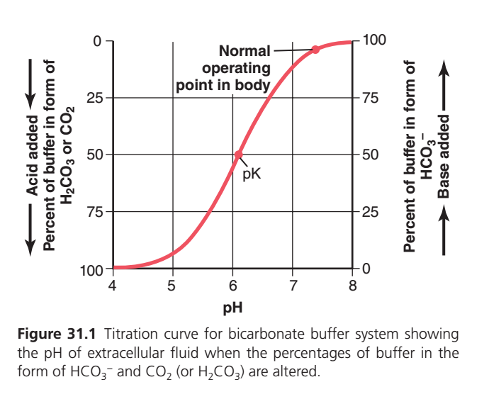
Figure 31.1: Bicarbonate Buffer Titration Curve (SUPERFAST Explanation)This graph explains how the bicarbonate buffer system (HCO₃⁻/H₂CO₃) keeps the blood pH stable when acid or base is added.
Step 1: Understand the Axes
X-Axis (Horizontal)
pH
This shows how acidic or alkaline the blood is.
- Left side (pH 4) = Very acidic
- Middle (pH 6.1) = Equal amounts of HCO₃⁻ and H₂CO₃
- Normal blood = pH 7.4
- Right side (pH 8) = Very alkaline (basic)
As you move right → pH increases → Blood becomes more alkaline.
Left Y-Axis
Percent of Buffer in the Form of H₂CO₃ (or CO₂)
This tells us:
How much of the bicarbonate buffer exists as acid (H₂CO₃).
Top = 0%
Bottom = 100%
Notice the arrow on the left:
⬇ Acid Added
Meaning:
When acid is added,
more buffer converts into H₂CO₃ (acid form).
Right Y-Axis
Percent of Buffer in the Form of HCO₃⁻
This tells us:
How much buffer exists as bicarbonate (base form).
Bottom = 0%
Top =100%
Arrow on the right:
⬆ Base Added
Meaning:
When base is added,
more buffer converts into HCO₃⁻ (base form).
The Red S-Shaped Curve
This curve is called the titration curve.
It tells us
At each pH, what percentage of buffer is acid (H₂CO₃) and what percentage is base (HCO₃⁻).
Start at pH = 4 (Far Left)
Look at the left end of the curve.
Here:
Left axis = nearly 100%
Right axis = nearly 0%
Meaning
Almost all buffer is H₂CO₃ (acid form).
Very little bicarbonate remains.
Why?
Because blood contains lots of acid.
The buffer has accepted hydrogen ions.
Reaction:
HCO₃⁻ + H⁺ → H₂CO₃
Almost everything becomes H₂CO₃.
Moving from pH 4 → 5
The curve begins rising slowly.
This means
Some H₂CO₃ changes back into HCO₃⁻.
Still,
Most buffer is acid.
pH = 6.1 (Point Marked “pK”)
This is the most important point.
The graph labels it pK.
Here,
Left axis = 50%
Right axis = 50%
Meaning
Exactly half the buffer is
H₂CO₃
and
Half is HCO₃⁻.
So,
HCO₃⁻ = H₂CO₃
This is why,
According to the Henderson–Hasselbalch equation:
When pH = pK, the ratio HCO₃⁻ : H₂CO₃ = 1 : 1.
Why is pK Important?
At pH = pK,
the buffer can
- accept acid
- accept base
equally well.
This is the point of maximum buffering efficiency.
From pH 6.1 → 7
The curve rises steeply.
Now,
More H₂CO₃ changes into HCO₃⁻.
The buffer becomes mostly bicarbonate.
Normal Operating Point (pH ≈ 7.4)
This dot is extremely important.
It represents the normal blood pH.
At this point,
Right axis ≈ 95%
Left axis ≈ 5%
Meaning
About
95% of buffer is HCO₃⁻ (base)
Only
5% is H₂CO₃ (acid).Why Does the Body Stay Here?
Because blood normally contains much more bicarbonate than carbonic acid.
Approximately,
HCO₃⁻ : H₂CO₃ = 20 : 1
This ratio gives
Normal blood pH ≈ 7.4.
From pH 7.4 → 8
The curve becomes flat again.
Almost all buffer is now bicarbonate.
Nearly
100%
is HCO₃⁻.
Very little acid remains.Understanding the Left Arrow (Acid Added)
Suppose acid enters the blood.
Example:
Lactic acid
Hydrochloric acid
Sulfuric acid
Immediately,
Hydrogen ions react with bicarbonate.
HCO₃⁻ + H⁺ → H₂CO₃ → CO₂ + H₂O
Result:
✅ HCO₃⁻ decreases
✅ H₂CO₃ increases
The graph moves
← toward the left.
Blood becomes more acidic.
Understanding the Right Arrow (Base Added)
Suppose a base enters blood.
Example:
NaOH
Extra bicarbonate
The base removes hydrogen ions.
Then,
H₂CO₃ breaks apart.
H₂CO₃ → H⁺ + HCO₃⁻
More bicarbonate is formed.
Result
✅ HCO₃⁻ increases
✅ H₂CO₃ decreases
The graph moves
→ toward the right.
Blood becomes more alkaline.
Why is the Curve S-Shaped?
The curve has three regions, each with a different meaning.
1. Left Flat Region (Low pH)
- Almost all buffer is H₂CO₃.
- There is very little HCO₃⁻ left to neutralize additional acid.
- Buffering capacity against further acid is low.
2. Middle Steep Region (Around pK)
3. Right Flat Region (High pH)
- Almost all buffer is HCO₃⁻.
- Very little H₂CO₃ remains to neutralize additional base.
- Buffering capacity against further base is low.
Easy Story to Remember
Imagine the buffer exists in two forms:
- H₂CO₃ = Acid Team
- HCO₃⁻ = Base Team
At different pH values:
- Low pH (acidic blood): Acid Team is much larger.
- pH 6.1 (pK): Both teams are equal (50% each).
- Normal pH 7.4: Base Team dominates (about 95%), while Acid Team is small (about 5%).
- High pH (alkaline blood): Nearly everyone is on the Base Team.
KEY CONCEPT
- X-axis: Blood pH (acidic → alkaline).
- Left Y-axis: Percentage of buffer present as H₂CO₃/CO₂ (acid form).
- Right Y-axis: Percentage of buffer present as HCO₃⁻ (base form).
- Red S-shaped curve: Shows how the proportions of H₂CO₃ and HCO₃⁻ change with pH.
- pK = 6.1: 50% H₂CO₃ and 50% HCO₃⁻ (1:1 ratio), giving maximum buffering efficiency.
- Normal blood pH = 7.4: About 95% HCO₃⁻ and 5% H₂CO₃, corresponding to the physiological 20:1 bicarbonate-to-carbonic acid ratio that keeps blood pH normal.
Bicarbonate Buffer System Is the Most Important Extracellular Buffer
- (Figure 31.1) shows the titration curve of the bicarbonate buffer system.
- From Figure 31.1, the bicarbonate buffer system does not appear to be a powerful buffer at first glance.
- There are two reasons why it seems weak.
Reason 1
- The normal extracellular fluid (ECF) pH is about 7.4.
- The pK of the bicarbonate buffer system is 6.1.
- This means there is about 20 times more bicarbonate (HCO₃⁻) than dissolved CO₂ (or H₂CO₃).
- Equation:
- HCO₃⁻ : CO₂ (or H₂CO₃) = 20 : 1
- Solved Ratio = 20 ÷ 1 = 20
- Therefore, HCO₃⁻ is 20 times greater than CO₂ (or H₂CO₃).
- Because of this 20:1 ratio, the bicarbonate buffer system works on the part of the titration curve where the slope is low.
- A low slope means the buffering power is poor.
Reason 2
- The concentrations of both components of the bicarbonate buffer system (CO₂ and HCO₃⁻) are not high.
- Therefore, this is another reason why the bicarbonate buffer system appears to be weak.
- Despite these characteristics, the bicarbonate buffer system is the most powerful extracellular buffer in the body.
- This seems like a paradox (an apparent contradiction).
- The main reason is that both components of the buffer system are continuously regulated.
- HCO₃⁻ is regulated by the kidneys.
- CO₂ is regulated by the lungs.
- Because the kidneys regulate HCO₃⁻ and the lungs regulate CO₂, the extracellular fluid pH can be controlled very precisely.
- The kidneys control pH by removing or adding HCO₃⁻.
- The lungs control pH by removing CO₂.
- Together, the kidneys and lungs maintain precise control of extracellular fluid pH.
KEY CONCEPT
- Figure 31.1 shows the titration curve of the bicarbonate buffer system.
- Normal ECF pH = 7.4 and pK = 6.1.
- HCO₃⁻ : CO₂ (or H₂CO₃) = 20 : 1, meaning bicarbonate is 20 times greater than dissolved CO₂.
- The bicarbonate buffer works on the low-slope part of the curve, so it appears to have poor buffering power.
- The concentrations of CO₂ and HCO₃⁻ are not high.
- Despite this, it is the most important extracellular buffer because HCO₃⁻ is regulated by the kidneys and CO₂ is regulated by the lungs.
- The kidneys and lungs together precisely regulate extracellular fluid pH.
PHOSPHATE BUFFER SYSTEM
- The phosphate buffer system is not a major extracellular fluid buffer.
- However, it plays an important role in buffering renal tubular fluid and intracellular fluid.
- The two main components of the phosphate buffer system are:
- H₂PO₄⁻ (dihydrogen phosphate)
- HPO₄²⁻ (hydrogen phosphate)
- When a strong acid (HCl) is added, the H⁺ ions are accepted by HPO₄²⁻ (the base form).
- The reaction is: HCl + Na₂HPO₄ → NaH₂PO₄ + NaCl
- Reaction Explanation:
- Strong acid (HCl) is converted into a weak acid (NaH₂PO₄).
- Therefore, the fall in pH is minimized.
- As a result, HCl is replaced by an additional amount of the weak acid NaH₂PO₄.
- Therefore, the decrease in pH becomes much smaller.
- When a strong base (NaOH) is added to the phosphate buffer system, the OH⁻ ions are buffered by H₂PO₄⁻.
- This forms more HPO₄²⁻ and water (H₂O).
- The reaction is: NaOH + NaH₂PO₄ → Na₂HPO₄ + H₂O
- Reaction Explanation:
- Strong base (NaOH) is converted into a weak base (Na₂HPO₄).
- Therefore, the increase in pH is only slight.
- The phosphate buffer system has a pK of 6.8.
- This pK is close to the normal body fluid pH of 7.4.
- Therefore, the phosphate buffer system works near its maximum buffering power.
- However, the concentration of phosphate in the extracellular fluid is low.
- It is only about 8% of the concentration of the bicarbonate buffer.
- Mathematical Calculation:
- Phosphate buffer concentration = 8% of bicarbonate buffer
- 8 ÷ 100 = 0.08
- Therefore, the phosphate buffer concentration is 0.08 times (8%) that of the bicarbonate buffer.
- Therefore, the total buffering power of the phosphate buffer in extracellular fluid is much less than that of the bicarbonate buffer system.
- In contrast, the phosphate buffer is especially important in the renal tubular fluid.
- There are two reasons for this.
Reason 1
- Phosphate becomes greatly concentrated in the renal tubules.
- Therefore, the buffering power of the phosphate buffer system increases.
Reason 2
- The tubular fluid usually has a much lower pH than the extracellular fluid.
- Therefore, the working pH of the buffer becomes closer to its pK (6.8).
- As a result, the phosphate buffer works more effectively in the renal tubules.
- The phosphate buffer system is also important inside cells (intracellular fluid).
- The concentration of phosphate inside cells is many times higher than in the extracellular fluid.
- The pH of intracellular fluid is lower than the pH of extracellular fluid.
- Therefore, the intracellular fluid pH is usually closer to the pK (6.8) of the phosphate buffer system.
- As a result, the phosphate buffer system is more effective inside cells than in the extracellular fluid.
KEY CONCEPT
- The phosphate buffer system mainly buffers renal tubular fluid and intracellular fluid.
- Its two components are H₂PO₄⁻ and HPO₄²⁻.
- Strong acid: HCl + Na₂HPO₄ → NaH₂PO₄ + NaCl, converting a strong acid into a weak acid.
- Strong base: NaOH + NaH₂PO₄ → Na₂HPO₄ + H₂O, converting a strong base into a weak base.
- The pK of the phosphate buffer system is 6.8, which is close to the normal body fluid pH.
- The phosphate buffer concentration in extracellular fluid is 8% (0.08 times) that of the bicarbonate buffer.
- Therefore, it has less buffering power in extracellular fluid.
- It is more effective in renal tubules and intracellular fluid because phosphate concentration is higher and the pH is closer to its pK (6.8).
MADE BY SELF LEARNING CEO AND FOUNDER DR SHEEN
