


Learning Objectives
After studying this topic, students will be able to:
- Identify the main divisions of the nervous system.
- Describe the basic structure of the central and peripheral nervous systems.
- Explain the organization and functions of cranial and spinal nerves.
- Differentiate between the somatic and autonomic nervous systems.
- Compare the sympathetic and parasympathetic divisions.
- Describe the role of visceral afferent nerves.
Introduction
- The nervous system is the body’s main control and coordination system.
- It works together with the endocrine system to control the activities of different body parts.
- It is divided into two main parts:
- Central Nervous System (CNS): Consists of the brain and spinal cord.
- Peripheral Nervous System (PNS): Consists of cranial nerves, spinal nerves, and their ganglia.
- Functionally, the nervous system has two divisions:
- Somatic Nervous System: Mainly controls voluntary activities of skeletal muscles.
- Autonomic Nervous System (ANS): Mainly controls involuntary activities of smooth muscle, cardiac muscle, and glands.
- The ANS has two main divisions:
- Sympathetic Division: Prepares the body for emergencies.
- Parasympathetic Division: Helps the body recover.
- Together, these systems control, coordinate, and regulate body activities to maintain normal body function.
- The nervous system and endocrine system work together to control and coordinate the activities of different body parts.
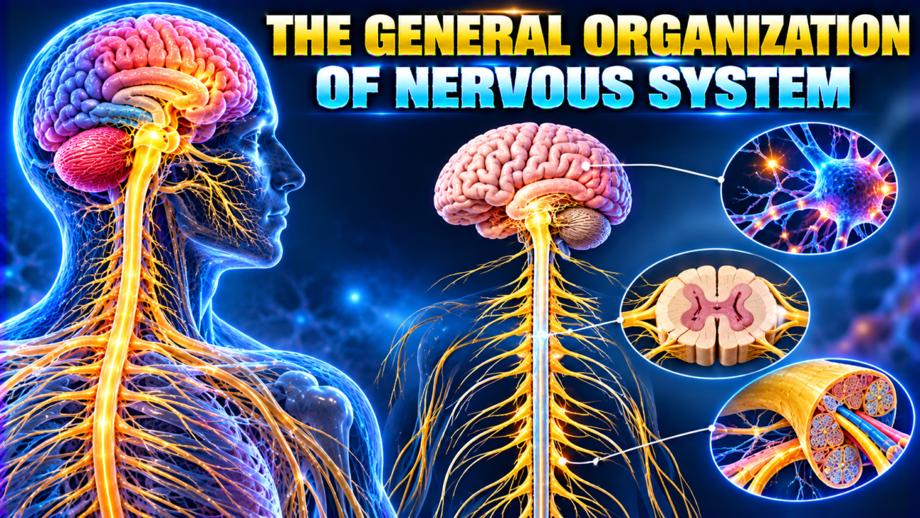
- The nervous system is divided into two main parts (Fig. 1.18):
- Central Nervous System (CNS): Consists of the brain and spinal cord.
- Peripheral Nervous System (PNS): Consists of paired cranial nerves, spinal nerves, and their associated ganglia.
- Functionally, the nervous system is divided into two parts:
- Somatic Nervous System:
- Controls the body’s interaction with the external environment.
- Mainly acts through skeletal muscles.
- Mostly produces voluntary responses to consciously felt sensations from the body wall and limbs.
- Autonomic Nervous System (ANS):
- Controls the body’s internal environment.
- Mainly acts through smooth muscle, cardiac muscle, and glands.
- Mostly produces involuntary responses to sensory signals that are not consciously felt.
- Somatic Nervous System:
CENTRAL NERVOUS SYSTEM
- The CNS contains many nerve cells and their processes.
- These cells are supported by specialized tissue called neuroglia.
- A neuron is a single nerve cell, including all its processes.
- Each neuron has three main components:
- Cell body: Main part of the neuron.
- Dendrites: Usually short processes that carry nerve impulses toward the cell body.
- Axon: Usually the longest process that carries nerve impulses away from the cell body and dendrites (Fig. 1.19A).
- Groups of neuron cell bodies within the CNS are called nuclei.
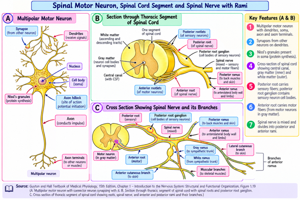
– The CNS contains two types of matter:
- Gray Matter:
- Mainly contains nerve cell bodies supported by neuroglia.
- Appears dull gray because it contains many nonmyelinated cell bodies.
- White Matter:
- Mainly contains axons and blood vessels supported by neuroglia.
- Appears white and shiny because it contains many myelinated nerve fibers.
- In the spinal cord, gray matter forms an H-shaped or butterfly-shaped pattern (Fig. 1.19B,C).
- This gray matter contains:
- Posterior (dorsal) gray horns: Extend along the spinal cord.
- Anterior (ventral) gray horns: Also extend along the spinal cord.
- Lateral gray horns: Present in the thoracic and upper lumbar regions.
- A central canal containing cerebrospinal fluid runs along the central part of the spinal cord.
- The entire CNS is surrounded by three protective membranes called meninges:
- Dura mater: Outermost membrane.
- Arachnoid mater: Middle membrane.
- Pia mater: Innermost membrane.
- The meninges:
- Protect the brain and spinal cord.
- Hold and stabilize the CNS.
- Form a surrounding sac containing cerebrospinal fluid.
PERIPHERAL NERVOUS SYSTEM
- The PNS consists of:
- Cranial nerves.
- Spinal nerves.
- Their associated ganglia.
- A ganglion is a group of neuron cell bodies located outside the CNS.
- Cranial and spinal nerves appear as grayish-white cords during dissection.
- These nerves consist of bundles of axons supported by delicate areolar connective tissue.
CRANIAL NERVES
- There are 12 pairs of cranial nerves.
- They arise from the brain and upper spinal cord and pass through openings in the skull called foramina.
- Most cranial nerves supply structures in the head and neck.
- The exception is cranial nerve X (vagus nerve), which also supplies structures in the thorax and abdomen.
- Cranial nerves are described in Chapter 12.
SPINAL NERVES
- There are 31 pairs of spinal nerves.
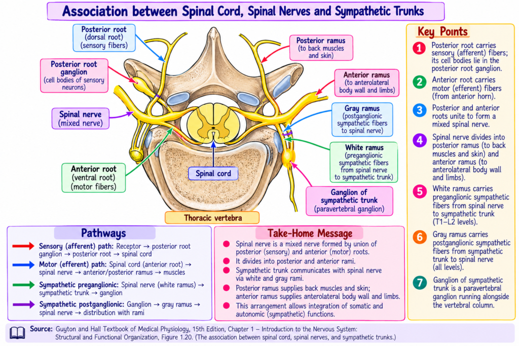
- They leave the spinal cord through openings between vertebrae called intervertebral foramina (Fig. 1.20; Fig. 1.18).
– Spinal nerves are named according to their vertebral regions:
- 8 cervical pairs
- 12 thoracic pairs
- 5 lumbar pairs
- 5 sacral pairs
- 1 coccygeal pair
– Each spinal nerve begins as two groups of small nerve fibers called rootlets:
- Anterior rootlets: Connected to the spinal cord near the anterior gray horn.
- Posterior rootlets: Connected near the posterior gray horn (Figs. 1.19 and 1.20).
- The rootlets join to form:
- An anterior root.
- A posterior root.
- The anterior and posterior roots unite within the intervertebral foramen to form a spinal nerve.
- Each posterior root contains a posterior root ganglion, located near the intervertebral foramen.
- After passing through the foramen, each spinal nerve divides into:
- Smaller posterior ramus
- Larger anterior ramus
– Anterior rootlets and roots (Motor/Efferent Fibers):
- Carry nerve impulses away from the CNS (Fig. 1.21; Fig. 1.19).
- Somatic motor neuron cell bodies are located in the anterior gray horn.
- These neurons supply skeletal muscles and cause contraction.
- Autonomic motor neuron cell bodies are located in the lateral gray horn.
- These neurons supply smooth muscle, cardiac muscle, and glands, causing contraction or secretion.
– Posterior rootlets and roots (Sensory/Afferent Fibers):
- Carry sensory impulses toward the CNS (Figs. 1.19 and 1.21).
- Carry information about touch, pain, temperature, and other sensations.
- The cell bodies of these sensory neurons are located in the posterior root ganglia.
– Mixed Spinal Nerves:
- Spinal nerves and their anterior and posterior rami contain both motor and sensory fibers.
- Therefore, damage to roots and damage to rami cause different problems.
- Anterior root injury: Affects motor fibers only.
- Anterior ramus injury: Affects both motor and sensory fibers.
– Anterior Rami:
- Pass forward and sideways.
- Supply muscles and skin of the anterolateral body wall.
- Also supply muscles and skin of the limbs.
– Posterior Rami:
- Pass backward around the vertebral column.
- Supply muscles and skin of the back.
– Other Branches of Spinal Nerves:
- Meningeal branch: Supplies vertebrae and spinal meninges.
- Rami communicantes: Found in thoracic and upper lumbar spinal nerves and are associated with the sympathetic nervous system.
– The anterior rami follow two main distribution patterns (Fig. 1.18):
1. Simple Segmentation
- Each anterior ramus remains separate from neighboring rami.
- Each nerve passes around the trunk independently.
- Example: Intercostal nerves.
- Each peripheral nerve remains identifiable by its spinal nerve of origin.
- Example: Intercostal nerve T5 comes from spinal nerve T5.
- Neighboring anterior rami may supply overlapping areas.
2. Plexuses
- Several nearby anterior rami join together.
- Their nerve fibers mix and form networks called nerve plexuses.
- Peripheral nerves arising from a plexus contain fibers from multiple spinal nerves.
- Therefore, these peripheral nerves cannot be identified as branches of only one spinal nerve.
- Example: The median nerve contains fibers from spinal nerves C5–T1.
- Major plexuses include:
- Cervical plexus: Neck.
- Brachial plexus: Root of the upper limb.
- Lumbosacral plexus: Root of the lower limb.
- The separation of the nervous system into CNS and PNS is mainly for easier description.
- In reality, nerve processes pass continuously between the CNS and PNS.
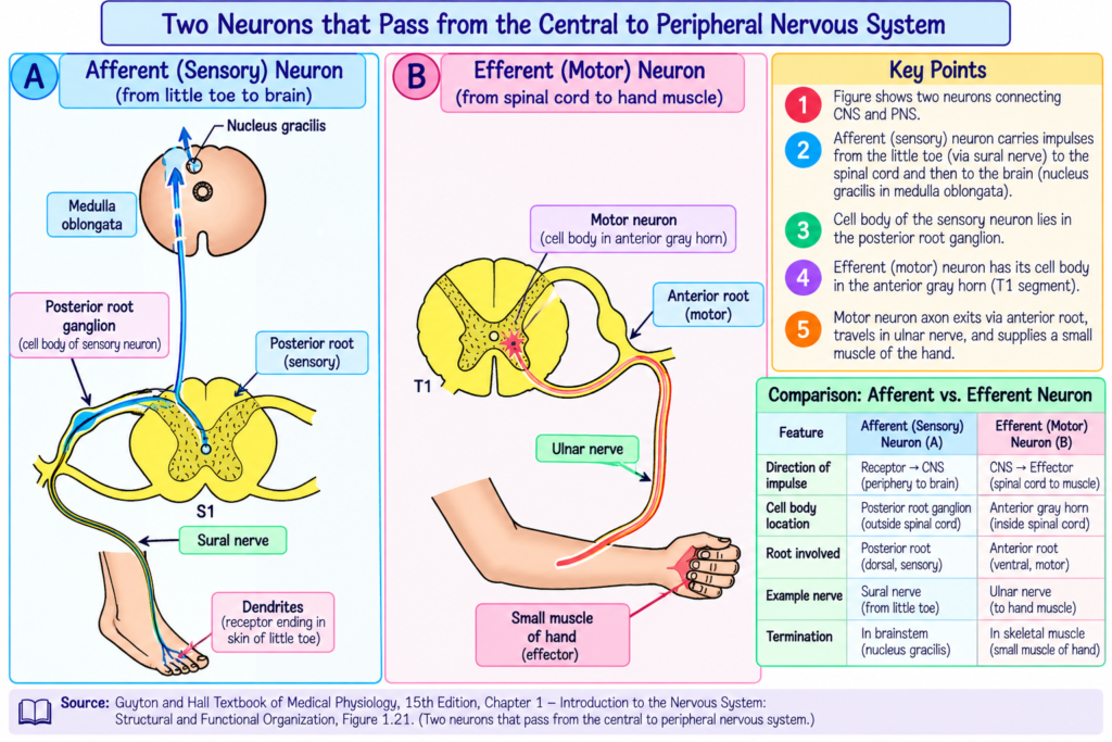
– Example 1: Motor Neuron
- A motor neuron begins in the anterior gray horn at spinal cord level T1.
- Its axon leaves through the anterior root of the first thoracic nerve (Fig. 1.21).
- It then passes through the brachial plexus.
- The axon travels through the arm and forearm in the ulnar nerve.
- Finally, it reaches the motor end plates of a small hand muscle.
- The total length may be about 90 cm (3 ft).
– Example 2: Sensory Neuron
- Touch sensation from the lateral side of the little toe is carried by a sensory neuron associated with spinal segment S1.
- Sensory fibers begin at receptors in the skin.
- The axon travels upward through:
- Sural nerve.
- Tibial nerve.
- Sciatic nerve.
- Lumbosacral plexus.
- It then passes through the posterior root of the first sacral nerve.
- Its cell body is located in the S1 posterior root ganglion.
- The central axon enters the posterior white column of the spinal cord.
- It travels upward to the nucleus gracilis in the medulla oblongata.
- The total distance may be about 1.5 m (5 ft).
- Thus, a single neuron may extend from the little toe to inside the skull.
– Both examples show that a single neuron can be extremely long.
AUTONOMIC NERVOUS SYSTEM
- The autonomic nervous system (ANS) controls the motor activities of:
- Smooth muscle.
- Cardiac muscle.
- Glands.
- The hypothalamus of the brain controls the ANS.
- It coordinates autonomic and neuroendocrine activities to maintain homeostasis.
- The ANS is distributed throughout the central and peripheral nervous systems.
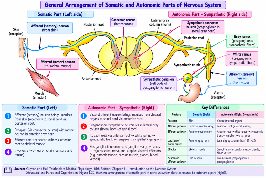
- The main differences between the somatic and autonomic nervous systems are summarized in Table 1.5 and Fig. 1.22.
– Main Anatomical Difference:
- Somatic Nervous System: Uses a one-neuron pathway from the CNS to skeletal muscle.
- Autonomic Nervous System: Uses a two-neuron pathway to reach its target organs.
- The ANS has two main divisions:
- Sympathetic Division: Prepares the body for emergency situations.
- Parasympathetic Division: Helps the body recover.
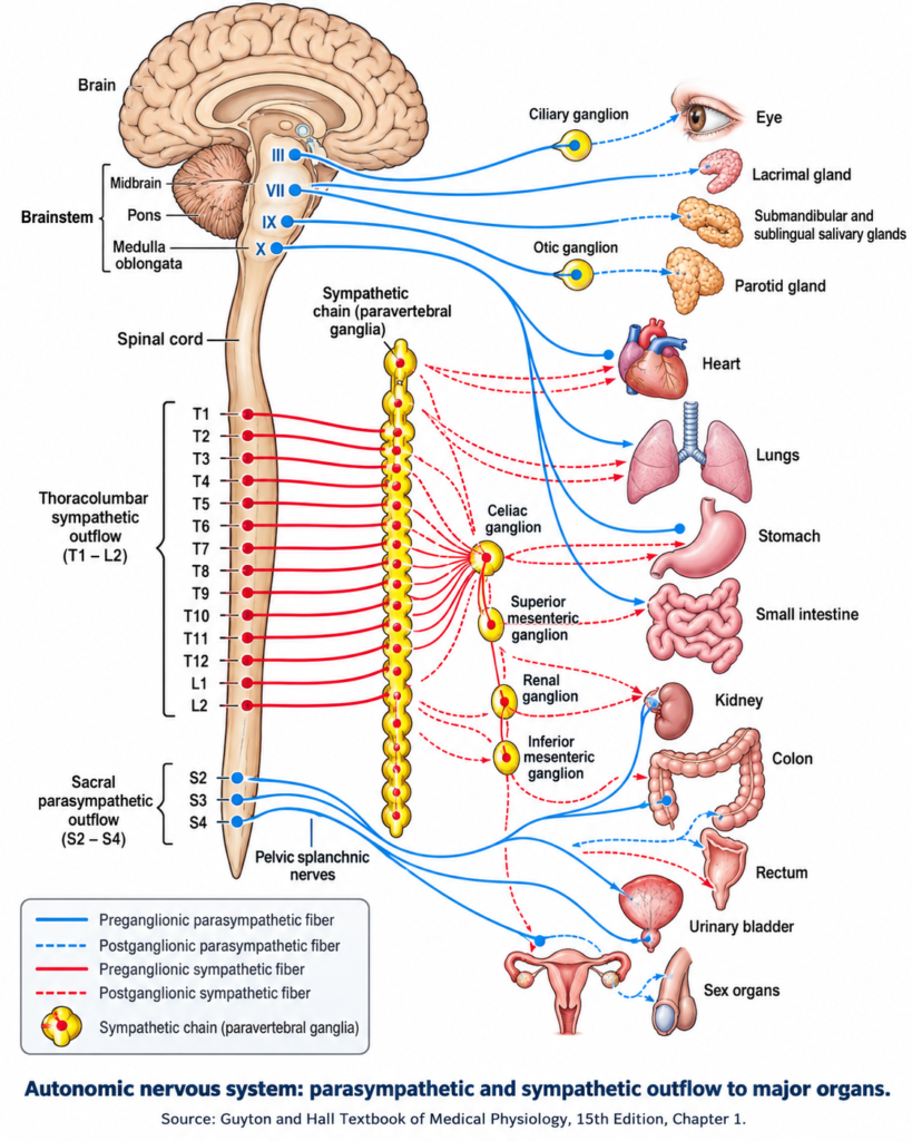
- Their general structure and functions are shown in Fig. 1.23 and Table 1.6.
– The sympathetic and parasympathetic divisions often produce opposite effects.
| Organ or activity | Sympathetic | Parasympathetic |
| Heart rate | Increases | Decreases |
| Bronchi | Dilate | Constrict |
| Intestinal peristalsis | Decreases | Increases |
| Sphincters | Close | Open |
| Bladder wall | Relaxes | Contracts |
| Pupils | Dilate | Constrict |
- Sometimes both divisions work together.
- Example: During normal sexual function:
- Parasympathetic activity: Produces erection.
- Sympathetic activity: Produces ejaculation.
- Sometimes one division works independently.
- Example:
- Sympathetic stimulation activates sweat glands.
- Parasympathetic nerves do not control sweat gland activity.
SYMPATHETIC DIVISION
– The sympathetic division supplies all parts of the body through a complex but organized nerve network.
– Origin of Sympathetic Fibers:
- Preganglionic neuron cell bodies are located in the lateral gray horn of spinal cord segments T1–L2 (Fig. 1.22).
- Their fibers leave through the anterior roots and spinal nerves at these levels.
- Therefore, the sympathetic division is traditionally called the thoracolumbar outflow.
– Sympathetic Trunks:
- Myelinated preganglionic fibers leave the spinal nerves through white rami communicantes.
- They enter the sympathetic trunks (Figs. 1.22 and 1.23).
- Two sympathetic trunks lie along the vertebral column, one on each side.
- Each trunk contains a chain of connected ganglia.
- These ganglia are called paravertebral ganglia because they lie beside the vertebral column.
– Distribution of Sympathetic Ganglia:
- The sympathetic trunks extend above and below T1–L2, along the vertebral column and sacrum (Fig. 1.23).
- They usually contain:
- 11–12 thoracic ganglia
- 3 cervical ganglia, formed by fusion of smaller embryonic ganglia.
- Variable numbers of lumbar and sacral ganglia, which may also fuse.
- At the lower end, a single midline coccygeal ganglion is present.
- It is called the ganglion impar.
- It forms from the fusion of two embryonic coccygeal ganglia.
– Sympathetic Pathways:
- Sympathetic fibers follow four main pathways to four target regions (Table 1.7; Fig. 1.23).
- Preganglionic fibers from T1–L2 enter the sympathetic chains.
- They synapse with postganglionic neurons in ganglia associated with their pathways.
- Synapses may occur in chain ganglia along the length of the sympathetic trunk.
- Postganglionic fibers leave the ganglia.
- They then follow branches of spinal nerves to reach the body wall and limbs.
- Further details of sympathetic distribution are described in later chapters.
PARASYMPATHETIC DIVISION
- The parasympathetic division has a more limited and less complex distribution than the sympathetic division.
- It mainly supplies:
- Head.
- Internal organs within body cavities.
- External genitalia.
- The body wall and limbs do not receive parasympathetic innervation.
– Origin of Parasympathetic Fibers:
- Preganglionic neurons arise from the brain and sacral spinal cord (Fig. 1.23).
– Cranial Origin:
- Parasympathetic neurons arise from nuclei associated with four cranial nerves:
- CN III: Oculomotor nerve.
- CN VII: Facial nerve.
- CN IX: Glossopharyngeal nerve.
- CN X: Vagus nerve.
- Their axons leave the brain through the corresponding cranial nerves.
– Sacral Origin:
- Preganglionic neurons arise from the lateral gray matter of spinal segments S2–S4.
- These segments do not contain a distinct lateral gray horn because the number of neurons is relatively small.
- Myelinated axons leave through the anterior roots of S2–S4.
- They then leave the spinal nerves to form the pelvic splanchnic nerves.
- Therefore, the parasympathetic division is traditionally called the craniosacral outflow.
– Parasympathetic Ganglia and Distribution:
- Preganglionic neurons synapse with postganglionic neurons in peripheral ganglia.
- These ganglia are usually located near the organs they supply.
– CN III, VII, and IX:
- Preganglionic fibers travel to autonomic ganglia in the head.
- Their distribution is limited to the head.
– CN X (Vagus Nerve):
- Preganglionic fibers have a wide distribution.
- They reach ganglia near internal organs in the neck, thorax, and much of the abdomen.
– Pelvic Splanchnic Nerves:
- Preganglionic fibers reach ganglia in the hypogastric plexuses.
- They also reach ganglia in the walls of lower abdominal and pelvic organs.
– Postganglionic Fibers:
- Are usually nonmyelinated.
- Are relatively short compared with sympathetic postganglionic fibers.
- Further details are described in later chapters.
– Recent View of Sacral Autonomic Outflow:
- Traditionally, sacral autonomic fibers have been classified as parasympathetic.
- However, recent studies discussed in the provided text suggest that these fibers may anatomically belong to the sympathetic nervous system.
- These fibers show a cholinergic phenotype, similar to sympathetic fibers supplying sweat glands.
- According to this proposed classification:
- Sympathetic outflow: Spinal outflow, T1–L2 and S2–S4.
- Parasympathetic outflow: Cranial outflow through CN III, VII, IX, and X.
- This proposed change is based on neurophysiological features and does not change the basic anatomy described above.
VISCERAL AFFERENTS
- The ANS is mainly a motor system.
- However, sensory neurons also travel closely with autonomic nerve fibers.
- These sensory neurons are called visceral afferents.
- They carry information from the body’s internal environment.
– Functions of Visceral Afferents:
- Carry sensory information for visceral reflexes.
- Help regulate blood pressure and blood chemistry.
- Help control:
- Heart rate.
- Respiratory rate.
- Blood vessel resistance.
– Important Visceral Sensory Receptors:
- Chemoreceptors
- Baroreceptors
- Osmoreceptors
– Normal Visceral Sensations:
- Normal intestinal movement and distension usually do not produce conscious sensations.
– Abnormal Visceral Sensations:
- Extreme stretching.
- Chemical irritation.
- Ischemia.
- These conditions may produce conscious sensations of pain or nausea.
– Pathways of Visceral Afferent Fibers:
- Myelinated visceral afferent fibers from thoracic, abdominal, and pelvic organs travel with sympathetic and parasympathetic nerves.
- They pass through autonomic plexuses and ganglia without forming synapses.
– Visceral Afferents Traveling with Sympathetic Nerves:
- Many visceral sensory fibers enter the sympathetic trunks.
- They then pass through the white rami communicantes into spinal nerves.
- From there, they enter the posterior roots of spinal nerves.
- Finally, they reach the spinal cord (Fig. 1.22).
- Their cell bodies are located in the posterior root ganglia.
– Visceral Afferents Traveling with the Vagus Nerve:
- Other visceral sensory fibers travel through the vagus nerve.
- They enter the brainstem.
- Their cell bodies are located in the vagal sensory ganglia at the base of the skull.
HIGH-YIELD POINTS – NERVOUS SYSTEM
1. Nervous System: Works with the endocrine system to control and coordinate body activities.
2. Two Main Divisions:
- CNS: Brain and spinal cord.
- PNS: Cranial nerves, spinal nerves, and ganglia.
3. Functional Divisions:
- Somatic Nervous System: Mainly controls voluntary skeletal muscle activities.
- Autonomic Nervous System: Mainly controls involuntary activities of smooth muscle, cardiac muscle, and glands.
4. Neuron: Basic nerve cell consisting of a cell body, dendrites, and an axon.
- Dendrites: Carry impulses toward the cell body.
- Axon: Usually carries impulses away from the cell body.
5. Nucleus vs. Ganglion:
- Nucleus: Collection of neuron cell bodies inside the CNS.
- Ganglion: Collection of neuron cell bodies outside the CNS.
6. Gray Matter: Mainly contains neuron cell bodies supported by neuroglia.
7. White Matter: Mainly contains axons, many of which are myelinated, giving it a white appearance.
8. Spinal Cord Gray Matter: Has an H-shaped or butterfly-shaped arrangement.
9. Three Meninges:
- Dura mater: Outer layer.
- Arachnoid mater: Middle layer.
- Pia mater: Inner layer.
10. Cranial Nerves: There are 12 pairs. The vagus nerve (CN X) also supplies thoracic and abdominal structures.
11. Spinal Nerves: There are 31 pairs:
- 8 cervical
- 12 thoracic
- 5 lumbar
- 5 sacral
- 1 coccygeal
12. Anterior Root = Motor (Efferent): Carries impulses away from the CNS.
13. Posterior Root = Sensory (Afferent): Carries sensory impulses toward the CNS.
14. Anterior Gray Horn: Contains cell bodies of somatic motor neurons supplying skeletal muscles.
15. Lateral Gray Horn: Contains autonomic motor neuron cell bodies in the thoracic and upper lumbar spinal cord.
16. Posterior Root Ganglion: Contains cell bodies of sensory neurons.
17. Spinal Nerves Are Mixed Nerves: They contain both motor and sensory fibers.
18. Nerve Injury:
- Anterior root lesion: Affects motor fibers only.
- Anterior ramus lesion: Affects both motor and sensory fibers.
19. Anterior Rami: Supply the anterolateral body wall and limbs.
20. Posterior Rami: Supply the muscles and skin of the back.
21. Major Nerve Plexuses:
- Cervical plexus — Neck.
- Brachial plexus — Root of upper limb.
- Lumbosacral plexus — Root of lower limb.
22. Somatic vs. Autonomic Motor Pathway:
- Somatic: One-neuron pathway.
- Autonomic: Two-neuron pathway.
23. Hypothalamus: Controls the ANS and coordinates autonomic and neuroendocrine functions to maintain homeostasis.
24. Sympathetic Division:
- Traditionally called thoracolumbar outflow (T1–L2).
- Prepares the body for emergencies.
- Preganglionic neuron cell bodies are located in the lateral gray horn.
25. Parasympathetic Division:
- Traditionally called craniosacral outflow.
- Arises from CN III, VII, IX, X and spinal segments S2–S4.
- Helps the body recover.
26. Sympathetic vs. Parasympathetic Effects:
| Sympathetic | Parasympathetic |
| Increases heart rate | Decreases heart rate |
| Dilates bronchi | Constricts bronchi |
| Decreases gut peristalsis | Increases gut peristalsis |
| Relaxes bladder wall | Contracts bladder wall |
| Dilates pupils | Constricts pupils |
27. Sympathetic Trunks: Two chains of connected paravertebral ganglia lie along the vertebral column.
28. White Rami Communicantes: Carry myelinated sympathetic preganglionic fibers from spinal nerves into sympathetic trunks.
29. Parasympathetic Ganglia: Usually lie close to the organs they supply. Their postganglionic fibers are relatively short and nonmyelinated.
30. Important Autonomic Actions:
- Parasympathetic activity produces erection.
- Sympathetic activity produces ejaculation.
- Sympathetic activity stimulates sweat gland secretion.
31. Sacral Autonomic Outflow: Traditionally classified as parasympathetic (S2–S4). The provided text also discusses a proposed sympathetic classification based on newer research.
32. Visceral Afferents: Carry sensory information from internal organs.
33. Important Visceral Sensory Receptors:
- Chemoreceptors.
- Baroreceptors.
- Osmoreceptors.
34. Visceral Sensations:
- Normal gut movements usually do not produce conscious sensations.
- Extreme stretching, chemical irritation, and ischemia may produce pain or nausea.
35. Visceral Sensory Pathways:
- Sensory fibers traveling with sympathetic nerves may reach the spinal cord through posterior roots.
- Their cell bodies lie in posterior root ganglia.
- Other visceral sensory fibers travel through the vagus nerve to the brainstem, with cell bodies in vagal sensory ganglia.
Most Important Concept: The nervous system receives sensory information, coordinates body activities, and sends motor signals. The somatic system mainly controls skeletal muscles, while the autonomic system regulates internal organs to maintain homeostasis.
